
Metarhabditis andrassyana Tahseen et al., 2004
|
publication ID |
https://doi.org/ 10.1080/00222933.2013.798702 |
|
DOI |
https://doi.org/10.5281/zenodo.5197899 |
|
persistent identifier |
https://treatment.plazi.org/id/038A87BE-FFEB-B856-E5F0-7CE8E915FB3B |
|
treatment provided by |
Felipe |
|
scientific name |
Metarhabditis andrassyana Tahseen et al., 2004 |
| status |
|
Metarhabditis andrassyana Tahseen et al., 2004
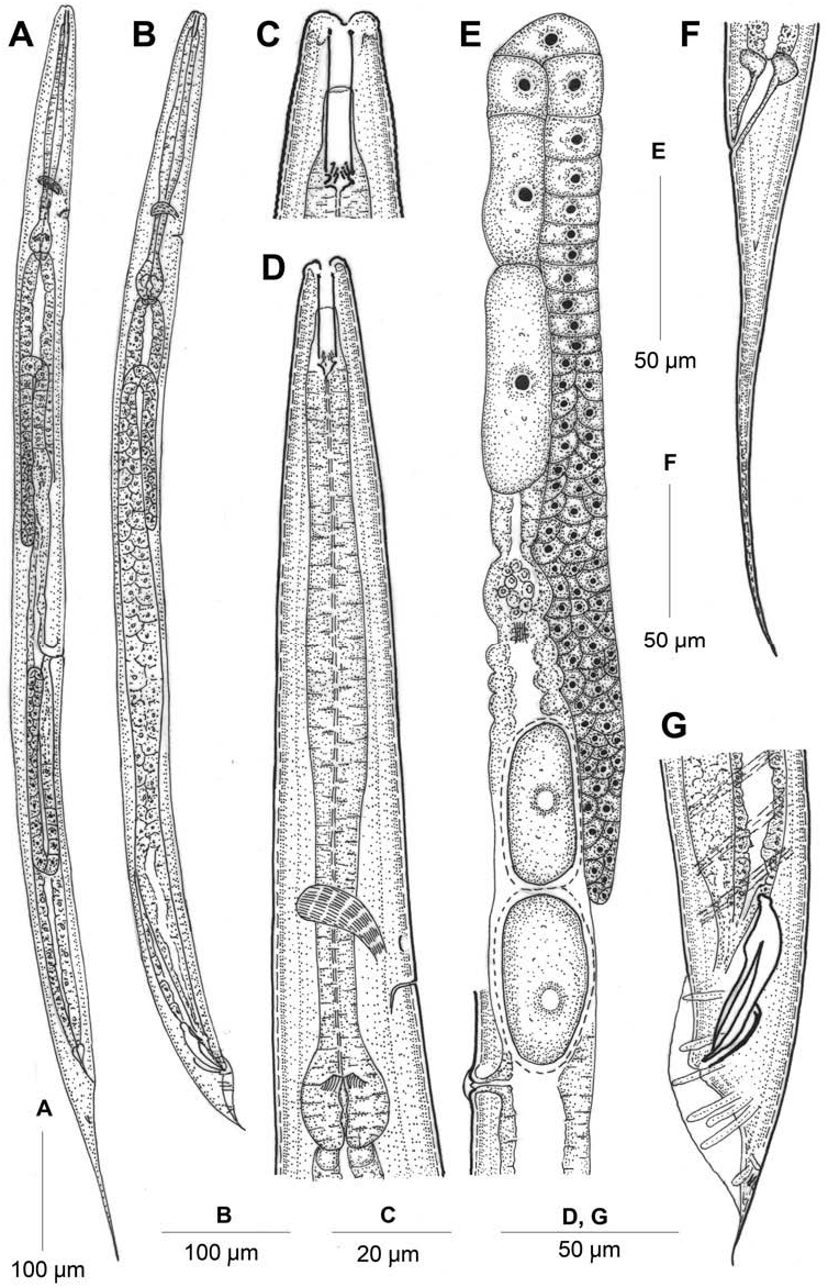
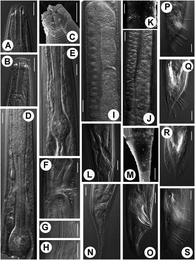
( Figures 3 View Figure 3 and 4 View Figure 4 )
Measurements
See Table 1.
Description
Female. Body straight or slightly arcuate, tapering at both extremities, more towards posterior end. Cuticle with both outer and inner layers striated. Longitudinal striae moderately to faintly developed. Lateral lines three in few specimens splitting into five to six in posterior body region. Lip region almost continuous to adjoining body. Lips round, in doublets forming three sectors (one subdorsal and two subventral). Amphids inconspicuous. Stoma rhabditoid type, long, narrow, 1.8–3.0 times the lip diameter in length. Cheilostom conspicuous, moderately cuticularized; gymnostom smaller than stegostom. Pharyngeal collar surrounding 55–60% of stoma. Metastegostom isomorphic and isotopic with each plate bearing four to six denticles having knobbed heads. Telostegostom isoglottoid. Pharynx differentiated into 120–143 µm long, cylindrical, weakly developed corpus, slightly narrower, 59–70 µm long isthmus and a round, basal bulb of 28–32 × 23–26 µm dimension with a prominent grinder and faintly double-chambered haustrulum. Nerve ring at 66–78% of pharyngeal length. Secretory–excretory pore slightly anterior to basal bulb or 75–81% of pharyngeal length with distally cuticularized duct. Hemizonid usually observed anterior to secretory–excretory pore. Pharyngeal corpus ca.1.3–1.7 times longer than isthmus and basal bulb together. Body at pharyngeal end 2.9–5.4 times the labial diameter in length. Cardial flaps conoid, 5–7 µm long. Intestine with refractive lumen lining. Rectum slightly longer than anal body diameter with dilated lumen; three conspicuous rectal glands present. Prerectum markedly differentiated in few specimens only. Reproductive system didelphic, amphidelphic with well-developed, dorsally reflexed ovaries. Anterior ovary on right and posterior on left side of intestine. Oviduct leading to offset, ovoid spermathecae containing rounded sperm, demarcated from uterus by distinct sphincters. Two to fifty intrauterine eggs observed in different stages of embryonation. Vagina thick-walled, at right angle to longitudinal body axis, about one-third of vulval body diameter in length. A pair of globular pieces associated with vagina reflect muscles in cross-section. Vulva a wide transverse slit, almost extending the full width of the body diameter at the level of the vulva, with an epiptygma and distinct cuticular flap. Tail long, filiform. Phasmids tubular, at base of conical part of tail or 28–36 µm posterior to anal level.
Male. Similar to female in general morphology except in pronounced posterior body curvature. Testis single, reflexed ventrally on left side of intestine. Vas deferens a broad tube, filled with sperm without demarcation of seminal vesicle. Ejaculatory glands absent. Spicules long, robust having round capitula with attenuated ventral and distally notched dorsal arm. Spicule lenght 1.2–1.7 times the anal body diameter. Gubernaculum slightly curved plate with curved proximal end, 40–47% of spicule length. Bursa fairly developed, open leptoderan type, not enclosing small tail spike. Bursal margins often crenate. Tail spicate with fine terminus. Genital papillae eight pairs in 1 + 1/1 + 3 + 2 + P configuration. GP1 , GP2 spaced, precloacal; GP3 shifted posterior to cloaca; out of other six postcloacal pairs – GP4 , GP5 and GP6 closely placed; GP5 , GP7 dorsally oriented; GP8 anterior to fine tubular phasmids. Copulatory muscles six-paired faintly visible bands extending anteriorly to spicules.
Habitat and locality
Moist soil samples collected from a ditch with decaying matter at Sasni (Geographic coordinates 27 ◦ 51 l 42.78 ll N, 78 ◦ 04 l 14.37 ll E), Aligarh, Uttar Pradesh, India.
Voucher material
Eight females and six males on slide Metarhabditis andrassyana NOS /1-7 deposited in the Nematode Collection, Department of Zoology , Aligarh Muslim University, Aligarh, Uttar Pradesh, India . One female and one male on slide Metarhabditis andrassyana NOS /8 deposited at the Laboratory of Nematology , Wageningen University and Research Center ( WUR), 6700 ES Wageningen, the Netherlands .
Remarks
The present population conforms to Metarhabditis andrassyana Tahseen et al., 2004 in most morphometric and morphological characteristics. However, some minor differences can be noted: distinct (versus indistinct) lateral fields; faint (versus distinct) longitudinal striae; smaller (versus larger) and four to six (versus seven to eight) metastegostomal denticles on each plate; nearly undifferentiated (versus slightly differentiated) corpus; and a relatively posteriorly located GP3 (versus adcloacal position of GP3 reported in Metarhabditis andrassyana Tahseen et al., 2004 ). Another important feature observed is the conspicuously dilated though short rectum uniformly present in all specimens.
No known copyright restrictions apply. See Agosti, D., Egloff, W., 2009. Taxonomic information exchange and copyright: the Plazi approach. BMC Research Notes 2009, 2:53 for further explanation.
|
Kingdom |
|
|
Phylum |
|
|
Class |
|
|
Order |
|
|
Family |
|
|
Genus |