
Metarhabditis amsactae ( Ali et al., 2011 ) Sudhaus, 2011
|
publication ID |
https://doi.org/ 10.1080/00222933.2013.798702 |
|
persistent identifier |
https://treatment.plazi.org/id/038A87BE-FFEE-B84E-E564-78F9EBB8FB28 |
|
treatment provided by |
Felipe |
|
scientific name |
Metarhabditis amsactae ( Ali et al., 2011 ) Sudhaus, 2011 |
| status |
|
Metarhabditis amsactae ( Ali et al., 2011) Sudhaus, 2011
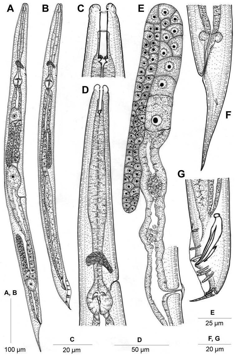
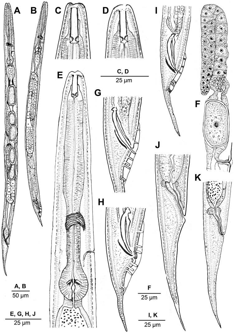
( Figures 5–8 View Figure 5 View Figure 6 View Figure 7 View Figure 8 )
Measurements
See Table 1.
Description
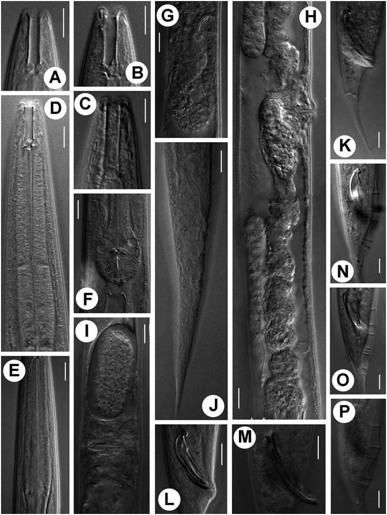
Hermaphrodite female. Body mostly straight rarely arcuate, tapering at both extremities, more towards posterior end. Cuticle with both outer and inner layers striated with striations 1–1.2 µm wide varying with body regions. Longitudinal striations obscure. Six lateral lines in most specimens. Lip region almost continuous to slightly offset from adjoining body. Lips round, arranged in doublets forming three sectors (one subdorsal and two subventral) around the triangular oral aperture; subdorsals slightly more elevated in few specimens. Amphids inconspicuous. Stoma rhabditoid type, ca.1.5–2.5 times the lip diameter in length. Cheilostom inconspicuous; gymnostom cuticularized, smaller than stegostom, ca.30–40% of stoma length. Pharyngeal collar surrounding 50–55% of stoma. Metastegostom isomorphic and isotopic with each plate bearing four to six setose denticles. Telostegostom isoglottoid. Pharynx differentiated into 93–136 µm long, cylindrical to weakly swollen corpus, slightly narrower 35–55 µm long isthmus and a rounded to ovoid, occasionally pyriform basal bulb of 25–35 × 22–26 µm dimension with a weak to moderately developed grinder and faintly double-chambered haustrulum. Nerve ring encircling the anterior half of pharynx, at ca.62–67% of pharyngeal length. Secretory–excretory pore at ca.71–83% of pharyngeal length, variable in position ranging from middle of isthmus to closely anterior to basal bulb. Secretory–excretory duct distally cuticularized for a length of 8–10 µm. Deirids and hemizonid not observed. Pharyngeal corpus ca.1.4–1.6 times longer than postcorpus (isthmus and basal bulb together). Cardia conoid, 6–7 µm long. Intestinal lumen wide, dilated posterior to the basal bulb and in prerectal region often containing bacterial aggregates. Rectum length ca.1.2–1.8 times anal body diameter, associated with rectal glands (obscure in most specimens) at its junction with prerectum. Prerectum distinguishable from intestine in lacking prominent cell nuclei, dilated in few specimens containing undigested bacteria. Reproductive system didelphic, amphidelphic. Ovaries moderately developed, dorsally reflexed; but with distal end not reaching to vulval level, anterior ovary slightly larger, located on right and posterior on left side of intestine. Usually one or two small rounded pseudocoelomocytes observed in close proximity to proximal end of ovaries. Oviduct proximally dilated, connected to offset, ovoid spermatheca containing sperm. Intrauterine eggs 1–19, in different stages of embryonation, dimensions 47–55 × 24–30 µm. Uteri well developed, differentiated into long glandular and muscular regions mostly filled with sperm. Vagina thick-walled, often cuticularized, at right angle to longitudinal body axis, with length equal to about one-third of vulval body diameter. Vulva a wide transverse slit, extending ca.75% of corresponding body diameter with protruding lips, inconspicuous or weak epiptygma but distinct cuticular flap. Tail elongate conoid, gradually tapering to a fine terminus. Phasmids tubular, located about one anal body diameter posterior to anus.
Male. Similar to female in general morphology except for smaller size with pronounced posterior body curvature and relatively fine cuticular striations. Testis single, reflexed ventrally on right side of intestine; length of reflexed part ca.123–210 µm long. Vas deferens a broad tube, filled with sperm without demarcation of seminal vesicle. Ejaculatory glands absent. Spicules long, arcuate, free with distinctly hooked capitula with ventral triangular process, attenuated ventral arm and distally prominent or flanged dorsal arm. Gubernaculum slightly curved plate with tapering and attenuated proximal end, 40–45% of spicule length. Bursa anteriorly open, narrow, leptoderan, not enclosing large tail spike. Bursal margins smooth. Tail conoid with posterior two-thirds sharply narrowing and attenuated. Genital papillae eight pairs in 1 + 1/1 + 3 + 2 + P configuration. GP1, GP2 spaced, precloacal; GP3 slightly posterior to cloaca in most specimens. Postcloacal pairs – GP4, GP5 and GP6 closely placed; GP5, GP7 dorsally directed; GP8 anterior to fine, tubular phasmids. Copulatory muscles bands mostly faint or weakly developed.
Habitat and locality
Population 1: Sampled from infected egg masses of yellow stem borer ( Scirpophaga incertulas ) collected from rice fields at Directorate Rice Research, Hyderabad, Andhra Pradesh, India.
Population 2: Sampled from naturally infected Galleria mellonella larvae collected from deserted honeycomb at Directorate Rice Research building, Hyderabad, Andhra Pradesh, India.
Population 3: Sampled from decaying debris at Shekha Jheel (Geographic coordinates 27 ◦ 51 l 26.14 ll N, 78 ◦ 13 l 13.94 ll E), Aligarh, Uttar Pradesh, India.
Voucher material
Eight females and eight males on slide Metarhabditis amsactae pop.1/1-6; Eight females and eight males on slide Metarhabditis amsactae pop.2/1-5 and nine females and nine males on slide Metarhabditis amsactae pop.3/1-6 deposited in the Nematode Collection, Department of Zoology , Aligarh Muslim University, Aligarh, Uttar Pradesh, India . One female and one male on slide Metarhabditis amsactae pop.3/7 deposited at the Laboratory of Nematology , Wageningen University and Research Centre ( WUR), 6700 ES Wageningen, the Netherlands .
Based on the morphological characteristics of the present collections, a revised diagnosis of M. amsactae is given below.
Remarks
The three populations under study resemble each other considerably besides conforming to M. amsactae in most morphological and morphometric characters. However, the present specimens were collected from different hosts (yellow stem borer and Galleria larvae) or location (decaying debris at Shekha wetland) from that reported for M. amsactae apud Ali et al. 2011 . Some other differences that have been noted can be accounted for as being inter-population variations. The cuticle of populations 1 and 2 has discernible striations whereas population 3 has a relatively smooth cuticle. The cheilostom is relatively noticeable in population 3 compared with the other populations. The stoma is narrower in population 1 along with a pharyngeal corpus that is more cylindrical compared with the other two populations, which possess a slightly swollen corpus. The basal bulb is rounded to ovoid in most individuals with the exception of a few individuals from population 3 where the basal bulb is pyriform. The proximal cuticularized end of the secretory–excretory duct is shorter in population 2 compared with the other two populations. Although hermaphroditism was not reported in the original population of M. amsactae , the present populations had sperm of two sizes in the female genital tracts. Moreover, the subsequent generations in the cultured medium exhibited a drastic decline in the number of males suggesting autofertilization as the dominant reproductive mode. Population 3 had wide, conspicuous bacterial pouches at the anterior and posterior ends of intestine. The maximum number (19) of intrauterine eggs was recorded in population 3 whereas in population 1 the maximum number was 12. Most gravid females of population 2 have only a pair of intrauterine eggs. The spicules in population 1 include rounded to hooked capitula, whereas the spicules of populations 2 and 3 have triangular capitula. Of the three populations the spicules are most slender in population 2. The precloacal pair of GP 1 in populations 1 and 2 is located at the anterior to middle level of spicules whereas it is located posterior to the middle level of spicules in population 3. The rectum was observed to be slightly dilated in specimens of population 2.
The original population of M. amsactae , isolated from the red hairy caterpillar, Amsacta moori from Mungbean field, has been reported to be necromenic. The present populations besides differing in their locality and host also differ in three characters. The separate, unfused lips; 10–11 lateral lines and pharyngeal collar surrounding 30% of stoma as reported in M. amsactae apud Ali et al., 2011 are the characters that need to be verified. However, as per Figure 1C View Figure 1 , the labial contour appears asymmetrical, possibly indicating fusion of lips; the pharyngeal collar seems to be inadequately drawn. Likewise the 10–11 lateral lines in Figure 2E View Figure 2 appear to be an erroneous interpretation as the lateral fields of both sides cannot be viewed at the same focus as shown. Nevertheless, M. amsactae has been found to be a valid species having marked differences with other congeners, which are enumerated below.
Emended diagnosis and relationship
Metarhabditis amsactae is characterized by medium-sized hermaphroditic females with males found in equal proportions in natural populations. The species is characterized by a slightly offset lip region; globular lips in doublets forming three sectors; each metastegostomal plate bearing setose denticles; weakly swollen corpus followed by a tapering isthmus; females with conical tails and males with small spicules with hooked capitula and prominent dorsal and ventral arms; trough-shaped gubernaculum; open leptoderan, reduced bursa, not enclosing tail spike posteriorly; free genital papillae in 1 + 1/1/3 + 2 + P configuration.
It differs from the type species M. andrassyana in having a relatively thick (versus extremely cylindrical) corpus and a proportionately longer tail in the male (c = 8.9–17.8 versus 20–25) and shorter tail in the female (c = 8.7–13.7 versus 6.5–8.1). Further differences have been noticed in the present populations in their relatively slen- der (versus stout) spicules and reduced, narrow (versus well developed) bursa in males not enclosing long (versus short) tail spike; from M. blumi ( Sudhaus, 1974) Sudhaus, 2011 in having relatively smaller individuals (631–1022 µm versus 912–1819 µm) with relatively greater ‘c’ value (8.7–13.7 versus 7.0–9.7) and small eggs (47–54 × 25–30 µm versus 57–78 × 31–44 µm) in females whereas relatively smaller ‘c’ value (8.9–17.8 versus 17.0–27.0) and smaller spicules (25.2–40 µm versus 45–51 µm); from M. rainai ( Carta and Osbrink, 2005) in invading different hosts (hairy caterpillar versus termite) and in having smaller females (631–1022.0 µm versus 884–1748 µm), and males with smaller ‘c’ value (8.9–17.8 versus 20–28), smaller spicules (25.2–40.0 µm versus 36–59 µm) and gubernaculum (10.3–20.0 µm versus 16–29 µm) and a leptoderan (versus peloderan) bursa; from M. costai ( Martins, 1985) Sudhaus, 2011 in inhabiting different host organisms (hairy caterpillar versus cattle) and in having smaller females (631.0–1022.0 µm versus 884–1748 µm) and smaller males (515–868 µm versus 844–1202 µm) with smaller ‘c’ value (8.9–17.8 versus 23.2–34.2), relatively smaller spicules (25.2–40.0 µm versus 33–50 µm) and gubernaculum (10.3–20.0 µm versus 20–24 µm); from M. freitasi ( Martins, 1985) it differs in having different hosts (hairy caterpillar versus cattle) and in having smaller females (631–1022 µm versus 1253–1714 µm) with smaller ‘b’ value (4.0–5.5 versus 6.5–8.5) and smaller males (515–868 µm versus 1035–1405 µm) having smaller ‘b’ (3.9–5.3 versus 6.7–9.1) and ‘c’ (8.9–17.8 versus 23.9–39.5) values, smaller spicules (25.2–40.0 µm versus 43–52 µm) and gubernaculum (10.3–20.0 µm versus 17–22 µm); and from M. adenobia ( Poinar, 1971) in having different hosts (hairy caterpillar versus rhinoceros beetle) besides having smaller females (631–1022 µm versus 1056–1296 µm) and relatively smaller males (515–868 µm versus 768–1248 µm) with smaller ‘c’ value (8.9–17.8 versus 19.2–24.0), smaller spicules (25.2–40 µm versus 40–53 µm) and gubernaculum (10.3–20.0 µm versus 20–26 µm).
The two species namely Oscheius ciceri Shaheen et al., 2011 and Oscheius hussaini Shaheen et al., 2011 described from India show considerable similarities with M. amsactae . Oscheius ciceri resembles the latter in having similar or overlapping ‘b’, ‘c’ and ‘ cl’ values in females and ‘c’, ‘ cl’ in males. Both species further show nearly overlapping values of ‘a’ and ‘b’ in males. The spicules of O. ciceri though stated to be relatively longer do not correspond with Figure 1E–G View Figure 1 , where they fall within the range reported in M. amsactae . In addition, the drawing ( Figure 1E View Figure 1 ) of genital papillae appears to be unusual, with GP3 much more anterior in the cloaca. On the basis of a great degree of similarity despite some inadequacies in figures, instead of placing O. ciceri as species inquirendum, we consider it as a synonym of M. amsactae .
Oscheius hussaini Shaheen et al., 2011 also lacks sound differentiating features from M. amsactae due to similar ‘a’, ‘c’, ‘ cl’ and ‘V’ values in females in addition to overlapping values of ‘c’, ‘ cl’ in males. The table data with some printing lapses as well as the structures in figures not conforming to the table values (length of spicules in Figure 2H, I View Figure 2 ; body width in Figure 2E View Figure 2 ; tail length in Figure 2J View Figure 2 apud Shaheen et al., 2011) further make its valid status doubtful. Seeing the great degree of similarity, the species O. hussaini is considered a junior synonym of M. amsactae . Another recently described species Oscheius gingeri Parvez et al., 2012 with duplication of figures [ Figure 1B, G View Figure 1 and 2F View Figure 2 exactly similar to Figure 1E, H View Figure 1 and 2J View Figure 2 of Metarhabditis amsactae (apud Ali et al., 2011), Figure 1C View Figure 1 exactly similar to Figure 1B View Figure 1 of Oscheius ciceri (apud Shaheen et al., 2011)] is, hereby, synonymized with M. amsactae .


No known copyright restrictions apply. See Agosti, D., Egloff, W., 2009. Taxonomic information exchange and copyright: the Plazi approach. BMC Research Notes 2009, 2:53 for further explanation.
|
Kingdom |
|
|
Phylum |
|
|
Class |
|
|
Order |
|
|
Family |
|
|
Genus |