
Peniophora inflata Burt (1925: 267)
|
publication ID |
https://doi.org/10.11646/phytotaxa.174.1.4 |
|
DOI |
https://doi.org/10.5281/zenodo.5150992 |
|
persistent identifier |
https://treatment.plazi.org/id/038B130D-8907-FFE2-FF42-FF48FC2115B5 |
|
treatment provided by |
Felipe |
|
scientific name |
Peniophora inflata Burt (1925: 267) |
| status |
|
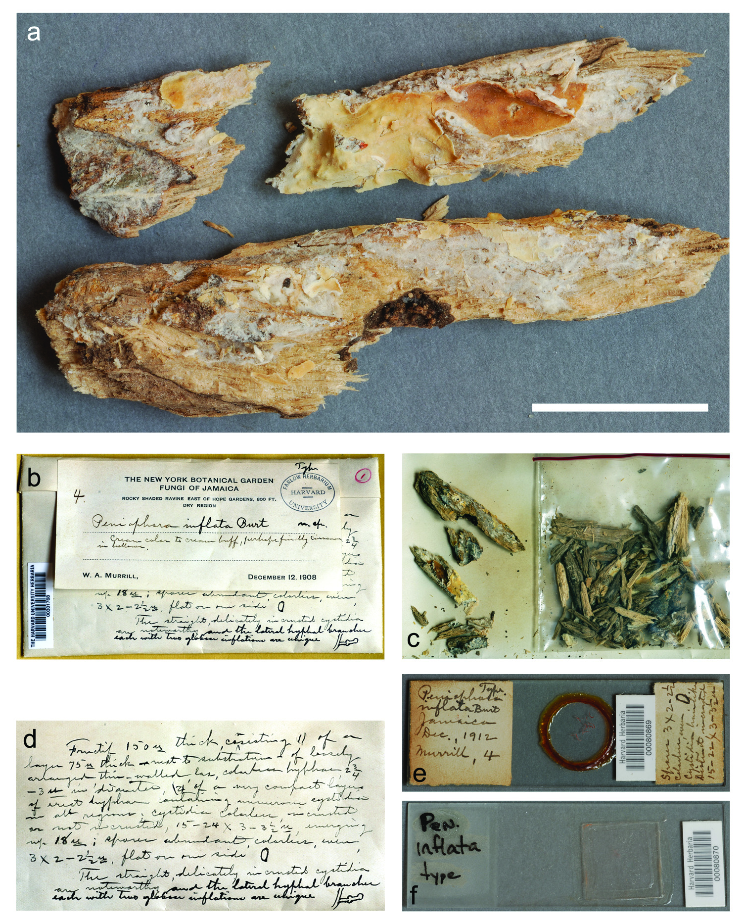
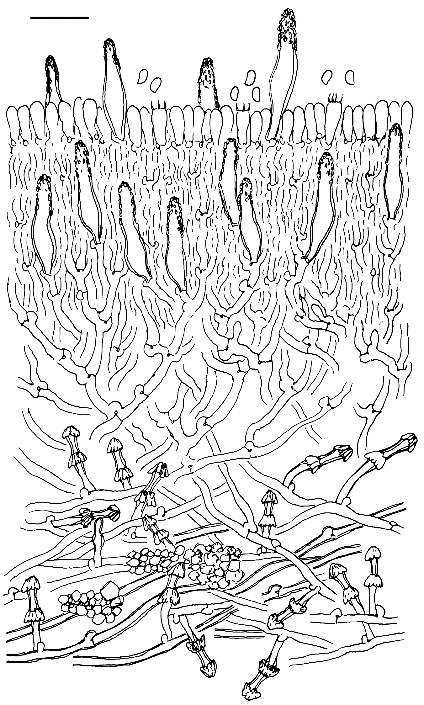
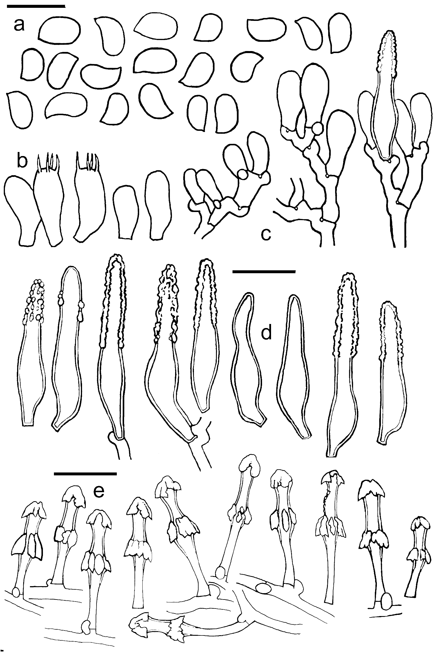
Peniophora inflata Burt (1925: 267) View in CoL Figs. 1–3 View FIGURE 1 View FIGURE 2 View FIGURE 3
Basidiomata annual, resupinate, effuse, pellicular, loosely adnate, fragile and brittle when dry; upon sectioning up to 250 µm thick, with loose, cottony or soft felty and white subiculum, and darker, dense upper layer; hymenial surface almost smooth, with a few indistinct and scattered, low tubercles; cream-buff to pale dirty orange when dry; margin not observed; rhizomorphs present in subiculum, under basidiomata and in wood, radially branched, white. Hyphal system monomitic, both in basidiomata and rhizomorphs; generative hyphae thin to thick-walled, hyaline, all septa with clamp-connections. Subicular layer up to 180 µm thick, consisting of hyphae 1.6–3.6 µm wide, with long cells, thin to thick-walled, with distinct, semicircular clamps, branched, hyaline, loosely arranged and interwoven, smooth or sometimes covered by subhyaline or yellowish (in KOH and CB), rhomboid and irregular crystals of various size forming agglomerations, rarely occurring singly. Subicystidia are borne laterally on hyphae, 16–27 × 1.6–2.4 µm, with basal clamp, with narrow, thin or thick-walled stalk and thick-walled upper part with two inflations, median and terminal, up to 4.4 µm in diameter or 5.6 µm when measured with encrustation, both inflations covered by subhyaline or yellowish crystalline matter, finally umbrella-like or more or less trapezoidal caps; encrustation is almost KOHresistant, dissolving very slowly and only partially in a 5% solution. Subhymenial layer up to 80 µm thick, forming a dense structure of vertically arranged and short-celled, thin-walled hyphae 2.0–3.0 µm wide, mixed with lamprocystidia scattered on all levels of this layer. Rhizomorphs (hyphal cords) present in lower part of the subiculum, under the basidiomata, in the wood, and probably, also on the margin (not observed), 15–80(–150) µm wide, white, branched, consisting of parallel and relatively densely arranged generative hyphae similar to subicular hyphae, with scattered subicystidia and agglomerations of rhomboid crystals. Hymenial and subhymenial lamprocystidia 20–32 × 2.8–4.8 µm, embedded between or projecting above basidia, subventricose or subcylindrical with inflated lower part, slightly tapering toward the obtuse apex, with somewhat short-stalked base, with indistinct basal clamp, thick-walled, with walls up to 1.5 µm, usually apically encrusted, very rarely naked. Basidia 9–13 × 3.8–4.6 µm, short-clavate, sometimes slightly constricted, with 4 sterigmata and basal clamp. Basidiospores 3.2–4.2 × 2–2.4 µm, narrowly ellipsoidal or sometimes almost lacrymoid, often with flattened or slightly concave adaxial side, with indistinct apiculus, smooth, thin-walled, hyaline, inamyloid, nondextrinoid and acyanophilous.
Specimen examined:― JAMAICA. Saint Andrew: rocky shaded ravine east of Hope Gardens , 800 ft. alt., on welldecayed wood remnants, 12 December 1908, W.A. Murrill 4 ( holotype FH!) .
| FH |
Fort Hays |
No known copyright restrictions apply. See Agosti, D., Egloff, W., 2009. Taxonomic information exchange and copyright: the Plazi approach. BMC Research Notes 2009, 2:53 for further explanation.
|
Kingdom |
|
|
Phylum |
|
|
Class |
|
|
Order |
|
|
Family |
|
|
Genus |
Peniophora inflata Burt (1925: 267)
| Karasiński, Dariusz 2014 |
Peniophora inflata
| Burt, E. A. 1925: ) |