
Monticulipora kolaluensis Jaroshinskaja, 1962
|
publication ID |
https://doi.org/10.5252/g2010n2a1 |
|
persistent identifier |
https://treatment.plazi.org/id/038C517D-C70F-F80F-E0EA-D32CFC2DFB57 |
|
treatment provided by |
Marcus |
|
scientific name |
Monticulipora kolaluensis Jaroshinskaja, 1962 |
| status |
|
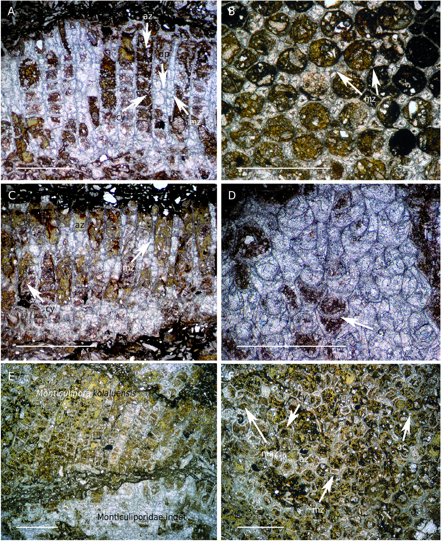
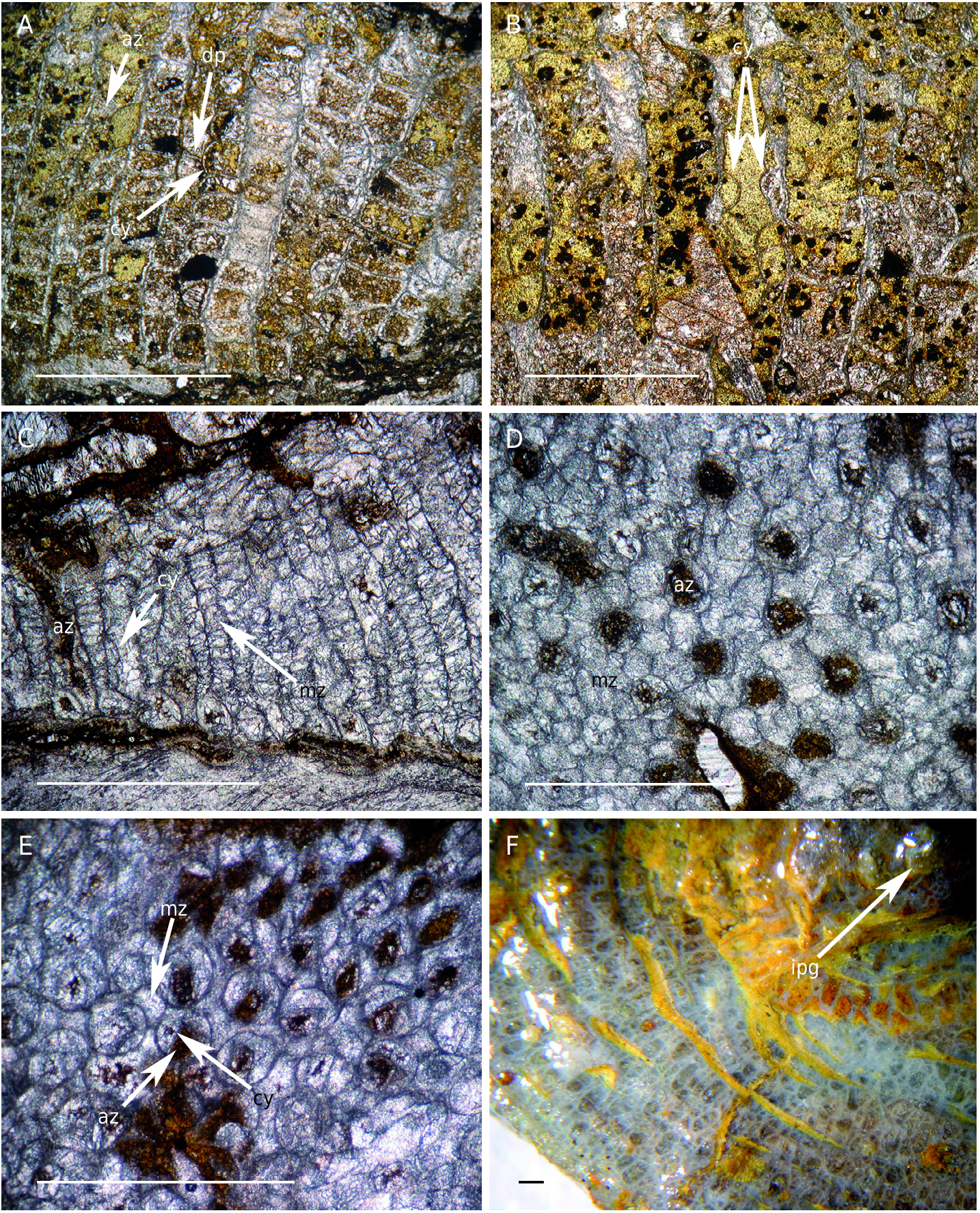
Monticulipora kolaluensis Jaroshinskaja, 1962 View in CoL ( Figs 3E, F View FIG ; 5A, B View FIG ; Table 2)
Monticulipora kolaluensis Jaroshinskaja, 1962: 147 View in CoL , pl. 1, fig. 3, text-fig. 4.
MATERIAL EXAMINED. — One complete zoarium ( MPZ 2006/109).
STRATIGRAPHIC AND GEOGRAPHIC RANGE. — Upper Ordovician of the Altai Mountains, Siberia ( Jaroshinskaja 1962); and in the La Peña Member from the Cystoid Limestone Formation, in the Valdelaparra section (Zaragoza, Spain).
EMENDED DIAGNOSIS. — Hexagonal autozooecial cross sections with one semicircular cystiphragm cross section inside; diaphragms straight and perpendicular to walls in most autozooecia; cystiphragms mainly arranged in single series lining one side of wall; acanthostyles regularly distributed; and walls with laminated microstructure.
DESCRIPTION
General characters
Zoarium massive with free growth habit, composed of two layers of irregular thickness and length, with maximum zoarial height of 4.5 mm, minimum height of 1.5 mm, and maximum apparent diameter of 35.0 mm.
Tangential section
Autozooecial apertures regularly and irregularly hexagonal with two distinct ranges of diameter. Smaller apertures with an average diameter of 0.27 mm, more regular in form than larger ones, and with fewer mesozooecia between them. Larger apertures have an average diameter of 0.44 mm and form the maculae with a group of mesozooecia. These maculae are separated from each other by 3.5 mm on average, measured from center to center. Without differentiating between macular and intermacular areas there are on average 7.5 autozooecia per mm2 and 2.6 per linear mm. All autozooecia have a maximum of 50% of their section occupied by one single circular or subcircular cystiphragm, commonly touching one or two sides of the hexagonal autozooecial aperture. Mesozooecial apertures polygonal and fitting in the spaces between autozooecia. Mesozooecia abundant in maculae, rare in the intermacular area. Mesozooecia vary greatly in size, from 0.09 to 0.22 mm, with an average of 0.14 mm. Without differentiating between macular and intermacular areas there are on average 2.2 mesozooecia per mm2 and one mesozooecium per linear mm. Acanthostyles located in the junction of three autozooecia; on average 4.2 acanthostyles per mm 2, without differentiating between macular and intermacular areas. Acanthostyles average diameter of 0.027 mm. Th e largest acanthostyles have two well differentiated zones: 1) a central one composed of a light-coloured core with massive amorphous, hyaline microstructure; and 2) an outer sheath formed by a series of dark-coloured laminae concentrically arranged around the core. The smallest acanthostyles circular with darker colour than zooecial walls. Autozooecial walls 0.024 mm thick on average, having laminated microstructure. Mesozooecial walls with the same microstructure and thickness as autozooecial ones. Zooecial boundaries not discernible.
Longitudinal section
Zoarium composed of two layers of variable thickness. Overgrowth surface marked by the development of new basal diaphragms. Autozooecia start to growth at an angle of 16° on average (measured only in two autozooecia), sharply bending to intersect the zoarial surface at an angle of 84°. Autozooecia tubular, containing cystiphragms and diaphragms. Cystiphragms generally arranged in a single series on one side of the autozooecial chamber, but can also be seen in both sides of the walls either as a single series or isolated, or as double series in a single side. They occupy from 25 to 50% of the autozooecial tube on average, taking up more space in the proximal part, where they are mainly arranged as a single series in one side of a wall. Most autozooecia lack cystiphragms in their distal part. Th ere is an average of seven complete cystiphragms per mm along the total length of the autozooecia. Diaphragms in the lower part of autozooecia either joining cystiphragms with the opposite wall or spanning the autozooecial tube where cystiphragms are absent; they are straight, perpendicular to the walls and less abundant (mean of 3.3 diaphragms per mm of autozooecial length). Mesozooecia scarce, tubular and smaller than autozooecia. Th ey are tabulated by perpendicular and concave diaphragms, averaging seven diaphragms per mm of length. Some mesozooecia have small cystiphragms attached to one side of the wall that curve and lean on a lower cystiphragm or diaphragm.Acanthostyles commonly recognized as a thickening of the autozooecial wall, but in some cases it is possible to observe the core longitudinally cut. Acanthostyles have variable length, most of them reaching the zoarial surface. In proximal zone autozooecial walls slightly thinner than in distal ones ( 0.024 mm on average). The difference in thickness increases when the wall contains acanthoslyles; in this case the simple laminated microstructure becomes a chevron laminated microstructure. Endozone-exozone boundary marked by disappearance of diaphragms.
REMARKS
The specimen described here has tangential sections identical to those of Monticulipora kolaluensis Jaroshinskaja, 1962 , from the Upper Ordovician of Siberia, displaying regular to irregular hexagonal autozooecial sections with a cystiphragm touching one or two sides of the autozooecial chamber, as well as scarce mesozooecia, mainly concentrated in maculae. Longitudinal sections are similar, with cystiphragms either arranged in series on one or both sides of the autozooecial walls, or isolated.However, the Siberian specimens have more abundant diaphragms in both autozooecia and mesozooecia, an average of 12 and 14 respectively, whereas the Iberian specimens have 3.5 and 7.0 diaphragms, respectively. In spite of this difference the studied material is assigned to Monticulipora kolaluensis because the identified similarities appear to predominate. In her description, Jaroshinskaja (1962) did not mention either the presence of acanthostyles or the wall microstructure, and the diagnosis of the species is here emended to include these characters.
Monticulipora kolaluensis is clearly distinguished from Monticulipora cystiphragmata n. sp. by having thicker walls with laminated microstructure, more regular autozooecial apertures, smaller autozooecial cystiphragms arranged in a single series or isolated on one or both sides of the wall, and less inclined autozooecia in the proximal zone of colony.
Monticulipora kolaluensis View in CoL is not an endemic species as it was thought, having a more extended geographic distribution. Th is new distribution implies a greater mobility for its larvae and a greater capacity for this species to adapt itself to different conditions of temperature and salinity, since the Mediterranean Region had a latitude of about 60°S ( Fortey & Cocks 2002) whereas Siberia was placed in tropical latitudes during the Upper Ordovician ( Fortey & Cocks 2002).
| MPZ |
Museo Paleontologico de la Universidad de Zaragoza |
No known copyright restrictions apply. See Agosti, D., Egloff, W., 2009. Taxonomic information exchange and copyright: the Plazi approach. BMC Research Notes 2009, 2:53 for further explanation.
|
Kingdom |
|
|
Phylum |
|
|
Class |
|
|
Order |
|
|
Family |
|
|
Genus |
Monticulipora kolaluensis Jaroshinskaja, 1962
| Jiménez-Sánchez, Andrea 2010 |
Monticulipora kolaluensis
| JAROSHINSKAJA A. M. 1962: 147 |