
Phoremia rolfsi Pereira, Sperber & Lhano
|
publication ID |
https://doi.org/ 10.5281/zenodo.206914 |
|
DOI |
https://doi.org/10.5281/zenodo.6187523 |
|
persistent identifier |
https://treatment.plazi.org/id/038C8787-FFAB-FF88-FAA3-062A54DB0CE9 |
|
treatment provided by |
Plazi |
|
scientific name |
Phoremia rolfsi Pereira, Sperber & Lhano |
| status |
sp. nov. |
Phoremia rolfsi Pereira, Sperber & Lhano , sp. nov.
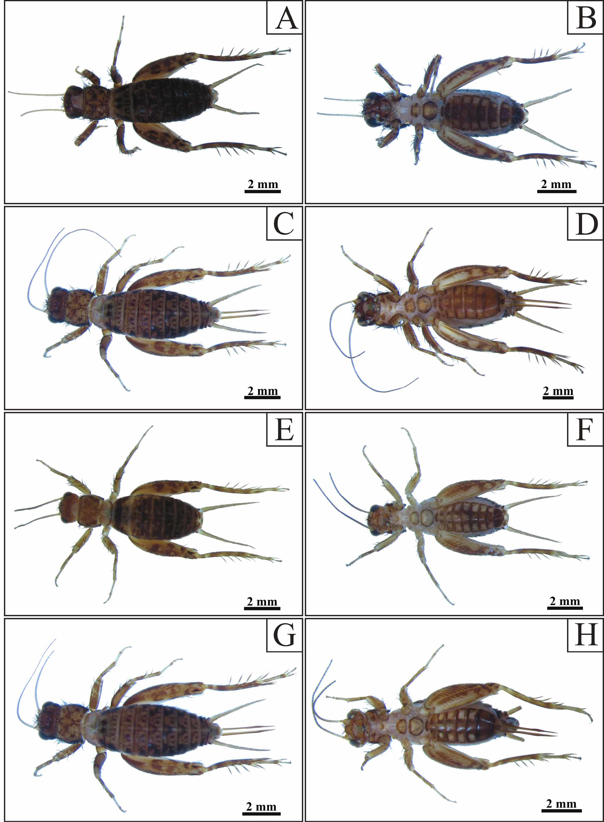
( Figs. 14 View FIGURE 14 E – H, 16, 19B, 24)
Phoremia sp. A in Sperber (1999)
Etymology. The specific epithet refers to Peter Henry Rolfs, first director of Escola Superior de Agricultura e Vet-erinária—ESAV, actually known as Universidade Federal de Viçosa—UFV.
Type. Holotype, male, Brasil, MG, Viçosa, Mata da Biologia, 30.x.2002 (Mendes, M. H leg.).
Diagnosis. This species may be distinguished from the other species of Phoremia by the following combination of characteristics: (i) pseudepiphallus showing strong invagination above median lobe, when in lateral view (Fig. 19C); (ii) central endophallic sclerite with proximal ends pointed (Fig. 19A, B) and (iii) rami and ectophallic apodeme extending with sub-straight shape, when in lateral view (Fig. 19C).
Description. Holotype, male, measurements (mm): BL 7,93; MID 1,00; LP 1,32; MWP 1,93; MLF 3,83; MLT 2,83. Head orangish, showing light brown spots covered with long black bristles on the median region; presence of fine and short light brown hairs covering all its extension; black eyes; three ocelli present, central ocellus surrounded on the superior and inferior border, by dark brown spot, lateral ocelli partially surrounded, on the internal border, by same color of the central; antennal scape enlarged and dark yellow, antennal articles varying from light yellow (first articles) to light brown (last articles); gena dark brown; clypeus dark brown on the superior portion and light yellow on the inferior; labrum light brown; mandibles coloration varying from dark yellow on the base to light brown on the apex; maxillary palpi coloration varying from light yellow to light brown with whitish not truncate apex. Pronotum dark yellow with diffuse dark brown spots, presence of the fine and short light brown hairs covering all its extension; latero-inferior lobe light brown; row of long bristles present on anterior and posterior pronotum border; mesonotum light yellow on the center with dark brown spots on lateral portion; metanotum coloration varying from light yellow on the center and posterior portion, to dark brown on lateral and anterior portions. Abdominal sternites 1 – 5 light brown with light yellow spots on center; sternites 2 – 8 with rectangular light yellow spots on laterals ( Fig. 14 View FIGURE 14 F); tergite 1 dark brown; tergites 2 – 3 dark brown covered by light brown spots; tergites 4 – 10 dark brown with central and laterals dark yellow spots; presence of short light brown hairs covering all tergites; cercus dark yellow; supra-anal plate little sclerotized and light yellow, presence of long light yellow hairs on lateral and posterior portions; subgenital plate dark brown with two small light yellow spots on distal portion. Fore and middle legs showing dark yellow femurs with some light yellow and light brown spots associated with long black bristles ( Fig. 14 View FIGURE 14 E); tibiae light brown to dark yellow with black bristles; tarsomeres light yellow; tympanum absent on the fore tibiae; hind legs femur with transversal spots light brown and some light and dark yellow diffuse in all extension; hind tibiae light brown with two light yellow spots, presence of three apical spurs and three inner and outer dorsal spurs; tarsomeres light brown to light yellow. Male genitalia: pseudepiphallic apical lobes little sclerotized and partially separate by a cleft when in ventral, dorsal and posterior view (Figs. 16A, B and D); pseudepiphallus showing strong invagination above median lobe, when in lateral view (Fig. 16C). Endophallus divided in three sclerites, being one central and two laterals (Figs. 16A, B and D); central endophallic sclerite with proximal ends pointed (Fig. 16A, B). Rami and ectophallic apodeme extending with sub-straight shape, when in lateral view (Fig. 16C). Female: Body shape very similar to male, showing only the following differences: head with dark brown spots; sternites 2 – 5 adorned with light yellow central spots and lateral spot of same coloration; subgenital plate dark yellow with central dark brown spot. Ovipositor varying from dark yellow on the proximal to light brown on distal portion ( Figs. 14 View FIGURE 14 G,H e 16E).
Observations. Some female specimens show lighter spots in abdominal sternites, mainly in central region.
Measurements. Males (n= 7, excluding holotype). BL 6,41 – 7,90 (7,45 ± 0,86); MID 0,90 – 1,0 7 (1,0 1 ± 0,06); LP 1,22 – 1,28 (1,26 ± 0,03); MWP 1,81 – 2,38 (2,17 ± 0,23); MLF 3,73 – 4,38 (3,98 ± 0,21); MLT 2,71 – 3,13 (2,86 ± 0,13). Females (n= 5). BL 6,90 – 9,33 (7,87 ± 1,05); MID 1,0 0 – 1,0 7 (1,0 7 ± 0,07); LP 1,27 – 1,32 (1,28 ± 0,03); MWP 2,0 0 – 2,42 (2,24 ± 0,22); MLF 3,83 – 4,38 (4,14 ± 0,24); MLT 2,75 – 3,0 8 (2,97 ± 0,15); OL 3,25 – 3,33 (3,28 ± 0,04).
Ocorrence. Species known only from Viçosa, MG, Brazil.
FIGURE 16. Phoremia rolfsi sp. nov. A —phallic complex ventral view, B —dorsal, C —lateral, D —posterior, E —female genitalia lateral view. Abbreviations: Ps. A. L—pseudepiphallic apical lobe; Ps. M. L—pseudepiphallic median lobe; Ps. Ppseudepiphallic parameres; Ps. S—pseudepiphallic sclerite; Ect. Ap—ectophallic apodeme; End. Esc—endophallic sclerite.
Material examined. Paratypes: 6 males, 3 females, same holotype data; 1 male, 2 females, Brasil, MG, Viçosa, Mata da Biologia, 27.x.2002 (Mendes, M. H leg.).
No known copyright restrictions apply. See Agosti, D., Egloff, W., 2009. Taxonomic information exchange and copyright: the Plazi approach. BMC Research Notes 2009, 2:53 for further explanation.