
Liostracina joyceae, Smith & Paterson & Brock, 2018
|
publication ID |
https://doi.org/ 10.11646/zootaxa.4396.1.1 |
|
publication LSID |
lsid:zoobank.org:pub:8EEBE6DE-0ECC-4B9C-AD14-01438291782B |
|
DOI |
https://doi.org/10.5281/zenodo.5980824 |
|
persistent identifier |
https://treatment.plazi.org/id/B7CEEF6E-8407-418F-A8AE-EA9FD353C687 |
|
taxon LSID |
lsid:zoobank.org:act:B7CEEF6E-8407-418F-A8AE-EA9FD353C687 |
|
treatment provided by |
Plazi |
|
scientific name |
Liostracina joyceae |
| status |
sp. nov. |
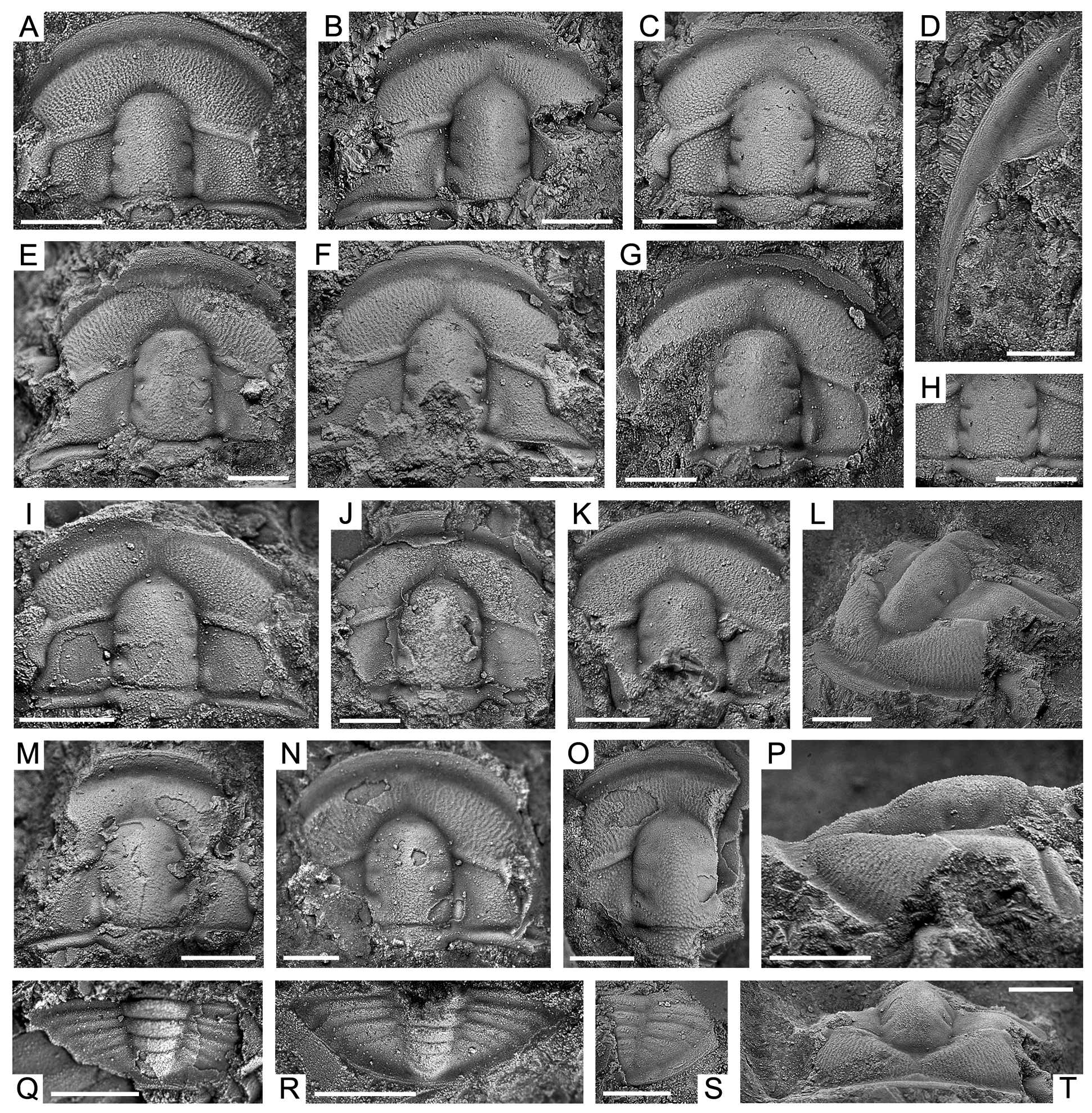
Liostracina joyceae sp. nov.
Fig. 20 View FIGURE 20
Etymology. In honour of Joyce Gilbert-Tomlinson, for her work on the Cambrian fossils of the Amadeus Basin.
Holotype. CPC42336, cranidium from the Goyder Formation sample GOY/97.0, 73.2 m above the base of the formation ( Fig. 20A View FIGURE 20 ).
Paratypes. Eleven cranidia; CPC42337 ( Fig. 20B View FIGURE 20 ); CPC42338 ( Fig. 20C, H View FIGURE 20 ); CPC42340 ( Fig. 20E, L, P, T View FIGURE 20 ); CPC42341 ( Fig. 20F View FIGURE 20 ); CPC42342 ( Fig. 20G View FIGURE 20 ); CPC42343 ( Fig. 20I View FIGURE 20 ); CPC42344 ( Fig. 20J View FIGURE 20 ); CPC42345 ( Fig. 20K View FIGURE 20 ); CPC42346 ( Fig. 20M View FIGURE 20 ); CPC42347 ( Fig. 20N View FIGURE 20 ); and CPC42348 ( Fig. 20O View FIGURE 20 ); one librigena, CPC42339 ( Fig. 20D View FIGURE 20 ); and three pygidia; CPC42349 ( Fig. 20Q View FIGURE 20 ); CPC42350 ( Fig. 20R View FIGURE 20 ); CPC42351 ( Fig. 20S View FIGURE 20 ); from sample GOY/97.0, 73.2 m above the base of the formation.
Other material. Twelve cranidia, one librigena, and three pygidia figured, CPC42336–CPC42351. 43 cranidia, fifteen librigenae and one pygidium not figured (mostly fragments).
Diagnosis. Cranidium outline transversely subelliptical. Glabella anteriorly truncate, oblong in outline, width:length ratio of 58% to 63%, occupying 62% to 71% of the cranidial length. S1 short (tr.). S2 short (tr.), slightly less deeper than S1. S3 short (tr.), shallow to almost completely effaced. Occipital ring of moderate length.
Moderately deep, narrow (sag.) SO. Anterior cranidial border 6% to 9% of sagittal cranidial length. Preglabellar field 26% to 29% of sagittal cranidial length, with faint preglabellar furrow. Palpebral lobes 20% cranidial length, reniform in outline. Eye ridge well defined. Ovate bacculae, moderately developed. Genal spine elongate, approximately 1.4 times the length of the librigena. Pygidium small, subtriangular, length:width ratio 33%. Axis prominent, width:length ratio 83%, occupying about 93% of sagittal length of pygidium. Four well defined axial rings and a small terminal piece. Pleural regions with three narrow (exsag.), shallow pleural and two shallow, narrow (exsag.) interpleural furrows. Frontal area evenly covered in small granules and network of genal caeca, except in preglabellar furrow. Glabella, occipital ring, eye ridges, palpebral lobes, palpebral areas, postocular fields and posterolateral projections of fixigena all densely covered in small granules. Librigenal field covered in faint small granules and a network of genal caeca. Pygidium prosopon faint small granules.
Description. Cephalon semicircular, up to 4 mm long (sag.). Cranidium outline transversely subelliptical, maximum width across posterolateral projections of fixigenae, narrowest point at anterior tip of palpebral lobes (γ– γ); slightly convex (sag., tr.). Anterior margin strongly rounded. Posterior margin slightly bowed forward. Anterior branches of facial suture (γ-β section) diverge strongly from one another at 75°, then curve abruptly towards the midline from β, slightly behind the border furrow. Glabella anteriorly truncate, parallel-sided, oblong in outline; moderately convex, with maximum convexity across midwidth; width:length ratio of 58% to 63% (mean 61%; n = 5), occupying 62% to 71% (mean 66%; n = 4) of the cranidial length. Axial furrow narrow (sag., exsag., tr.) and deep. S1 short (tr.), deep and narrow (exsag.), intersecting axial furrow slightly anterior to ε, directed transversely. S2 short (tr.), slightly less deep than S1, narrow (exsag,), intersecting axial furrow at γ, directed transversely. S3 short (tr.), shallow to almost completely effaced, intersecting axial furrow slightly anterior of where the eye ridges intersect the glabella, directed anteromedially. Occipital ring of moderate length (sag.), becoming distinctly narrower abaxially, posterior margin bowed strongly backwards, faint median node surmounting the posterior of occipital ring. Moderately deep, narrow (sag.) SO, directed transversly. Anterior cranidial border moderately narrow (sag., exsag.), occupying about 6% to 9% (mean 8%; n = 3) of sagittal cranidial length, narrowing abaxially. Anterior border furrow slightly wide (sag., exsag.) and deep. Preocular field moderately convex, sloping toward the anterior border furrow. Preglabellar field is slightly concave forming a narrow (tr.), moderately shallow preglabellar furrow. Preglabellar field 26% to 29% (mean 28%; n = 4) of sagittal cranidial length. Palpebral lobes (tr.), 20% cranidial length, reniform in outline, defined by a narrow (tr.), deep palpebral furrow; anterior tip situated level with S2, posterior tip level with the point between the midlength of S1 and SO. Eye ridge well defined and prominent, extending posterolaterally from the axial furrow opposite S 3 in a straight line towards the anterior tip of palpebral lobe. Palpebral area of fixigena transverse, maximum width (tr.) is 100% to slightly greater than the adjacent glabellar width. Postocular field short (exsag.), and slightly downsloping toward the posterolateral projections; ovate bacculae, moderately developed near axial furrow, anterior tip situated slightly posterior S1, posterior tip opposite SO. Posterolateral projections of fixigena narrow (exsag.), strongly downsloping towards lateral corners. Posterior border narrow (exsag.), separated from fixigenal field by deep, moderately narrow (exsag.) border furrow.
Librigena up to 2 mm in length excluding spine. Lateral margin, including that of genal spine, evenly curved. Genal field subtrapeziform, 53% of librigenal width (tr.) along posterior border, slightly convex. Lateral border well defined. Lateral border furrow wide (tr.) and deep, lateral furrow becomes wider posteriorly and continues onto genal spines for a short distance. Genal spine flattened, elongate, blade-like, length approximately 140% librigenal length.
Pygidium small, up to 2 mm long (sag.), subtriangular, slightly convex, wide (tr.), length:width ratio of 33%. Anterior margin slightly curved, with the articulating half-ring being slightly convex. Posterior margin rounded. Axis prominent, narrow (tr.), tapered posteriorly, width:length ratio of 83%, occupying about 93% of sagittal length of pygidium. Articulating half-ring narrow (sag.), defined by narrow (sag.), moderately deep articulating furrow. Four well defined axial rings present, separated by moderately deep to shallow, narrow (sag.) inter-ring furrows, becoming fainter posteriorly. Terminal piece small. Axial furrow shallow and wide (tr.), fading around terminal piece. Pleural regions only slightly convex, with three narrow (exsag.), shallow pleural furrows that terminate before reaching margin, becoming shorter and fainter posteriorly. Shallow, narrow (exsag.) interpleural furrows on first and second pleural ribs, extending the entire length, dividing pleural rib in two equal parts. Border narrow (sag., exsag.), defined by change in convexity from the pleural region.
Prosopon consists of granules varying in size. Frontal area evenly covered in small granules and network of genal caeca, except in preglabellar furrow. Glabella, occipital ring, eye ridges, palpebral lobes, palpebral areas, postocular fields and posterolateral projections of fixigena all densely covered in small granules. Librigenal field covered in faint small granules and an anastomosing network of genal caeca. Anterior and lateral borders covered in horizontal terrace ridges. Pygidium prosopon faint small granules.
Hypostome, rostral plate and thorax unknown.
Discussion. The taxon from the Goyder Formation is comparable to at least three other species of Liostracina Monke, 1903 , all of which share a prosopon covered in granules of varying sizes. The most similar species is Liostracina bella Lin & Zhou in Lin et al., 1983 , from the eponymous zone (late Guzhangian–early Paibian) of southeastern China. The type material of L. bella includes three poorly preserved cranidia with an associated librigena from drillcore material in the Tuanshan Formation ( Lin et al., 1983). However, better-preserved specimens of L. bella were figured by Peng et al. (2004a, pl. 61, figs 1–14, pl. 62, figs 1–13) from the Huaqiao Formation of southeast China. Both taxa share an identical prosopon, as well as a cranidium with a transversely subelliptical outline, short (tr.) S1, S2 and S3 glabellar furrows (with S3 almost effaced), prominent eye ridges, moderately developed ovate (exsag.) bacculae, pygidia with a length:width ratio of approximately 33%, and a posterior margin which is moderately rounded. The Goyder Formation specimens differ in having the following characters: a much wider (tr.) and slightly longer (sag.) glabella; a more transverse occipital ring; a somewhat less upturned anterior border; shorter (tr.), thicker eye ridges; longer (exsag.) palpebral lobes; and more distinct pleural and interpleural furrows on the pygidium. The librigenae of L. bella are similar to those from the Goyder Formation, except for a narrower (tr.) and more prominent lateral border.
Liostracina joyceae sp. nov. is also similar to L. nolens Öpik, 1967 from the Mindyallan Georgina Limestone in the Georgina Basin. Liostracina nolens effectively shares the same features as L. bella , with which it is likely synonymous (compare Öpik 1967, pl. 35, fig. 6, 7 to Peng et al. 2004a, pl. 61, figs 1–5, 7, 8, 12, pl. 62, fig. 1–5, 8, 12).
The only other species with a granulose prosopon is Liostracina simesi Jago & Cooper, 2005 from the Mindyallan of Antarctica. Liostracina joyceae is different from L. simesi in having a much wider (tr.) glabella with more distinct S1–S3 furrows, a more transverse occipital ring, shorter (tr.), thicker and more prominent eye ridges, less distinct bacculae, and a more densely granulose prosopon with genal caeca on the frontal area.
All other taxa currently assigned to the genus, particularly well-known species such as Liostracina bilimbata Zhang in Qui et al., 1983 (= L. suixiensis Bi in Qui et al., 1983 ; see Park et al. 2014), L. kaulbacki Shergold, Laurie & Shergold, 2007 , L. krausei Monke, 1903 , L. qingyangensis Qian in Qui et al., 1983 , L. tangwangzhaiensis Park, Kihm, Kang & Choi, 2014 , and L. volens Öpik, 1967 , lack a granulose prosopon. These other taxa also have more effaced glabellar furrows and eye ridges, as well as a glabella that is distinctly narrower (tr.), with the exception of L. kaulbacki that may be slightly wider (tr.).
There are at least five other taxa which are assigned to Liostracina (in some cases tentatively), including: L. bifurcata Zhang in Qui et al., 1983 ; L. (?) pauper Resser & Endo in Endo & Resser, 1937; L. (?) paupiforme Endo, 1944; and two described below (see L. cf. kaulbacki and Liostracina sp.). These species are based on either a single specimen or small collections of poorly preserved cranidia, making further comparison difficult.
Occurrence. GOY section horizons 49.4, 73.2 and 83.9 m ( Fig. 3 View FIGURE 3 ). Also recovered from GOYWEST.
Distribution. Goyder Formation, Amadeus Basin, Northern Territory. Cambrian Series 3, Guzhangian (Mindyallan) in age.

No known copyright restrictions apply. See Agosti, D., Egloff, W., 2009. Taxonomic information exchange and copyright: the Plazi approach. BMC Research Notes 2009, 2:53 for further explanation.