
Hebeia stewarti, Smith & Paterson & Brock, 2018
|
publication ID |
https://doi.org/ 10.11646/zootaxa.4396.1.1 |
|
publication LSID |
lsid:zoobank.org:pub:8EEBE6DE-0ECC-4B9C-AD14-01438291782B |
|
DOI |
https://doi.org/10.5281/zenodo.5980814 |
|
persistent identifier |
https://treatment.plazi.org/id/2E1DE850-3523-4A48-A579-CC7A2928A21B |
|
taxon LSID |
lsid:zoobank.org:act:2E1DE850-3523-4A48-A579-CC7A2928A21B |
|
treatment provided by |
Plazi |
|
scientific name |
Hebeia stewarti |
| status |
sp. nov. |
Hebeia stewarti sp. nov.
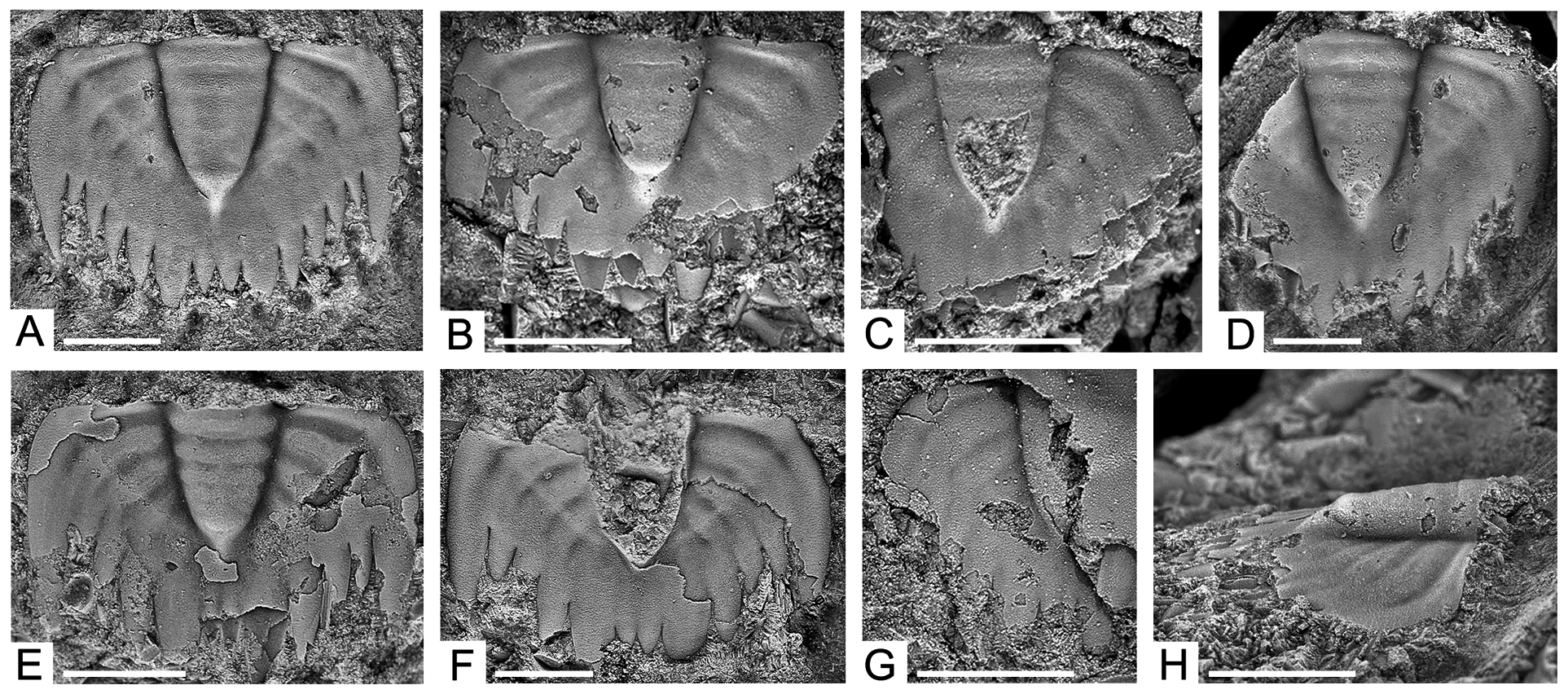
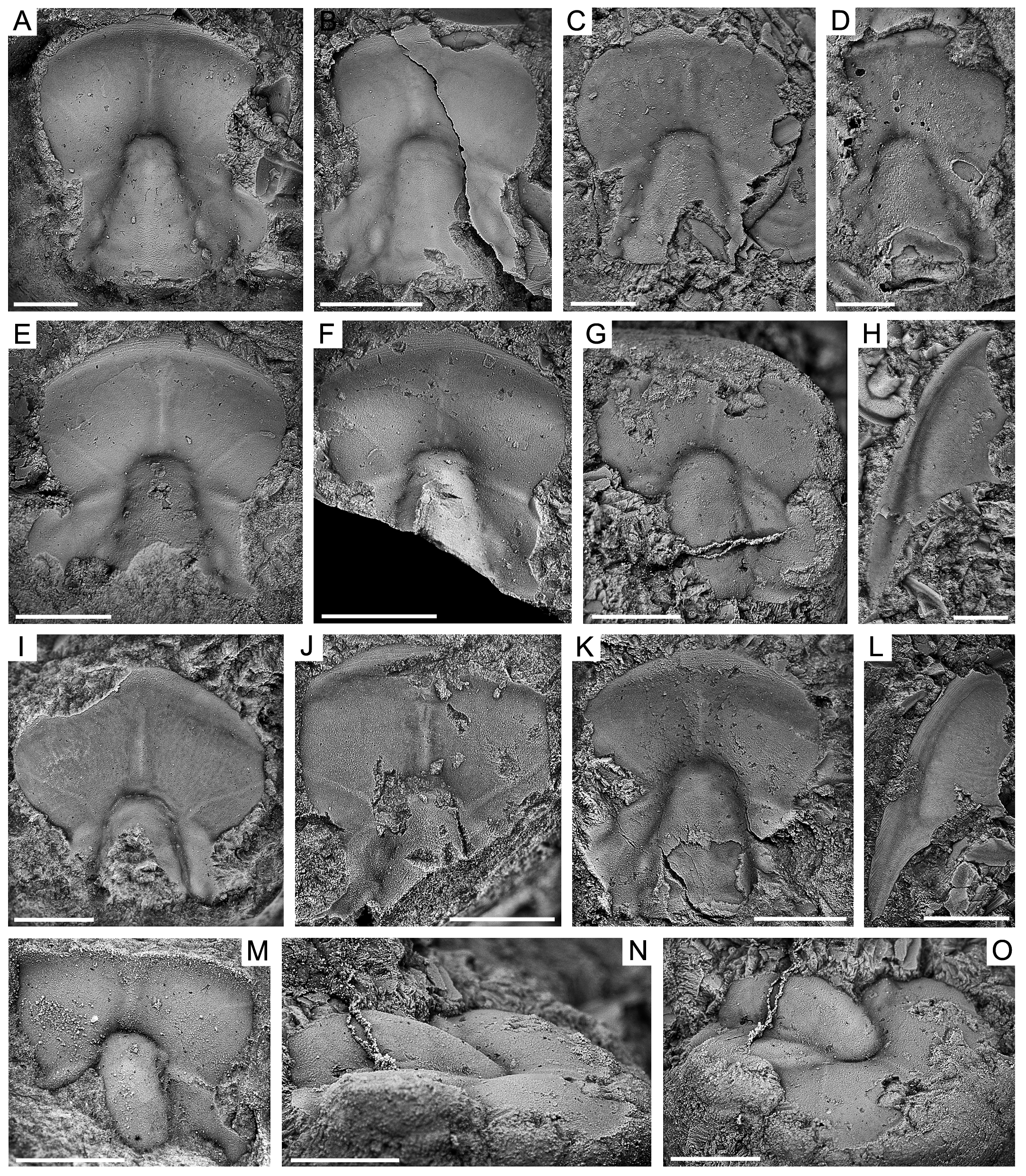
Figs 16 View FIGURE16 , 17 View FIGURE 17
Etymology. In honour of A.J. Stewart, for his geological investigations in the Amadeus Basin. Holotype. CPC42308, cranidium from the Goyder Formation at sample GOY/97, 73.2 m above the base of the formation ( Fig. 16A View FIGURE16 ).
Paratypes. One cranidium: CPC42317 ( Fig. 16J View FIGURE16 ), from sample GOY/86, 64.9 m above the base of the formation. Nine cranidia: CPC42309 ( Fig. 16B View FIGURE16 ); CPC42310 ( Fig. 16C View FIGURE16 ); CPC42311 ( Fig. 16D View FIGURE16 ); CPC42312 ( Fig. 16E View FIGURE16 ); CPC42313 ( Fig. 16F View FIGURE16 ); CPC42314 ( Fig. 16G, N, O View FIGURE16 ); CPC42316 ( Fig. 16I View FIGURE16 ); CPC42318 ( Fig. 16K View FIGURE16 ); and CPC42320 ( Fig. 16M View FIGURE16 ); two librigenae: CPC42315 ( Fig. 16H View FIGURE16 ); CPC42319 ( Fig. 16L View FIGURE16 ); and seven pygidia: CPC42321 ( Fig. 17A, H View FIGURE 17 ); CPC42322 ( Fig. 17B View FIGURE 17 ); CPC42323 ( Fig. 17C View FIGURE 17 ); CPC42324 ( Fig. 17D View FIGURE 17 ); CPC42325 ( Fig. 17E View FIGURE 17 ); CPC42326 ( Fig. 17F View FIGURE 17 ); and CPC42327 ( Fig. 17G View FIGURE 17 ), from sample GOY/97.0, 73.2 m above the base of the formation.
Other material. Eleven cranidia, two librigenae, and seven pygidia figured, CPC42308–CPC42326. 51 cranidia, thirteen librigenae, and sixteen pygidia not figured (mostly fragments).
Diagnosis. Glabella strongly tapered at 24° to 30°, elongate trapezoid to subtriangular in outline; width:length ratio of 81%, occupying 56% of the cranidial length. All lateral glabellar furrows except SO effaced. Shallow, wide (exsag.), faint SO. Anterior border furrow wide (sag., exsag.) and shallow. Preocular field slightly convex. Preglabellar field concave forming a wide (tr.) moderately deep preglabellar furrow. Preocular field and preglabellar field elongate, 33% of sagittal cranidial length. Two faint, narrow (tr.), facial ridges cross preocular field and one faint, narrow (tr.), median ridge crosses preglabellar field. Palpebral lobes wide (tr.). Eye ridge weakly defined. Genal spine flattened, blade-like. Pygidium subtrapezoidal in outline, sagittal length:width ratio of 59% excluding spines. Axis moderately narrow (tr.), width:length ratio of 87%, short occupying about 63% of sagittal length of pygidium. Three faintly defined and a fourth indistinct axial ring present. Pleural regions with three to four faint, wide (exsag.), shallow pleural furrows. Border with seven pairs of broad-based spines (six in smaller specimens), first and sixth pair (fifth in smaller specimens) are slightly longer (exsag.) and wider (tr.), first pair is much longer than the others; seventh pair (sixth on smaller specimens) short (exsag.). Prosopon minutely granulose.
Description. Cephalon semicircular, up to 8 mm long (sag.). Cranidium outline transversely subelliptical, maximum width across posterolateral projections of fixigenae based on librigena outline, narrowest point at anterior tip of palpebral lobes (γ–γ); slightly convex (sag., tr.). Anterior margin strongly rounded. Posterior margin incompletely preserved. Anterior branches of facial suture (γ-β section) diverge strongly from one another at 60°, then curve abruptly towards the midline from β before they reach the anterior margin. Glabella elongate, strongly tapered at 24° to 30° (mean 26°; n = 4), elongate trapezoid to subtriangular in outline; width (tr.) across anterior 29% maximum width; slightly convex; maximum convexity across midwidth, lateral slopes gently convex; width:length ratio of 81%, occupying 56% of the cranidial length; axial furrow moderately narrow (sag., exsag., tr.) and only slightly deep. All lateral glabellar furrows except SO effaced. Occipital ring of moderate length (sag.), becoming slightly wider abaxially, posterior margin slightly bowed backwards. Shallow, wide (exsag.), faint SO; bowed backwards medially. Anterior cranidial border moderately narrow (sag., exsag.) and slightly convex, occupying about 5% of sagittal cranidial length, narrowing abaxially. Anterior border furrow wide (sag., exsag.) and shallow. Preocular field slightly convex, moderately downsloping toward the anterior border furrow. Preglabellar field concave, forming a wide (tr.), moderately deep preglabellar furrow. Preocular field and preglabellar field elongate, 33% of sagittal cranidial length. Two faint, narrow (tr.), facial ridges cross preocular field and one faint, narrow (tr.), median ridge crosses preglabellar field; facial ridges extend from the proximal part of the eye ridges to just posterior of β; median preglabellar ridge traces the sagittal line between the glabellar anterior and anterior border furrow, nested within the preglabellar furrow. Palpebral lobes wide (tr.), reniform in outline, defined by a wide (tr.), shallow palpebral furrow; anterior tip situated 60% of cranidial length from the anterior border, posterior tip just posterior of SO. Eye ridge weakly defined, extending posterolaterally from the axial furrow just anterior of γ in a straight line towards the anterior tip of palpebral lobe. Palpebral area of fixigena transverse, maximum width (tr.) is 100% the adjacent glabellar width; elongate (exsag.) bacculae, width:length of 50%, moderately developed near axial furrow, anterior tip situated opposite midlength of palpebral lobe, posterior tip opposite anterior of SO. Postocular field short (exsag.) and strongly downsloping toward the posterior border furrow. Posterolateral projections of fixigena incompletely preserved. Posterior border narrow (exsag.), separated from fixigenal field by shallow, moderately narrow (exsag.) border furrow.
Librigena up to 8 mm in length excluding spine. Lateral margin, including that of genal spine, evenly curved. Genal field subtrapeziform, 77% of librigenal width (tr.) along posterior border, slightly convex. Lateral border well defined. Lateral and posterior border furrows wide (exsag.) and shallow, lateral furrow becomes wider (tr.) posteriorly and continues onto genal spines for a short distance. Facial ridges continue from fixigena onto librigenal field, extending anterolaterally from just posterior to the β point to the lateral border furrow. Genal spine flattened, blade-like, length approximately 50% librigenal length.
Pygidium up to 5 mm long (sag.), subtrapezoidal in outline excluding spines, slightly convex, sagittal length:width ratio of 59% excluding spines. Anterior margin slightly curved, with the articulating half-ring being slightly convex. Axis prominent, moderately narrow (tr.), tapered posteriorly, width:length ratio of 87%, short occupying about 63% of sagittal length of pygidium. Narrow (sag.) articulating half-ring, faintly defined by a shallow, narrow (sag.) articulating furrow. Three faintly defined axial rings and a fourth indistinct axial ring present, separated by faint, shallow, narrow (sag., exsag.) inter-ring furrow. Terminal piece small, up to 33% of axial length (sag.); small, faint, postaxial ridge present. Axial furrow moderately deep and narrow (tr.), becoming shallower and broader posteriorly until fading out completely. Pleural regions only slightly convex, with three to four faint, wide (exsag.), shallow pleural furrows. Furrows roughly transverse, directed slightly more backwards and becoming fainter posteriorly, terminate slightly before reaching margins. Border with seven pairs of broadbased spines on largest specimen (and six on smaller specimens), first and sixth (fifth on smaller specimens) respective pairs are slightly longer (exsag.) and wider (tr.), first pair is much longer than the others; seventh pair of spines (sixth on smaller specimens) short (exsag.); all spines directed posteriorly.
Prosopon over cephalon, librigena and pygidium minutely granulose. Anterior border of cranidium and lateral borders of librigena covered by horizontal terrace ridges.
Discussion. Cranidia from the Goyder Formation are similar to the type species, H. conica from the Guzhangian of northeastern China (compare Fig. 16A–G, I–K, M View FIGURE16 with Guo & Duan 1978, pl. 2, figs 17–19). Only a slightly wider (tr.) γ–γ section of the cranidium, less distinct eye ridges, a wider (tr.) palpebral area of the fixigena, and more prominent bacculae separate the Goyder Formation material. Hebeia pingquanensis and H. huainanensis , both from the Guzhangian of northeastern China, are more distinctive in having short glabellar furrows, a narrower γ–γ section of the cranidium, and either a wider (tr.) or absent preglabellar ridge. A shorter preglabellar field in H. pingquanensis also separates it from H. stewarti .
Of the two Chinese species that have associated pygidia, H. stewarti is most comparable to H. huainanensis (compare Fig. 17A–G View FIGURE 17 with Qiu in Qiu et al., 1983, pl. 48, fig. 11; and Zhang & Wang 1986, pl. 3, fig. 8, 9, 11 = H. pulchera ). The pygidia have the same subtrapezoidal outline, shallow pleural furrows, no defined border, and a long first pair of spines. Slight differences separate H. stewarti , including a slightly longer (sag.) axis and a more prominent seventh pair of spines (in larger specimens) at the posterior margin. The associated pygidia of H. pingquanensis (Zhang & Wang 1986, pl. 3, fig. 4–5) are quite different, especially in possessing two broad-based spines at the posterior margin, and appear to have more in common with pygidia of Shantungia species. Hence, it is possible that the pygidia of H. pingquanensis have been misidentified.
Pygidia of H. stewarti have a varying number of pygidial spines, with the largest specimen having seven pairs ( Fig. 17A View FIGURE 17 ) and smaller specimens having six pairs ( Fig. 17B, C–G View FIGURE 17 ). Given the size difference between the pygidia, this variation likely represents an ontogenetic trend.
Occurrence. GOY section horizons 64.9 and 73.2 m ( Fig. 3 View FIGURE 3 ). Specimens also recovered from GOYWEST.
Distribution. Goyder Formation, Amadeus Basin, Northern Territory. Cambrian Series 3, Guzhangian (Mindyallan) in age.

No known copyright restrictions apply. See Agosti, D., Egloff, W., 2009. Taxonomic information exchange and copyright: the Plazi approach. BMC Research Notes 2009, 2:53 for further explanation.