
Trophomera pacifica, Miljutin & Miljutina, 2009
|
publication ID |
https://doi.org/ 10.11646/zootaxa.2096.1.11 |
|
DOI |
https://doi.org/10.5281/zenodo.5334656 |
|
persistent identifier |
https://treatment.plazi.org/id/038D817D-FFB4-FFEF-A69F-FB21FBCFFD41 |
|
treatment provided by |
Felipe |
|
scientific name |
Trophomera pacifica |
| status |
sp. nov. |
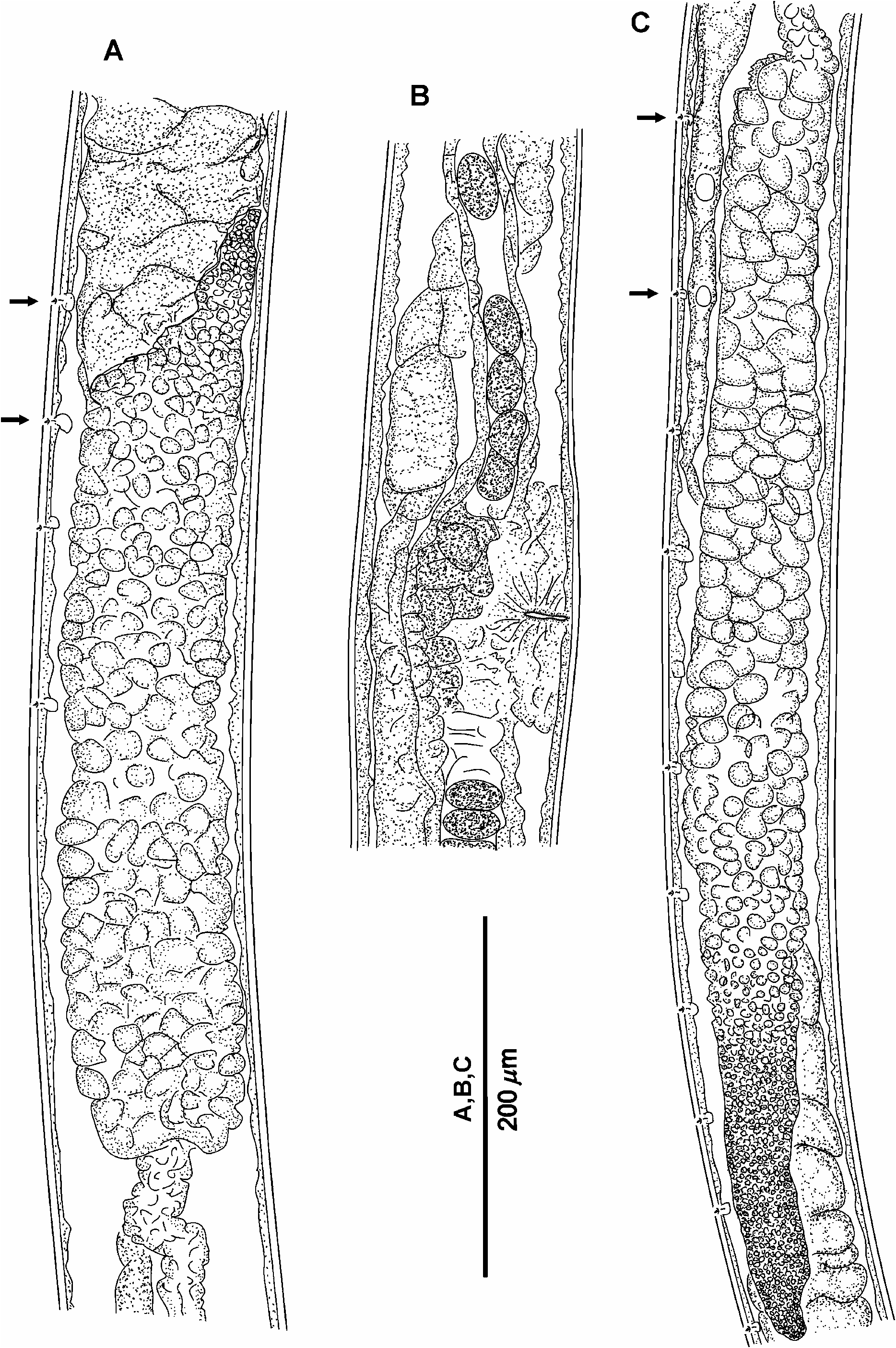
Trophomera pacifica sp. n.
Figs 6–7 View FIGURE 6 View FIGURE 7 ; Table 2
Type material: Holotype: one gravid female, collection number MNHN-BN496 .
Type locality: Polymetallic nodule area, 14°02.52’N, 130°07.98’W, 4950 m depth, 07.06.2004, multicorer #18, corer #2.
Etymology: “ Pacifica ” means “from the Pacific Ocean”.
Description: Female. Body cylindrical, with thickest body part at anterior half of body. Anterior end rounded, posterior end conical, with thick conical terminal spine 81 µm long, showing granular core. Cuticle with very thin and hardly visible transverse striations along whole body length; striations being more pronounced at caudal region. Cuticle thickness 5.5 µm at anterior tip, 4 µm at level of amphids, and 3 µm in the rest of the body parts. Amphids located at 0.86 c.b.d. from anterior end; amphidial apertures pore-like, 1 µm in diameter. Four submedian cephalic setae 2.5 µm long inserted in tiny pits. Somatic sensilla in shape of small papillae 1–1.5 µm long arranged in 4 longitudinal lateromedian rows from postamphidial region to anal region. Mouth opening reduced to a thin channel in cuticle. Pharynx a non-muscular string devoid of an internal lumen. Several large hyaline cells situated at posterior pharynx. Cardia absent. Midgut an oligocellular trophosome without visible internal lumen and obviously consisting of 1 row of cells. Borders between trophosomal cells distinct. Rectum reduced to an indistinctly visible tube. Female reproductive system didelphic, amphidelphic, occupying approximately 2/3 body length in the gravid specimen. Ovaries outstretched, 530 µm long in anterior branch, 715 µm in posterior branch. Oviducts very short. Uterus very long, occupying approximately 3/5 body length. Mature eggs 34x20 µm in diameter situated in one row in uterus except vulva region. Neither morphologically differentiated spermatheca nor spermatozoa observed. Vulvar glands present. Caudal glands absent.
Host unknown.
Male, juvenile stages unknown.
Differential diagnosis: The female of T. pacifica sp. n. possesses outstretched ovaries. Only three Trophomera species have outstretched ovaries: T. laubieri ( Petter 1987) , T. pseudominuta ( Miljutin 2004) , and, possibly, T. senckenbergi sp. n.
T. pacifica sp. n. differs from T. pseudominuta by its larger body length (5.4 mm vs. 2.8 mm), and its tail shape (long conical spine vs. rounded).
T. pacifica sp. n. differs from T. senckenbergi sp. n. by its larger body length (5.4 mm vs. 1.5 mm), and its tail shape (long conical spine vs. cone-shaped with rounded tip).
In its general appearance, T. pacifica sp. n. strongly resembles T. laubieri . It differs from T. laubieri by: presence of a granular core inside the caudal terminal spine; a thickened cuticle at the cephalic apex; a difference in the ratio “distance from anterior end to amphid / c.b.d.” (0.86 vs. 0.72); trophosome structure (one row of cells at the longitudinal optical section vs. several rows); a longer female reproductive system (occupying approximately 0.7 body length vs. 0.45–0.50); egg size (34x20 µm vs. from 38x38 to 40x40 µm); and a different egg layout inside oviduct (only one row of eggs vs. several ones).

No known copyright restrictions apply. See Agosti, D., Egloff, W., 2009. Taxonomic information exchange and copyright: the Plazi approach. BMC Research Notes 2009, 2:53 for further explanation.
|
Kingdom |
|
|
Phylum |
|
|
Class |
|
|
Order |
|
|
Family |
|
|
Genus |