
Trophomera marionensis (Petter, 1983)
|
publication ID |
https://doi.org/ 10.11646/zootaxa.2096.1.11 |
|
DOI |
https://doi.org/10.5281/zenodo.5334650 |
|
persistent identifier |
https://treatment.plazi.org/id/038D817D-FFB6-FFE8-A69F-FA8DFD54FD80 |
|
treatment provided by |
Felipe |
|
scientific name |
Trophomera marionensis (Petter, 1983) |
| status |
|
Trophomera marionensis (Petter, 1983)
Fig. 4 View FIGURE 4 ; Table 2
Additional data and illustrations as Benthimermis marionensis in Petter (1983b): 9–10, Fig. 5 View FIGURE 5 ; in Chesunov (1988b): 15–17, Figs 3–4 View FIGURE 3 View FIGURE 4 ; in Bussau (1993): 570–571, Fig. 247; and in Miljutin (2004): 32–34, Fig. 7 View FIGURE 7 .
Material: One virgin female.
Locality: 13°55.63’N, 130°12.20’W, 4800 m depth, 2–5 cm sediment layer, 26.05.2004, submersible “Nautile”, station 1595-3 CL 05.
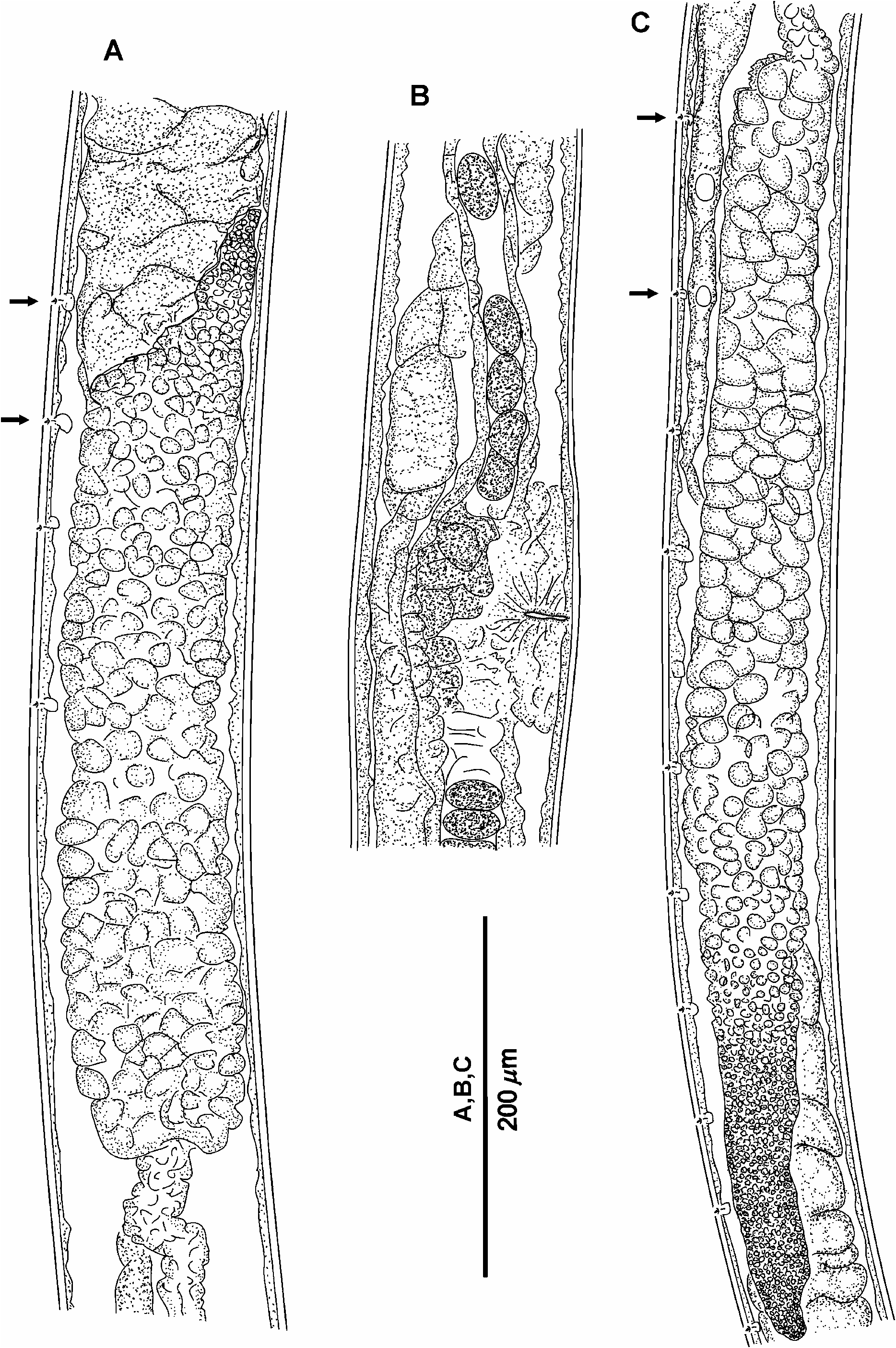
Description: Female. Body thread-like, cylindrical. Anterior end in shape of a rounded cone. Posterior end conical, with a terminal spine 110 µm long, possessing a cytoplasmic core with granular inclusions. Mouth opening vestigial, looking like an obscure channel at apical part of head end. Four submedian papilloid cephalic setae approximately 3.5 µm long inserted in tiny pits. Amphids not observed. Pharynx a nonmuscular, parenchymal string without an internal lumen. Nerve ring visible between pharynx and trophosome. Midgut, a multicellular trophosome without visible internal lumen, consisting of several rows of cells in optical section. Anterior trophosomal cells located ahead of posterior part of pharynx. Borders between trophosomal cells distinct. Rectum and anus present. Female reproductive system amphidelphic.
Remarks: B. marionensis was described upon eight gravid females from the southern part of the Indian Ocean, 46°52′S, 37°51′E, 31–110 m depth ( Petter 1983b). Other specimens were described later by Chesunov (1988b) from the Southern Atlantic (57°09′S, 26°09′W, depth 1729 m), by Bussau (1993) from the Eastern Pacific (07°04.04′S, 88°27,41′W, depth 4154 m), and by Miljutin (2004) from the Western Atlantic (8°07.88′ N, 49°03.71′ W, 4440 m depth). The new finding is the second record for the Pacific Ocean but it is far away from the previous region i.e. at a distance of about 5200 km. The new specimen agrees with the original description of B. marionensis in tail shape with terminal spine possessing a cytoplasmic core and structure of trophosome; body length and “V” value (10.2 mm and 57.3% vs. 9.3–14.8 mm and 46.5–74.0% respectively). However, the terminal spine of the new specimen is half as long as in the original description (110 µm vs. 200–320 µm) but closely resembles the specimen described in Miljutin (2004) (130 µm). Given the distance of the new specimen from the sites of the previous records, the slightly different morphological features may be attributed to differences between populations.


No known copyright restrictions apply. See Agosti, D., Egloff, W., 2009. Taxonomic information exchange and copyright: the Plazi approach. BMC Research Notes 2009, 2:53 for further explanation.
|
Kingdom |
|
|
Phylum |
|
|
Class |
|
|
Order |
|
|
Family |
|
|
Genus |