
Adicella makaria Malicky & Chantaramongkol 2002
|
publication ID |
https://doi.org/ 10.11646/zootaxa.3635.1.3 |
|
publication LSID |
lsid:zoobank.org:pub:25EBAAB4-4745-4C57-A922-06D071E58A87 |
|
DOI |
https://doi.org/10.5281/zenodo.6150777 |
|
persistent identifier |
https://treatment.plazi.org/id/038D8472-FFEC-6C4B-FF67-0A4AFD4266D5 |
|
treatment provided by |
Plazi |
|
scientific name |
Adicella makaria Malicky & Chantaramongkol 2002 |
| status |
|
Adicella makaria Malicky & Chantaramongkol 2002
( Figs. 1 View FIGURE 1 , 6 View FIGURE 6 )
Adicella makaria Malicky & Chantaramongkol 2002 in Malicky et al. 2002: 27, fig. 27, male, “ Thailand ”; Malicky 2006: 1514, correction: the type locality is not Thailand but is Taiwan; Shimura 2010: 50, 54, photo of adult, Japan (Ryûkyû Islands, Yonaguni-jima).
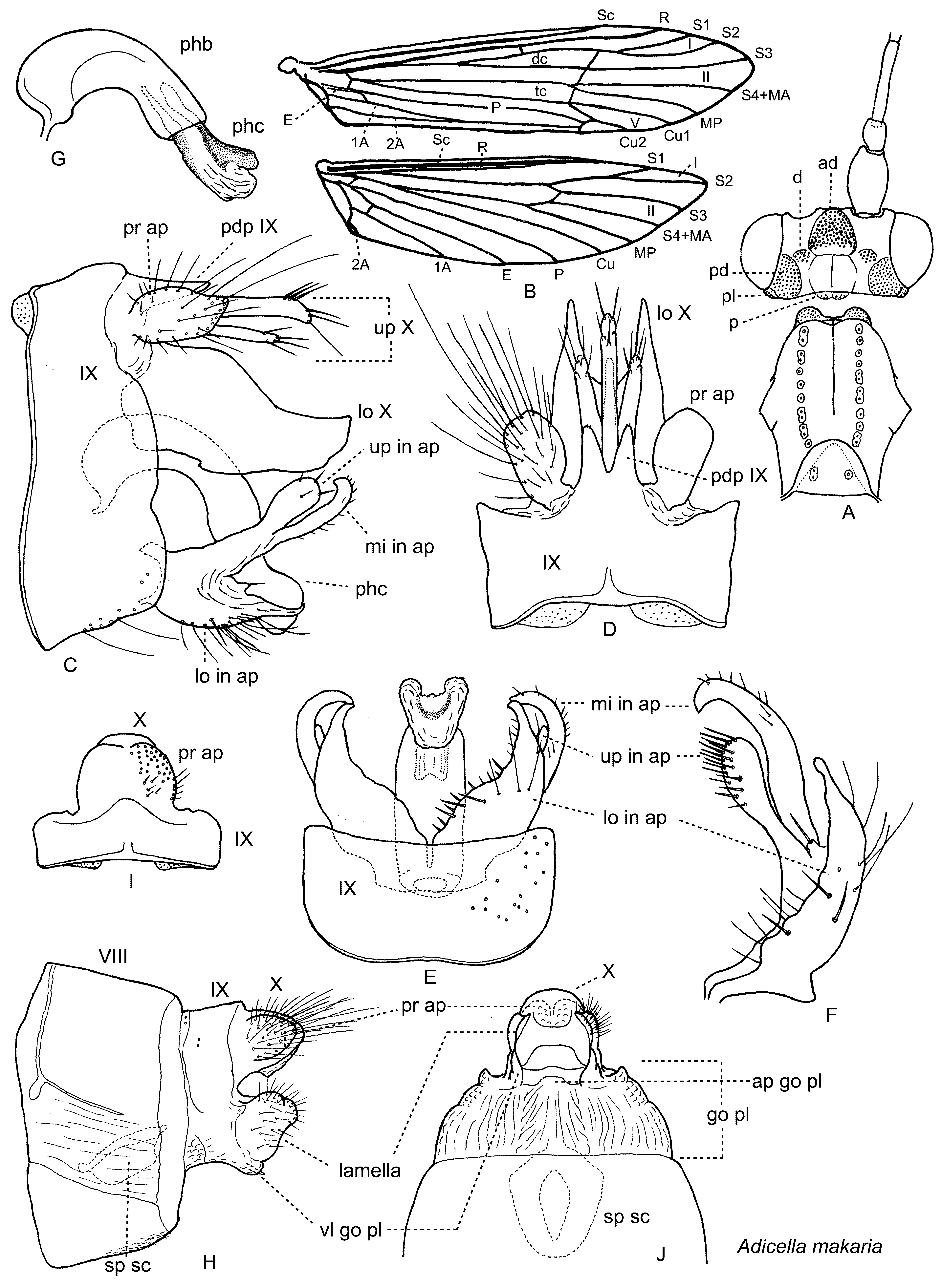
Adult ( Fig. 1 View FIGURE 1 ). Light brown, body length (from front of head to end of abdomen) 4.5–6.0 mm in male (n=7) and 4.5–6.0 mm in female (n=9). Antennae 4.0–4.5 times as long as body in male (n=3) and 3.5–4.0 times in female (n=2); scapes thick, long, each about 4 times as long as its pedicel. Maxillary palpi each 5-segmented, total length about 2.2 mm in male (n=2) and 2.0 mm in female (n=2); labial palpi each 3-segmented, total length 0.5–0.8 mm in male (n=4) and 0.55–0.60 mm in female (n=2); all segments of both palpi cylindrical and covered with fine setae. Head with large anterodorsal setal wart (ad; anteromesal setal wart in Wiggins & Currie 1996; vertexal medioantennal compact setal wart in Oláh & Johanson 2007) and 4 pairs of warts posterior from it, including dorsal (small, d; anterior setal warts in Wiggins & Currie 1996; vertexal ocellar compact setal warts in Oláh & Johanson 2007), postero-dorsal (large, pd; posterior setal warts in Wiggins & Currie 1996; occipal setal warts in Oláh & Johanson 2007), postero-lateral (small, pl; posterolateral setal warts in Wiggins & Currie 1996; postgenal setal warts in Oláh & Johanson 2007) and postoccipital (small, p; unnamed in Wiggins & Currie 1996 and Oláh & Johanson 2007) setal warts ( Fig. 1 View FIGURE 1 A). Pronotum with 1–2 pairs of warts, mesoscutum with pair of longitudinal setal lines, and mesoscutellum sometimes with pair of very small setal warts ( Fig. 1 View FIGURE 1 A).
Wings narrow ( Fig. 1 View FIGURE 1 B). Forewings mostly covered with brown hairs. Hind wings subacute apically, covered with brown hairs, with long fringes at posterior margins. Venation similar in both sexes. Forewings with apical forks I, II and V, fork I with stalk, fork II sub-rectangular, fork V sessil; discoidal cell narrow and long, about 1/3 as long as wing, thyridial cell narrow and very long, about 1/2 as long as wing; r - s crossvein present in some specimens; Cu2 and P connected by cu - p crossvein, then P fused with E+1A+2A to form single curved vein ending at arculus. Hind wings with apical forks I and II, fork II sub-rectangular; discoidal and thyridial cells absent; R fine (often invisible without staining) and fused to Sc near midlength of wings. Lengths of forewings and hind wings each 5.0–7.0 mm and 4.0–5.0 mm respectively in males (n=7), 4.5–6.0 mm and 3.5–4.5 mm respectively in females (n=9).
Male genitalia ( Figs. 1 View FIGURE 1 C–G). Segment IX (IX) rectangular, posterolateral margins slightly convex in lateral view ( Fig. 1 View FIGURE 1 C); in dorsal view posterodorsal margin produced with two triangular lobes ( Fig. 1 View FIGURE 1 D, pdp IX). Upper part of tergum X (up X) trifurcate, setose apically, middle process longer than lateral processes ( Figs. 1 View FIGURE 1 C, D). Lower part of tergum X (lo X) tall, hood-like, composed of two large vertical lobes fused dorsally in basal half, directed ventrocaudad in basal half then gently curved and directed caudad ( Fig. 1 View FIGURE 1 C), apex of each lobe subacute in dorsal view ( Fig. 1 View FIGURE 1 D). Preanal appendages (pr ap) oval, subacute apically in lateral view, round apically in dorsal view ( Figs. 1 View FIGURE 1 C, D). Inferior appendages each with three branches ( Figs. 1 View FIGURE 1 C, E, F): Upper branch (up in ap) clublike with many short setae mesally; middle branch (mi in ap) longest, bar-like, gently curved mesad, acute apically with few short setae on apical outer surfaces; lower branch (lo in ap) thick at base, gradually tapered with many long and short setae on ventral surface, subacute apically. Phallobase (phb) tubular, curved 90Ο; paramere spines absent; phallicata (phc) tubular, almost straight, about 2/3 as long as phallobase ( Fig. 1 View FIGURE 1 G), with U- or V-shaped phallotremal sclerite.
Female genitalia ( Figs. 1 View FIGURE 1 H–J). Segment IX (IX) short, tergum IX produced into subtriangular lobe posteromedially and fused with tergum X and preanal appendages ( Figs. 1 View FIGURE 1 H, I). Preanal appendages (pr ap) represented as broad setose mounds ( Figs. 1 View FIGURE 1 H, I). Segment X (X) forming longitudinally short tube with semimembranous ventral surface ( Figs. 1 View FIGURE 1 H, J). Lamellae irregularly shaped, vertical, setose lobes, each with small round flap on dorsolateral margin ( Fig. 1 View FIGURE 1 H). Gonopod plate (go pl) rugose, broadly semicircular or subquadrate; pair of small, round, vertical lobes (vl go pl) emerging from posterolateral ends, about 1/4 as large as lamellae in lateral view ( Fig. 1 View FIGURE 1 H), triangular in ventral view ( Fig. 1 View FIGURE 1 J). Apicomesal process of internal part of gonopod (ap go pl) slightly convex ( Fig. 1 View FIGURE 1 J), sometimes trapezoidal. Spermathecal sclerite (sp sc) subcircular in ventral view ( Fig. 1 View FIGURE 1 J), trapezoidal in lateral view ( Fig. 1 View FIGURE 1 H).
Specimens examined. JAPAN: Ryûkyû Islands. Ishigaki-jima: Hakusui, Nagura-gawa, small tributary, 12–21.x.1999, K. Konishi, M, 3 females (TI); same data except 11–12.iii.2009, TI, L & S, 3 females (TI); same data except 11–13.iv.2011, TI, L & S, 1 male, 2 females (TI); same data except 25–31.X.2012, TI, S & M, 1 male, 1 female (TI); Miyara-gawa, Nagura-damu-ue, 12.iv.2011, TI, L & S, 2 males (TI). Iriomote-jima: Sonai, 13–17.iii.2002, T. Yoshida & G. Sugaya, M, 1 male, 1 female (TI); small stream near boat station of Urauchi-gawa, 18.x.2005, TI, S, 1 male (TI). Yonaguni-jima: Hikawa-suigen, 24.iii.2009, N. Shimura, 1 female (N. Shimura); same data except 30.iii–1.iv.2010, N. Shimura, 3 males, 1 female (N. Shimura); Bôsai-rin, small stream, 27.iii.2009, N. Shimura, 1 male (N. Shimura).
Remarks. Malicky & Chantaramongkol (2002, in Malicky et al. 2002) did not recognize any species similar to Adicella makaria . However, it apparently belongs to the A. biramosa Group (Kimmins 1963, Schmid 1994) and its male most closely resembles that of A. trigitata Yang & Morse 2000 in the shape of the inferior appendages; the male of A. makaria differs from that of A. trigitata , however, by the absence of paramere spines. The female of this species is described here for the first time. In this species group, only the females of A. starmuehlneri Mallicky 1979 (Malicky 1979) , A. biramosa Martynov 1936 (Schmid 1958) and A. capitata Yang & Morse 2000 (Yang & Morse 2000) have been described previously. Among these, tergum X is much shorter in A. makaria and apically rounded in dorsal and ventral view, versus truncate ( A. starmuehlneri , A. capitata ) or with an acute dorsomesal projection ( A. biramosa ).
Habitat. Most adults were collected near small streams in forests.
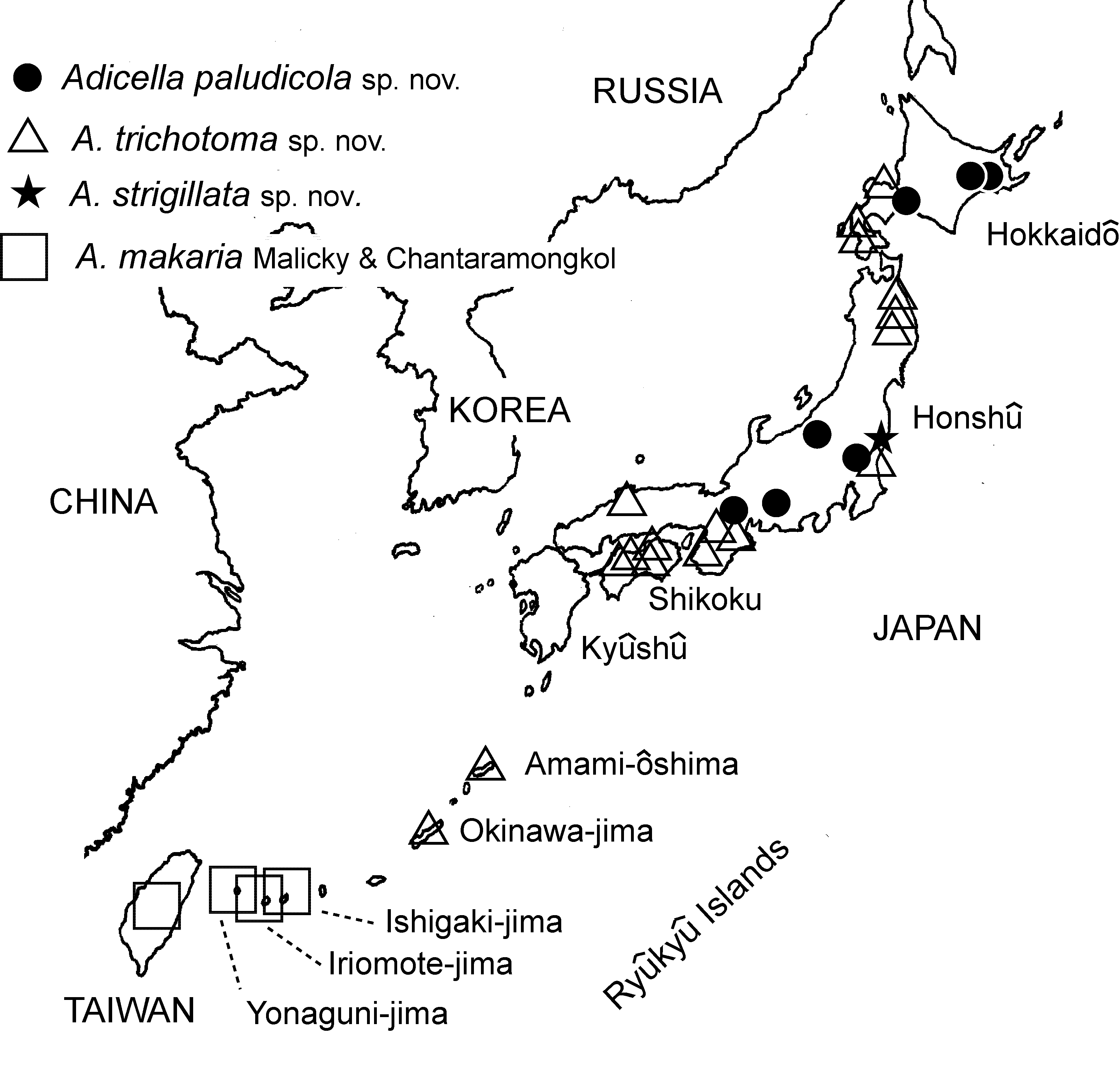
Distribution ( Fig. 6 View FIGURE 6 ). Taiwan: Nantou County, and Japan: Ryûkyû Islands (Ishigaki-jima, Iriomote-jima, Yonaguni-jima).
Japanese name. Taiwan-ko-higenaga-tobikera.
Adicella trichotoma Ito & Kuhara sp. nov. ( Figs. 2 View FIGURE 2 , 3 View FIGURE 3 , 6 View FIGURE 6 )
Adicella sp.: Kuhara 1997: 62, Japan (Hokkaidô); Kuhara 2001: 20, Japan (Hokkaidô); Ito et al. 2010: 64, Japan (Hokkaidô).
Diagnosis. Also a species of the Adicella biramosa Group (Kimmins 1963, Schmid 1994), the male of this species resembles that of Adicella mita Yang & Morse, 2000 , described from southeastern China, in having a trifurcate upper part of tergum X with shortest middle process and phallus without paramere spines. However, it clearly differs from A. mita as follows. Adicella trichotoma has (1) preanal appendages oval with no acute posterior margin in dorsal and lateral aspects, and (2) inferior appendages tribranched. On the other hand, A. mita has (1) preanal appendages orbicular with subacute posterior margin in dorsal and lateral aspects, and (2) inferior appendages mitten-like, with broad, short lower branch.
Among the A. biramosa Group for which the female is known, the female of this species resembles that of A. makaria in its short tergum X, but is distinguishable from the latter as follows: In A. trichotoma , vertical lobes of the gonopod plate are rather large, about 4/5 as large as lamellae in lateral view; in A. makaria , they are small, 1/4 as large as lamellae in lateral view.
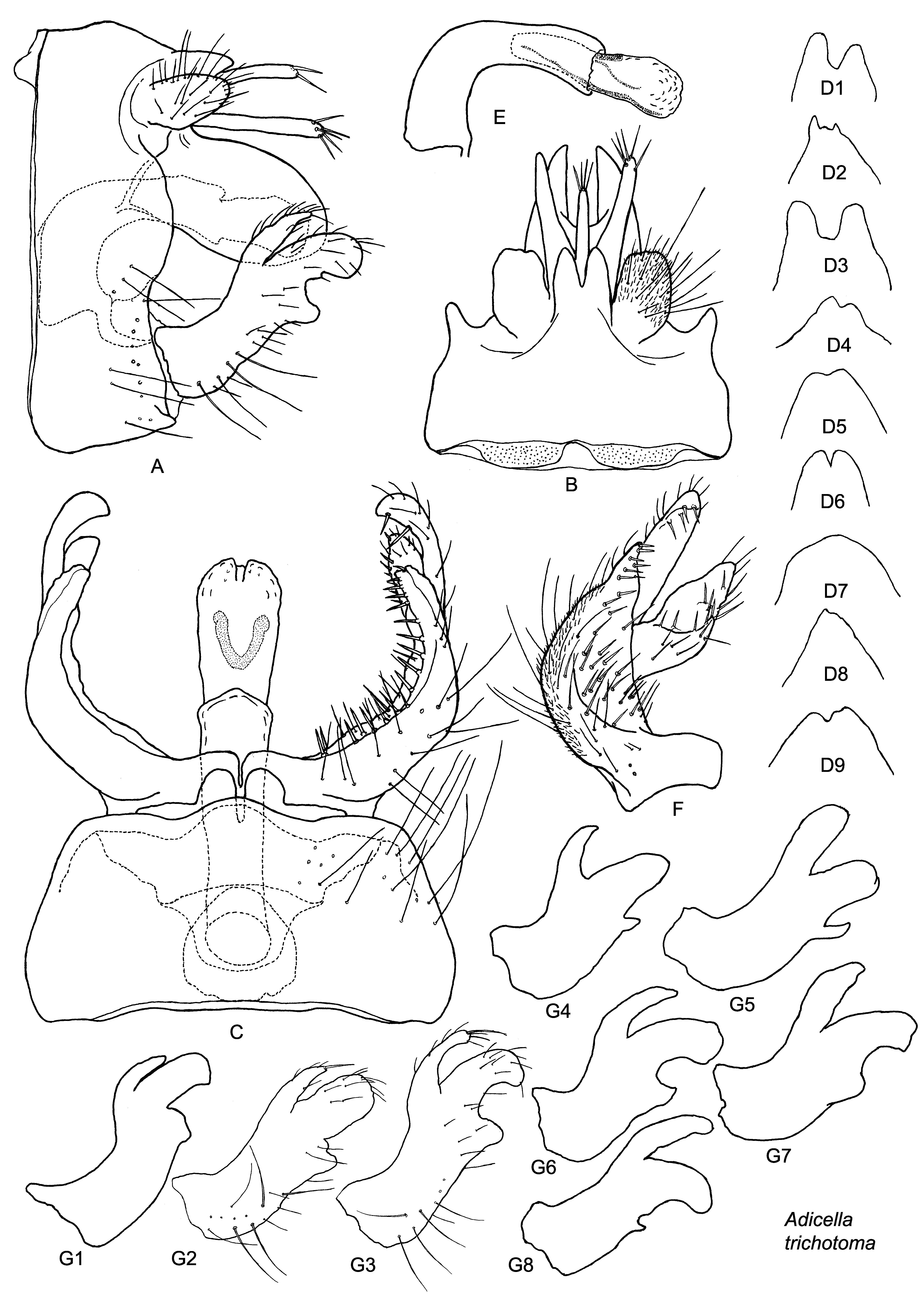
Adult ( Figs. 2 View FIGURE 2 , 3 View FIGURE 3 ). Light brown, body length 5.0– 6.5 mm in male (n=6) and 4.0– 6.5 mm in female (n=8). Antennae 3.1–3.6 times as long as body in male (n=4), 2.8–3.1 times in female (n=4); scapes thick, long, each about 4 times as long as its pedicel. Maxillary palpi each 5-segmented, total length 1.5–1.7 mm in male (n=2), 1.8–2.0 mm in female (n=5); labial palpi each 3-segmented, total length 0.5 mm in male (n=2), 0.5–0.8 mm in female (n=5); all segments of both palpi cylindrical and covered with fine setae. Warts on head and thorax as in A. makaria .
Wings: Color and venation as in A. makaria . Lengths of forewings and hind wings each 6.0–7.0 mm and 5.0– 5.5 mm respectively in males (n=7), 4.5–6.5 mm and 3.5–5.0 mm respectively in females (n=5).
Male genitalia ( Fig. 2 View FIGURE 2 ). Segment IX rectangular in lateral view with posterolateral margins slightly convex ( Fig. 2 View FIGURE 2 A), in dorsal view posterodorsal margin produced triangularly often with two lobes variable in size and shape individually ( Figs. 2 View FIGURE 2 B, D1–9), mostly asymmetrical ( Figs. 2 View FIGURE 2 D1–4, 8, 9), sometimes absent ( Figs. 2 View FIGURE 2 D7, 8). Preanal appendages oval in dorsal and lateral views ( Figs. 2 View FIGURE 2 A, B). Upper part of tergum X trifurcate, setose apically, middle process shorter than lateral processes ( Figs. 2 View FIGURE 2 A, B). Lower part of tergum X tall, hood-like, composed of two large vertical lobes fused dorsally in basal half, directed ventrocaudad ( Figs. 2 View FIGURE 2 A, B), apex of each lobe broad in lateral view ( Fig. 2 View FIGURE 2 A), subacute in dorsal view ( Fig. 2 View FIGURE 2 B). Inferior appendages each with three branches and variable in shape among sites and also individually in each site, with numerous short thick setae mesally and long slender setae ventrally and laterally ( Figs. 2 View FIGURE 2 A, C, F, G1–8): Upper branch finger-like, slightly bent ventrad ( Figs. 2 View FIGURE 2 A, G1–8); in lateral view, gently tapered with subacute apices in most specimens ( Figs. 2 View FIGURE 2 A, G2, G4, G5, G7), but parallel-sided with round apices in few specimens ( Figs. 2 View FIGURE 2 G1, G6, G8); middle branch thick, bar-like ( Figs. 2 View FIGURE 2 A, F, G), strongly curled mesad apically with subacute apex in ventral view ( Fig. 2 View FIGURE 2 C), in lateral view, thickest among three branches ( Figs. 2 View FIGURE 2 A, G), longest among three branches in most specimens ( Figs. 2 View FIGURE 2 A, G1–G4, G7), but slightly shorter than upper branch in few specimens ( Fig. 2 View FIGURE 2 G6), almost parallel sided with round or subquadrate apex in most specimens ( Figs. 2 View FIGURE 2 G1, G2, G5, G6, G7, G8), but apex slightly tapered ( Fig. 2 View FIGURE 2 G4) or broadened ( Figs. 2 View FIGURE 2 A, G3) in few specimens. Ratio of widths of middle part of middle branch to widths of middle part of upper branch 1.8–2.0 in Hokkaidô (n=7) ( Figs. 2 View FIGURE 2 A, G1), 2.0– 2.5 in Iwate (n=2) ( Fig. 2 View FIGURE 2 G2), about 3 in Ibaraki (n=4) ( Fig. 2 View FIGURE 2 G3), 1.8–2.3 in Mie (n=5) ( Fig. 2 View FIGURE 2 G4), 1.1–2.9 in Shikoku (n=9) ( Figs. 2 View FIGURE 2 D5, 7), 1.1 in Shimane (n=1) ( Fig. 2 View FIGURE 2 G6) and 1.5 in Okinawa (n=2) ( Fig. 2 View FIGURE 2 G8), with geographical cline not found in ratio; lower branch ( Figs. 2 View FIGURE 2 A, F, G) shortest, variable in size even at each site, often only protuberance ( Figs. 2 View FIGURE 2 G1, 3G7). Phallobase tube curved 90˚; paramere spines absent; phallicata tubular, almost straight, about 2/3 as long as phallobase, with U- or V-shaped phallotremal sclerite ( Figs. 2 View FIGURE 2 C, E).
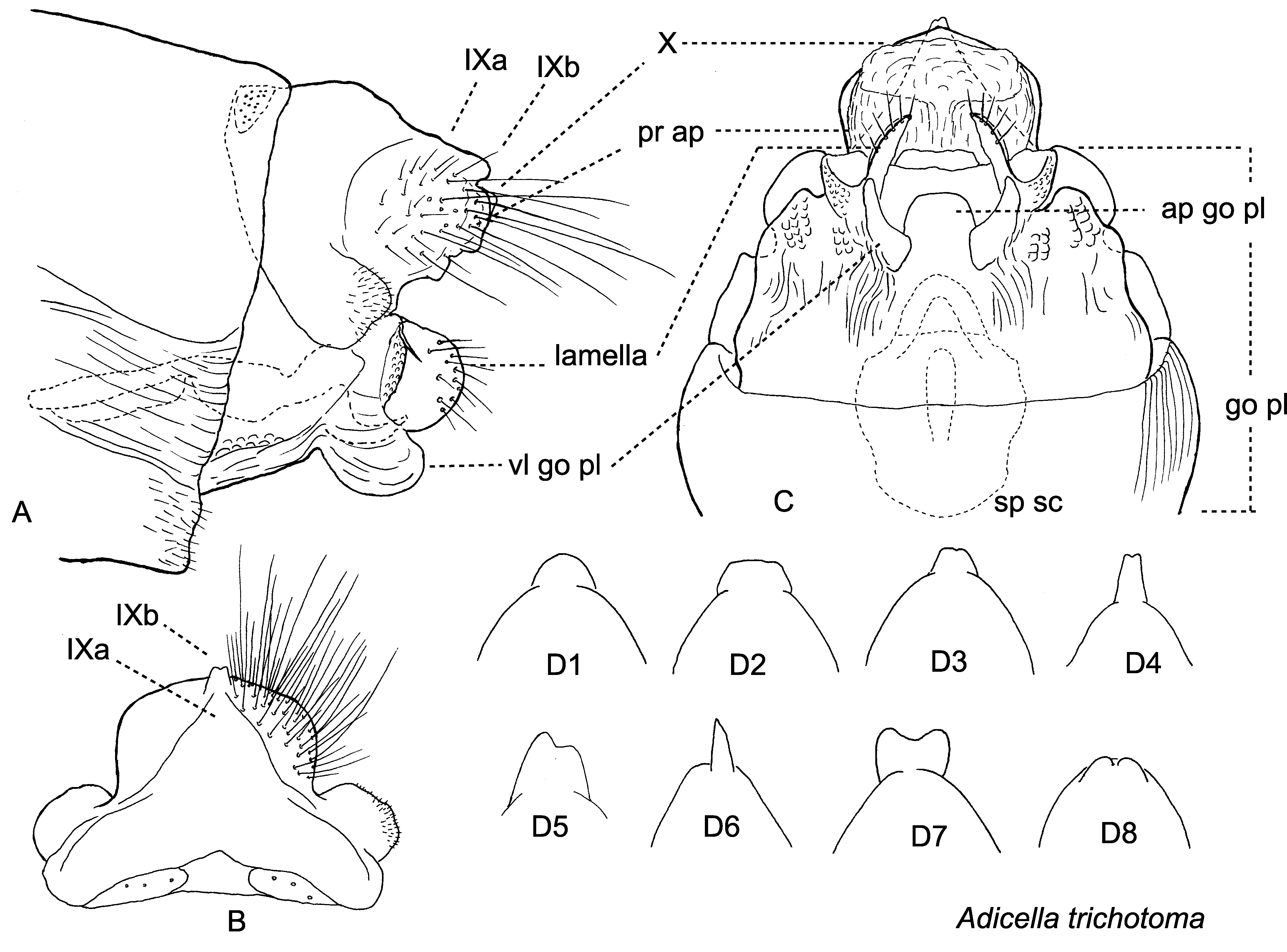
Female genitalia ( Fig. 3 View FIGURE 3 ). Segment IX short, fused with tergum X and preanal appendages ( Fig. 3 View FIGURE 3 A); terga IXa (IXa) and IXb (IXb) conspicuous in dorsal view ( Fig. 3 View FIGURE 3 B); IXb variable in size and shape individually even at each site ( Fig. 3 View FIGURE 3 D1–8), round ( Fig. 3 View FIGURE 3 D1) or subtrapezoidal ( Fig. 3 View FIGURE 3 D2), with ( Figs. 3 View FIGURE 3 B, D3–5, D7, D8) or without ( Figs. 3 View FIGURE 3 D1, 2) middle notch, asymmetrical in some specimens ( Figs. 3 View FIGURE 3 D5, 6), very short in few specimens ( Fig. 3 View FIGURE 3 D8). Preanal appendages (pr ap) represented as round, setose mounds ( Fig. 3 View FIGURE 3 A), semicircular in dorsal and ventral views ( Figs. 3 View FIGURE 3 B, C). Segment X (X) membranous, short, tube-like ( Fig. 3 View FIGURE 3 C), mesoventral slit present in few specimens. Lamellae round, vertical, setose lobe, each with rugose flap basolaterally ( Figs. 3 View FIGURE 3 A, C). Gonopod plate (go pl) broad, rugose laterally, with round vertical lobes (vl go pl) posterolaterally ( Figs. 3 View FIGURE 3 A, C); vertical lobes large, about 4/5 as large as lamellae in lateral view ( Fig. 3 View FIGURE 3 A), triangular in ventral view ( Fig. 3 View FIGURE 3 C). Apicomesal process of internal part of gonopod (ap go pl) semimembranous, subquadrate in ventral view ( Fig. 3 View FIGURE 3 C), acute in lateral view ( Fig. 3 View FIGURE 3 A). Spermathecal sclerite (sp sc) large sub-pentagonal in ventral view ( Fig. 3 View FIGURE 3 C), trapezoidal in lateral view ( Fig. 3 View FIGURE 3 A).
Holotype male: JAPAN: Hokkaidô; Otaru-shi, Okusawa-suigenchi (43˚09’N, 140˚58’E, 220 m), 26.viii.1996, Y. Sasaki & F. Takahashi, M (CBM-ZI 146707).
Paratypes: type locality, but 29.vii.1996, Y. Sasaki & F. Takahashi, M, 1 male, 2 females (CBM-ZI 146708–146710).
Other specimens. JAPAN: Hokkaidô: Otaru-shi, Okusawa-suigenchi, Anataki, 26.vii.1995, NKU, 1 female (NKU); Otaru-shi, Okusawa-suigenchi, Anataki, small stream, 26.vii.1995, NKU, 1 males, 1 female (NKU); Otaru-shi, Okusawa-suigenchi, 29.vii.1996, Y. Sasaki & F. Takahashi, M, 2 males, 5 females (1 male, 1 female, TI; 3 females, NKU); Otaru-shi, Okusawa-suigenchi, Shiraisawa, 2.viii.1996, Y. Sasaki & F. Takahashi, M, 2 males, 29 females (NKU); Otaru-shi, Okusawa-suigenchi, 9.viii.1996, Y. Sasaki & F. Takahashi, M, 10 females (NKU); Otaru-shi, Okusawa-suigenchi, 19.viii.1996, Y. Sasaki and F. Takahashi, M, 1 female (NKU); Shimamaki-mura, Chihase-gawa, Nagumono-sawa, 160 m, 5.viii.2008, TI, S, 1 male (TI); Shiriuchi-chô, Idesu-gawa, 180 m, 12.vii.2008, TI, S, 1 female (TI). Honshû. Iwate: Iwaizumi-chô, Mitagai-gawa, headwater, 18.vii.2004, NKU, 3 males, 1 female (NKU); Iwaizumi-chô, Osada, tributary of Ôkawa, 12.vii.1997, NKU, 1 male, 3 females (NKU); Iwaizumi-chô, small tributary of Akka-gawa, 650 m, 19.vii.2004, NKU, 1 male, 2 females (NKU); Miyako-shi, Kadoma, tributary of Miyama-gawa, 13.vii.1997, NKU, 1 male (NKU); Kuji-shi, Kassemba, 12.vii.1997, NKU, 1 male (NKU). Ibaraki: Hitachi-ômiya-shi, Shimoisehata, mountain stream, 25.vi.2005, NKA, 4 males, 5 females (TI); Hitachi-ôta-shi, Okami, small marsh, 8.viii.2009, NKA, 1 male, 2 females (NKA). Mie: Daian-chô, Ishigureminami, small stream, 13.vii.2001, H. Morita, M, 1 male, 3 females (TI); same data except 30.vii.2001, H. Morita, M, 1 male, 1 female (NKU); same data except 25.vii.2001, H. Morita, M, 1 male, 1 female (TI); Yokkaichi-shi, Suizawa-chô, small stream, 19.vi.2009, H. Morita, M, 1 male, 5 females (TI); same data except 5.vii.2009, H. Morita, M, 1 male (TI). Shimane: Okuizumi-chô, Sentsuzan, 5–26.vii.2006, M. Hayashi, M, 1 male (MKNM). Shikoku. Ehime: Uchiko-chô, Odamiyama, Namakusa-dani, 29.vi.2000, E. Yamamoto, M, 1 male, 1 female (TI); Uchiko-chô, Odamiyama, Odamiyama-keikoku, small stream, 15.viii.2000, E. Yamamoto, M, 5 males, 5 females (TI). Kochi: Kôchi-shi, Tosayama, Kuishiyama, 8.vi.2005, M. Takai, 1 male (TI); Kôchi-shi, Tosayama, Kuishiyama, 2.ix.2005, M. Takai, 2 males (TI); Kami-shi, Monobe, Nishikumabeppu-rindô, 25.vii.2004, M. Takai, 1 males (TI); Ino-chô, Teragawa, Yosakoi-tôge, 19.vi.2006, M. Takai, 1 male (TI); same data except 3.viii.2001, I. Yamashita, 1 male (MKNM). Ryûkyû Islands. Amami-ôshima: Amami-shi, Setouchi, Miyama-gawa, Dainimiyama-bashi, 21.iv.2008, TI, S, 3 females (TI). Okinawa-jima: Nago-shi, Genka-kawa, hygropetric habitat near Hogen-hashi, 8.iv.2011, TI, S, 2 males, 2 females (TI); Ôgimi-son, Takasato-gawa, small tributary at end of road, 17.iii.2012, TI, S, 1 male, 4 females (TI); Kunigami-son, Oguni, small stream, 21.iii.1999, TI & A. Ohkawa, S, 1 female (TI); Kunigami-son, Oku, small stream, 22.iii.1999, TI & A. Ohkawa, S, 1 female (TI); Kunigami-son, Ie, 11–15.iv.2001, K. Uesugi, M, 1 female (TI).
Etymology. The specific epithet is a latinized version of the Greek adverb “τρίχα” (=in 3 parts) and a variant of the Greek adjective “τομαϊος, -α, -οv” (=cut), referring to the shape of the three-branched upper part of tergum X. Habitat. Most adults were collected near streams in mountain area.
Distribution ( Fig. 6 View FIGURE 6 ). Japan: Hokkaidô, Honshû, Shikoku, Ryûkyû Islands (Amami-ôshima, Okinawa-jima). Japanese name. Mitsumata-ko-higenaga-tobikera.
Adicella paludicola Ito & Kuhara sp. nov. ( Figs. 4 View FIGURE 4 , 6 View FIGURE 6 )
Adicella sp. 1: Ito et al. 2007: 153, Japan (Hokkaidô); Ito & Kosugi 2007: 55, Japan (Hokkaidô).
Diagnosis. A species of the Adicella pulcherrima Group (Schmid 1994), the male of this species resembles that of A. papillosa Yang & Morse 2000 , distributed in southwestern China, in having the upper part of tergum X with seta-bearing papillae, a large lower part of tergum X and inferior appendages without branches. However, it clearly differs from the latter as follows: This species has (1) the upper part of tergum X is without a deep and wide dorsomesal slit and is never longer than the preanal appendages, (2) the lower part of tergum X is not extending beneath the phallus, and (3) the phallicata is without a phallotremal sclerite. On the other hand, A. papillosa has (1) the upper part of tergum X with a deep and wide dorsomesal slit and is much longer than the preanal appendages (2) the lower part of tergum X is extending mesad beneath the phallus, and (3) the phallicata has a U-shaped phallotremal sclerite.
The female of this species resembles that of A. trichotoma but is distinguishable from the latter as follows: In A. trichotoma , the vertical lobes of the gonopod plate are relatively large, about 4/5 times as large as the lamellae in lateral view; in A. paludicola , the vertical lobes of the gonopod plate are rather small, about 2/3 as large as the lamellae and each is largely fused to its lamella along its dorsal edge in lateral view.
Adult ( Fig. 4 View FIGURE 4 ). Light brown, body length 4.1–5.9 mm in male (n=11) and 4.0– 5.5 mm in female (n=11). Antennae 3.0–4.6 times as long as body in male (n=8), 2.8–3.5 times in female (n=5); scapes thick, long, each 2.0 times as long as its pedicel. Maxillary palpi each 5-segmented, total length 1.8–2.0 mm in male (n=3), 1.8 mm in female (n=2); labial palpi each 3-segmented, total length 0.5–0.6 mm in male (n=2), 0.7 mm in female (n=2); all segments of both palpi cylindrical and covered with fine setae. Warts on head and thorax as in A. makaria .
Wings: Shape, color and venation as in A.makaria , long hair pencil of hind wing absent. Lengths of forewings and hind wings each 6.0– 7.1 mm and 4.5–5.4 mm respectively in males (n=11), 5.3–6.2 mm and 4.3–5.1 mm respectively in females (n=11).
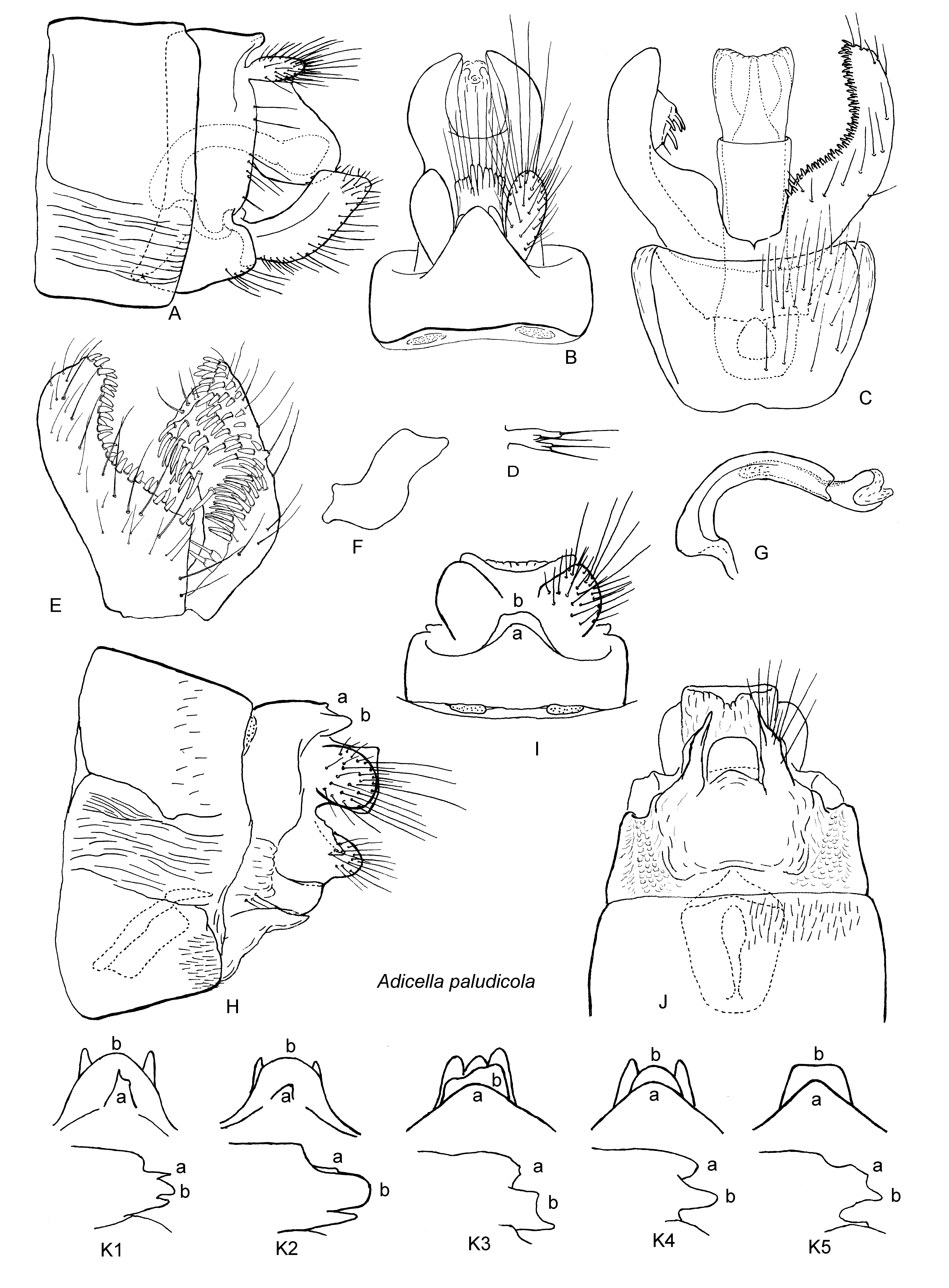
Male genitalia ( Figs. 4 View FIGURE 4 A–G). Segment IX rectangular, posterior margins almost straight or very slightly convex in lateral view ( Fig. 4 View FIGURE 4 A), small excision present near base of inferior appendages in some specimens ( Fig. 4 View FIGURE 4 A); in dorsal view posterodorsal margin produced backward at center in broad triangular process, with small lobes beneath; lobes variable in size individually and sometimes asymmetrical ( Fig. 4 View FIGURE 4 B). Preanal appendages oval ( Figs. 4 View FIGURE 4 A, B). Upper part of tergum X forming broad plate extending to, but never exceeding, tips of preanal appendages, with 10 seta-bearing papillae apically and with very narrow dorsomesal slit in few specimens ( Figs. 4 View FIGURE 4 B, D). Lower part of tergum X tall, hood-like composed of two large vertical lobes fused dorsally in basal half, each lobe directed ventrocaudad ( Figs. 4 View FIGURE 4 A, B), apices broad in lateral view ( Fig. 4 View FIGURE 4 A) and subacute in dorsal view ( Fig. 4 View FIGURE 4 B). Inferior appendages upcurved, each about 3 times as long as wide, without branches ( Figs. 4 View FIGURE 4 A, C, E, F); in lateral view, almost parallel-sided, truncate apically ( Fig. 4 View FIGURE 4 A) or truncate with protruding ventrocaudal corner ( Fig. 4 View FIGURE 4 F); at 3/5 from base small dorso-mesal protuberance present with numerous spines ( Figs. 4 View FIGURE 4 C, E). Phallobase slender tube curved 90˚; paramere spines absent; phallicata tubular, sinuate, about 4/5 times as long as phallobase, without phallotremal sclerite ( Fig. 4 View FIGURE 4 G).
Female genitalia ( Figs. 4 View FIGURE 4 H–K). Segment IX short; tergum IXa (a) produced into triangular or subtriangular lobe posteromedially ( Figs. 4 View FIGURE 4 H, I, K1–5), sometimes asymmetrical ( Figs. 4 View FIGURE 4 K1, K2); tergum IXb (b) round ( Figs. 4 View FIGURE 4 K2, K4), subtriangular ( Figs. 4 View FIGURE 4 K1) or subtrapezoidal ( Figs. 4 View FIGURE 4 I, K3, K5) in dorsal view, often with pair of small lobes ( Figs. 4 View FIGURE 4 K1, K2, K4), sometimes asymmetrical ( Fig. 4 View FIGURE 4 K3). Preanal appendages represented as broad setose mounds fused with tergum X ( Figs. 4 View FIGURE 4 H, I). Segment X forming longitudinally short tube with semimembranous ventral surface ( Fig. 4 View FIGURE 4 J). Lamellae round lobes, flattened laterally, each with oblique ridge on outer surface, posterior 2/5 covered with setae ( Figs. 4 View FIGURE 4 H, J). Gonopod plate broad, subsquadrarte, lateral regions rugose with vertical lobes on posterolateral margins ( Figs. 4 View FIGURE 4 H, J); vertical lobes round, rather small, about 2/3 as large as lamellae and each fused to its lamella along its dorsal edge in lateral view ( Fig. 4 View FIGURE 4 H), thin and triangular in ventral view ( Fig. 4 View FIGURE 4 J). Apicomesal process of internal part of gonopod subquadrate or semicircular ( Fig. 4 View FIGURE 4 J). Spermathecal sclerite pentagonal in ventral view ( Fig. 4 View FIGURE 4 J), trapezoidal in lateral view ( Fig. 4 View FIGURE 4 H).
Holotype male: JAPAN: Hokkaidô, Kushiro Shitsugen, Shibecha-chô, Kayanuma, Shirarutoro-ko, Ikoi-noie, (43˚11’N, 144˚30’E, 8 m), 28.vii.2012, TI, L (CBM-ZI 146711).
Paratypes: Same data as holotype, 2 males, 2 females (CBM-ZI 146712–146715).
Other specimens. JAPAN: Hokkaidô. Same data as holotype, 5 males, 16 females (TI); same data except 22.vii.2003, TI, L, 1 male (TI); same data except 7.viii.2006, TI, L, 1 male, 1 female (NKU); same data except 28.vii.2012, TI, L, 5 males, 7 females (TI); Kushiro Shitsugen, Shibecha-chô, Kayanuma, Shirarutoro-etoro-gawa, Tômi-bashi, 4.xi.2008, TI, L, 2 males, 2 females (TI); same data except 25.vii.2008, TI, L, 1 female (TI); Kushiro Shitsugen, Kushiro-shi, Kirakotan-misaki, 16.vii.2005, T. Kosugi, L, 1 male (TI); Yûfutsu Shitsugen, Tomakomaishi, Uenae, Bibi-gawa, 4 m, 16.vii.2010, TI, L, 1 female (TI). Honshû. Ibaraki: Hitachi-ôta-shi, Okami, small marsh, 24.vi.2006, NKA, 13 males, 3 females (11 males, 2 females, TI; 2 males, 1 female, NKU); same data except 8.viii.2009, NKA, 1 male, 17 females (NKA); same data except 16.vii.2012, NKA, 2 males, 3 females (NKA). Niigata: Asahi-mura, Miomote-gawa, Futagoshima, Shinrin-kôen, 10.ix.2003, TI, L, 1 male (TI). Aichi: Toyotashi, Asuke, Tanoshiri Shitsugen, 16.vii.2000, N. Kawase, 1 male (MKNM). Shiga: Kôka-shi, Kôka, Aburahi, Okunoin-shicchi, 6.vii.2008, N. Kawase, 1 male (MKNM).
Etymology. The specific epithet is from the Latin noun “ palus, - udis ” (=marsh) and Latin suffix “- colus, - a, - um ” (=living in), indicating that this species is a dweller of marshes.
Habitat. Most adults were collected in marshes.
Distribution ( Fig. 6 View FIGURE 6 ). Japan: Hokkaidô, Honshû.
Japanese name. Numa-ko-higenaga-tobikera.
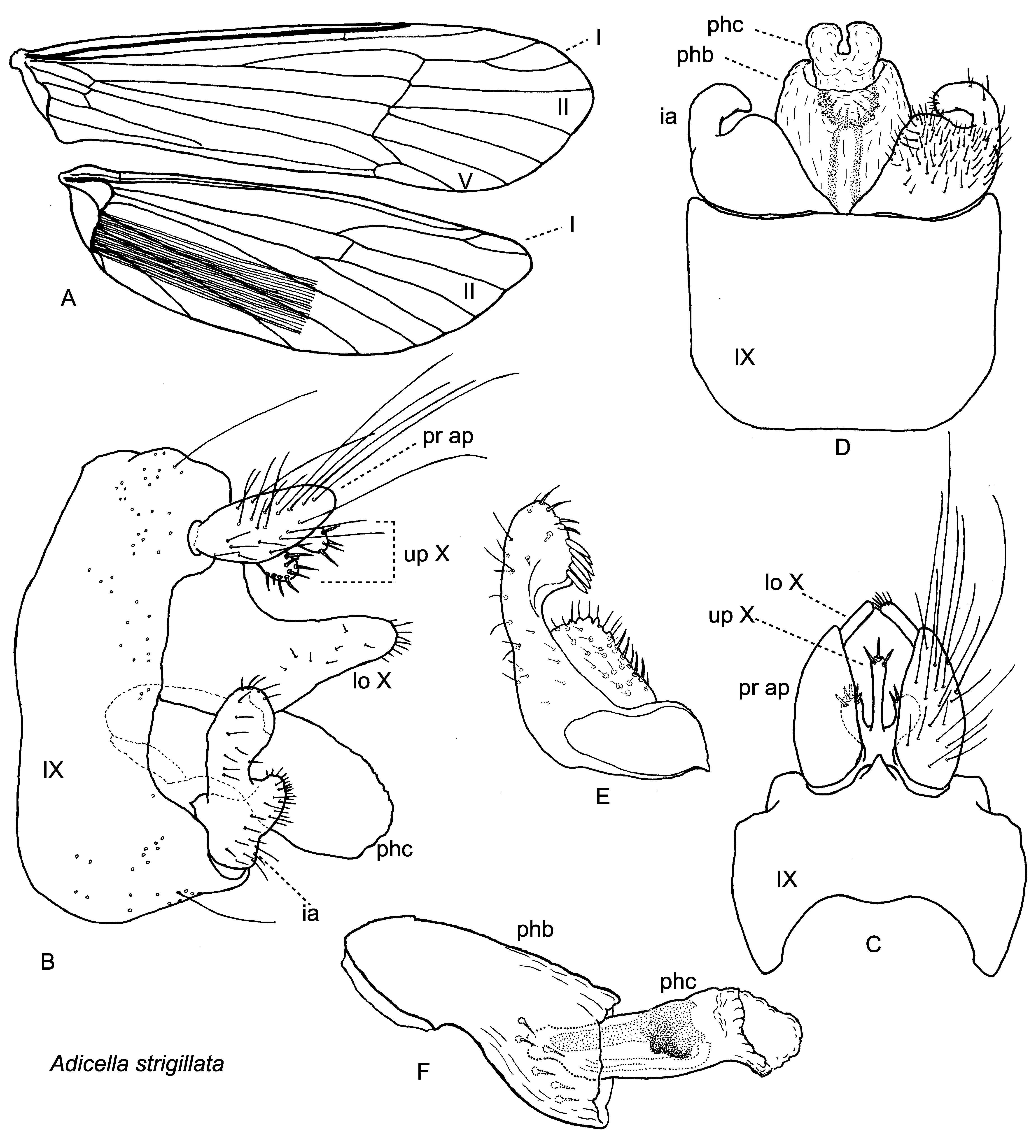
Adicella strigillata Katsuma & Ito sp. nov. ( Figs. 5–6 View FIGURE 5 View FIGURE 6 )
Adicella sp. 1: Katsuma 2011: 68, Japan (Honshû). Adicella sp. 3: Katsuma 2012: 67, Japan (Honshû).
Diagnosis. Also a species of the Adicella pulcherrima Group (Schmid 1994), the male of this species resembles that of A. penicillaris Yang & Morse 2000 , described from southeastern China, in having short inferior appendages and long hair-brushes of the hind wings. However, it differs from A. penicillaris by (1) the upper part of tergum X having only a single process in A. penicillaris , but with three processes in A. strigillata and (2) the lower part of tergum X being strongly sclerotized on its ventral margins and with its apices each produced into an acute dorsolateral projection and not bearing setae in A. penicillaris , but the lower part of tergum X is not sclerotized on its ventral margins and has rounded apices bearing apical setae in lateral view in A. strigillata .
Adult male ( Fig. 5 View FIGURE 5 ). Pale brown, body 4.2–5.0 mm long in male (n=3). Male antennae 3.6 times as long as body (n=1); scapes thick, long, each 2.0 times as long as its pedicel. Maxillary palpi each 5-segmented, 2.6 mm in total (n=2); labial palpi each 3-segmented, 0.8 mm in total (n=3); all segments of both palpi cylindrical and covered with fine setae. Warts on head and thorax as in A. makaria .
Wings ( Fig. 5 View FIGURE 5 A). Forewings broader than those of other Japanese Adicella species with round apical margin, mostly covered with brown hairs. Hind wings broader than other Japanese Adicella species, each with slightly acute apical margin, covered with brown hairs, with long fringes at posterior margins; dark brown or black hairbrushes conspicuous, long, 2/3 as long as hind wings, arising from jugal lobe. Forewings each with apical forks I, II and V, fork I broad with long (as long as fork I) stalk, fork II sub-rectangular, fork V sessil; discoidal cell long, 1/3 as long as wing; thrydial cell very long, about 1/2 as long as wings; presence of cross vein sc - r variable individually even in opposite sides of single specimen; Cu2 and P connected by cu - p crossvein, then P fused with E+1A+2A to form single curved vein ending at arculus; 1A often not reaching 2A. Hind wings each with apical forks I and II, fork I short and broad, with very long stalk (about 2.5 times as long as fork I), fork II subrectangular; discoidal and thyridial cells absent; Sc and R fine, fused into single vein at midlength of wing, Sc+R slightly sinuate apically. Lengths of forewings and hind wings each 6.2–6.6 mm and 4.7–5.0 mm respectively in male (n=3).
Male genitalia ( Fig. 5 View FIGURE 5 ). Segment IX (IX) rectangular, anterior margins slightly convex and posterior margins widely concave in lateral view ( Fig. 5 View FIGURE 5 B); in dorsal view posterodorsal margin produced backward at center in triangular process ( Fig. 5 View FIGURE 5 C). Preanal appendages (pr ap) oval ( Figs. 5 View FIGURE 5 B, C). Upper part of tergum X (up X) trifurcate, setose apically, middle process longer than lateral processes ( Figs. 5 View FIGURE 5 B, C). Lower part of segment X (lo X) tall, hood-like, composed of two large vertical lobes fused dorsally at base, each lobe broad in basal half and narrow in apical half, round apically in lateral view, bar-like in dorsal view ( Figs. 5 View FIGURE 5 B, C). Inferior appendages thick and short, each about 2 times as long as basal width, with humplike ventral branch ( Fig. 5 View FIGURE 5 B); basal half broad and setose, apical half narrower, round, with many spines along subapicomesal margin in ventral and dorsal views ( Figs. 5 View FIGURE 5 D, E). Phallobase (phb) thick almost straight, with about 10 short setae arranged in circle on inner surface of apical half; paramere spines absent; phallicata (phc) tubular, with phallotremal sclerite; phallotremal sclerite Ushaped in ventral view and subquadrate in lateral view ( Figs. 5 View FIGURE 5 D, F).
Female. Unknown.
Holotype male: JAPAN: Honshû, Ibaraki, Takahagi-shi, Kami-kimida, Takinokura-shitsugen (36˚47’N, 140˚32’E), 18.vii.2010, NKA, L (CBM-ZI 146716).
Paratypes: Same data as holotype, 1 male (CBM-ZI 146717).
Other specimens. Honshû, Ibaraki: Same data as holotype, 1 male (TI); Hitachi-ôta-shi, Okami-shitsugen, 19.viii.2006, NKA, 2 males (NKA).
Etymology. The specific epithet is from the Latin noun “ strigil ” (=scraper) and the Latin suffix “- atus, - a, - um ” (=possessing), referring to the long hair-brushes of the hind wings.
Habitat. Specimens were collected near small marshes.
Distribution ( Fig. 6 View FIGURE 6 ). Japan: Honshû.
Japanese name. Chômô-ko-higenaga-tobikera.
No known copyright restrictions apply. See Agosti, D., Egloff, W., 2009. Taxonomic information exchange and copyright: the Plazi approach. BMC Research Notes 2009, 2:53 for further explanation.