
Drepanosticta rahmani, Dow & Choong & Ng, 2017
|
publication ID |
https://doi.org/10.11646/zootaxa.4338.1.2 |
|
publication LSID |
lsid:zoobank.org:pub:84880B4B-2BA6-48FA-B1B8-5232EC1C38A3 |
|
DOI |
https://doi.org/10.5281/zenodo.5998899 |
|
persistent identifier |
https://treatment.plazi.org/id/038D87BA-FFB6-FF9E-C3ED-659DEB610631 |
|
treatment provided by |
Plazi |
|
scientific name |
Drepanosticta rahmani |
| status |
sp. nov. |
Drepanosticta rahmani View in CoL sp. nov.
( Figs 1 – 15, 17 View FIGURES 1 – 2 View FIGURES 3 – 6 View FIGURES 7 – 12 View FIGURES 13 – 17 )
Holotype. ♂ (PM16_PST16), steep tributary to stream in hills between Baling and Gulai, north west Kedah, Malaysia, 15 xi 2016, leg. R.A. Dow, to be deposited in the Natural History Museum, London.
Paratypes. 7 ♂ (PM16_PST9–15), data as holotype; 1 ♂ (PM16_PST7), 1 ♀ (PM16_PST8; associated with males by supposition) data as holotype except 16 ix 2016. Paratypes to be deposited in the Centre for Insect Systematics at Universiti Kebangsaan Malaysia, the Forest Research Institute Malaysia, and the Naturalis Biodiversity Centre.
Etymology. The species is named for Dato' Sri Dr. Hj. Abd. Rahman bin Hj. Abd. Rahim former Director- General of Forestry Peninsular Malaysia, in gratitude for granting us permission to conduct research in the Forest Reserves of Peninsular Malaysia; rahmani , a noun in the genitive case.
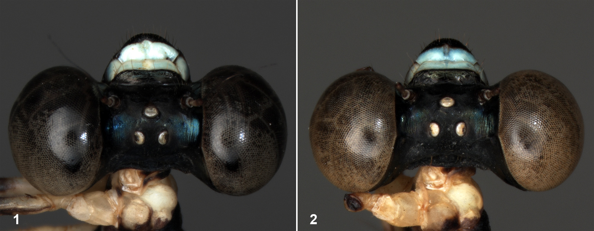
Description of holotype. Head ( Fig. 1 View FIGURES 1 – 2 ). Labium almost entirely pale. Anteclypeus, upper parts mandible bases, basal ca two-thirds labrum pale blue, most of remainder of head black except antennae whitish at top of scape, much of pedicel pale greyish white, brown at base and apex, rest missing. Ocelli whitish. Ratio of width of compound eye to width of vertex measured at level of lateral ocelli a little less than 0.9. Transverse occipital carina prominent.
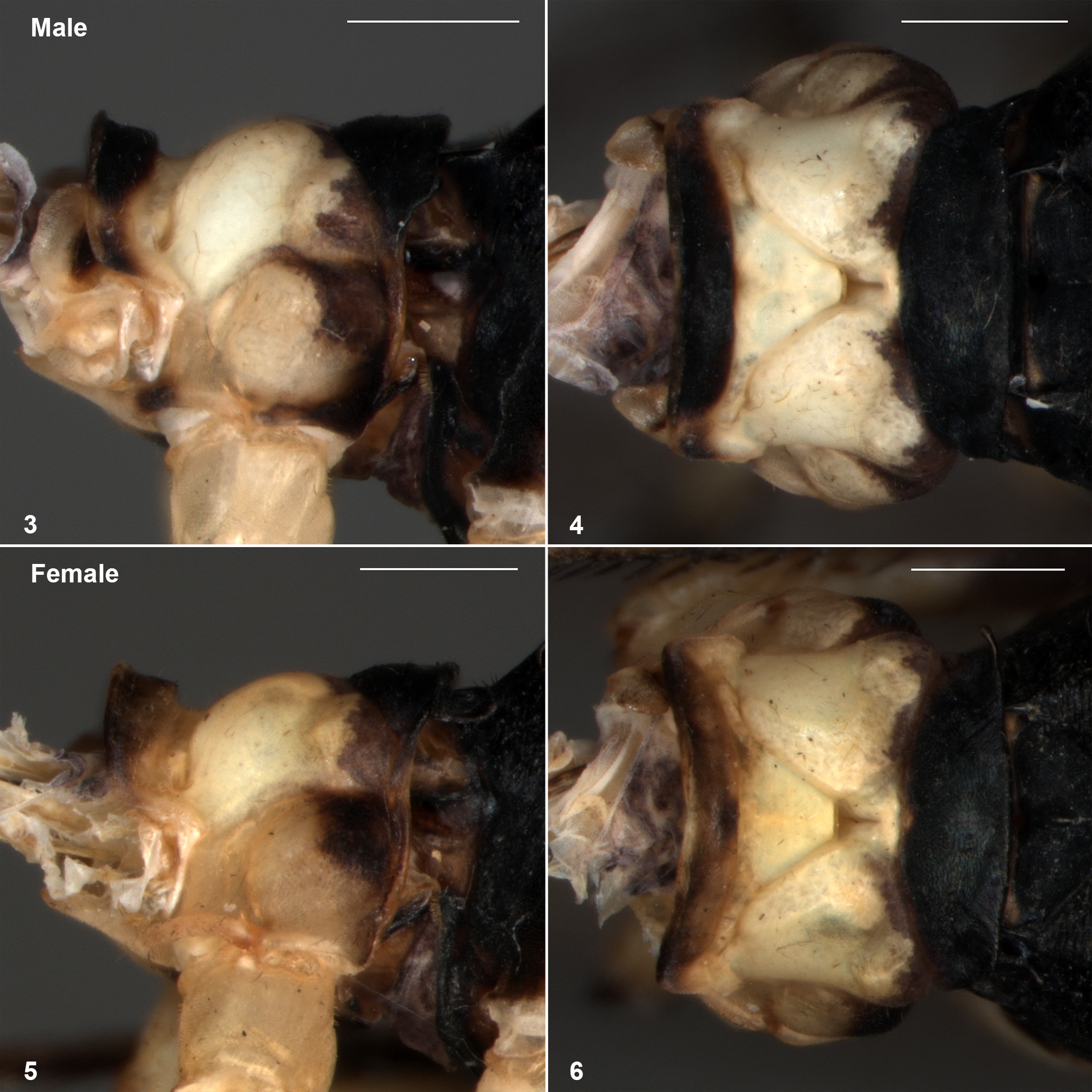
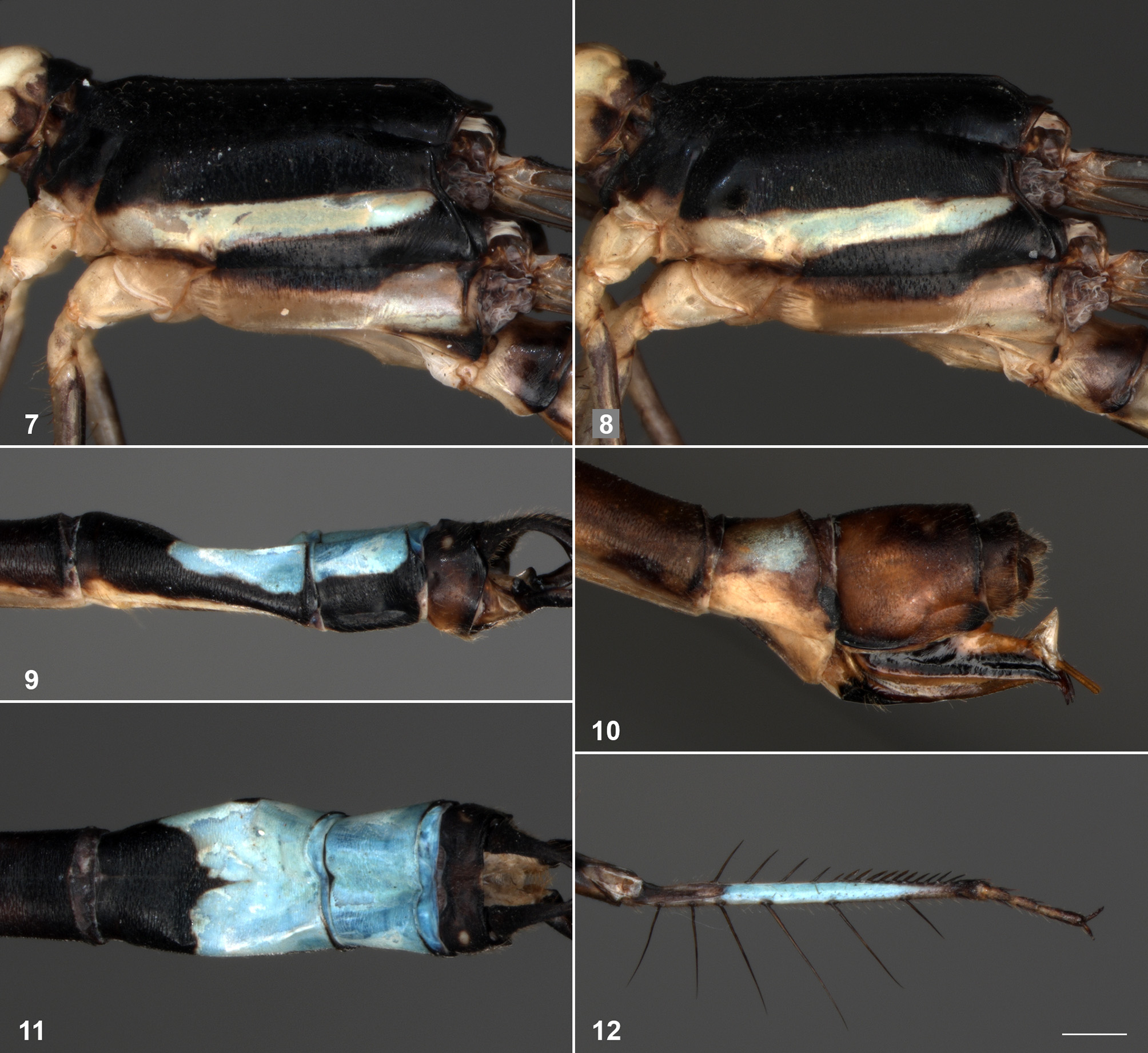
Thorax ( Figs 3–4 View FIGURES 3 – 6 , 7 View FIGURES 7 – 12 ). Propleuron ( Fig. 3 View FIGURES 3 – 6 ) largely whitish, with black and brown areas to rear, continued along much of notopleural suture. Raised part anterior pronotal lobe mostly black with whitish areas laterally, rest whitish. Middle pronotal lobe almost entirely pale except narrow grey marks to rear. Posterior lobe black, sub rectangular in dorsal view ( Fig. 4 View FIGURES 3 – 6 ), widest centrally, corners of free margin slightly produced. Synthorax ( Fig. 7 View FIGURES 7 – 12 ): mesepisternum entirely bronzy black with slight metallic greenish reflection; mesepimeron black; mesokatepisternum black and dark brown apart from rear corner above coxa where obscurely pale; antealar triangles brown; metepisternum black with bluish white band under interpleural suture, narrowing from legs to antealar carina ( Fig. 7 View FIGURES 7 – 12 ); metepimeron dirty white with black mark under metepleural suture for much of its length; venter of synthorax dirty white. Legs: each with coxa and trochanter whitish, small, obscure dark marks rear of trochanters; femurs whitish, mottled black and pale on extensor surfaces, large black marks flexor surfaces; anterior tibiae with pale extensor surfaces with poorly defined dark longitudinal stripe, much of flexor surface vivid pale blue (as in Fig. 12 View FIGURES 7 – 12 , which shows a paratype), remainder mostly dark; tibiae of other legs without blue marks, extensor surface greyish, darker near femur, flexor surface blackish; tarsi on all legs pale brownish, dark at joints, claws brown.
Wings. 14 (left) or 13 (right) Px in Fw, 12 Px in Hw. Vein Ab present, separating from Ac at or just after origin. Arculus slightly distal to Ax2. R4 arising at subnodus (very slightly proximal to it in right Fw, very slightly distal to it in right Hw), IR3 joined to it by a short stalk. Pterostigma trapezoidal with costal side shorter than anal side, slightly higher than long, black with narrow incomplete pale border, covering ca one underlying cell, a vein running slantwise from near its lower distal corner to wing margin except on right Hw.
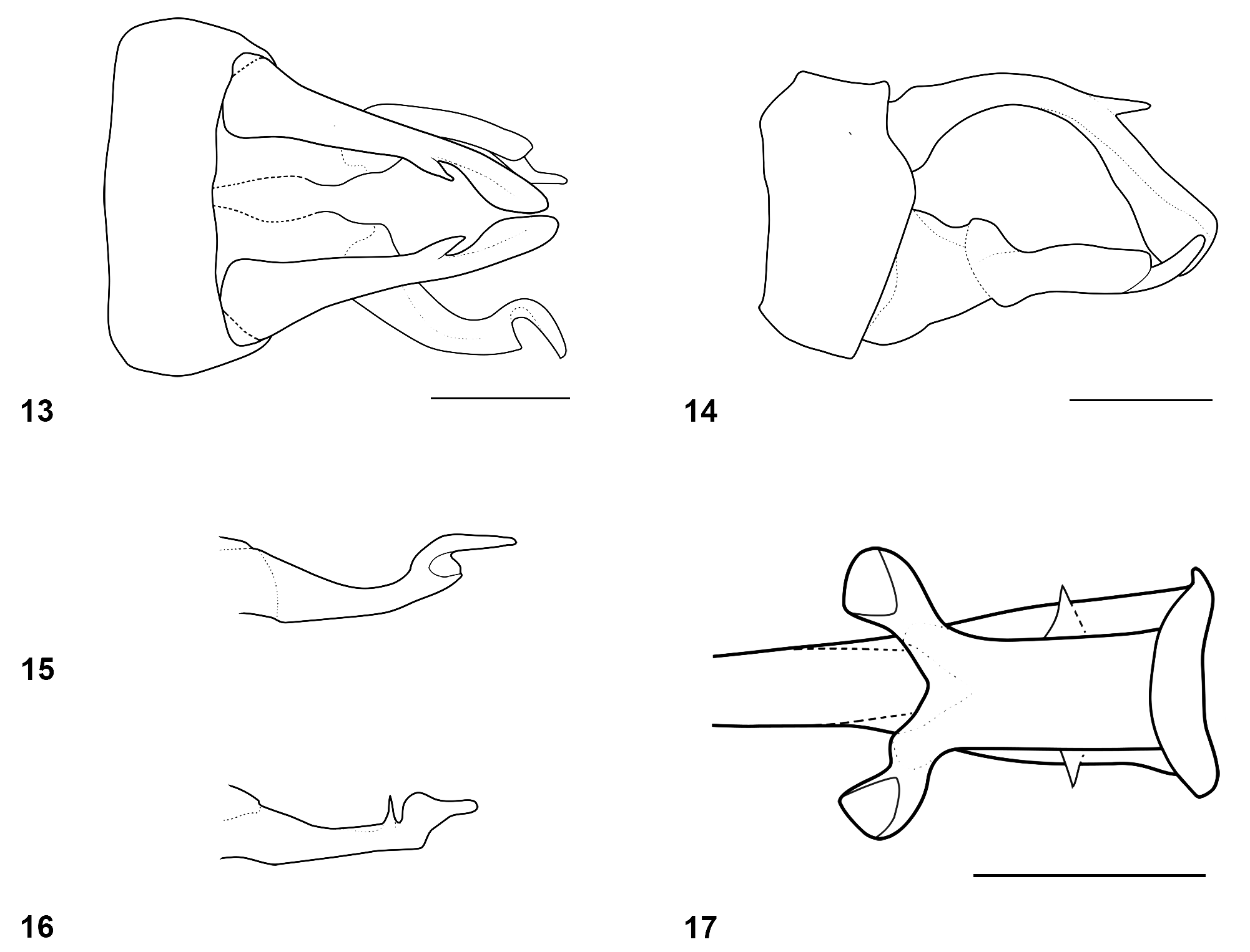
Abdomen. S1 whitish along lower margin tergite, somewhat extended up at base, brown above becoming darker apically. S2 brown above, laterally with whitish wedge shaped mark, highest basally, narrowing toward apex. S3–S7 dark brown with basal lower lateral whitish marking continued narrowly at base as incomplete basal annulus, large subapical pale area lower laterally; both these pale markings becoming smaller and less clear on successive segments. S8–S10 widened, S8, S9 with somewhat squashed appearance ( Figs 9, 11 View FIGURES 7 – 12 ). S8 black, pale stripe along lower margin tergite, dorsally with apical slightly more than half blue, S9 black, blue dorsally, S10 black, brown lower laterally. Anal appendages brown and black ( Figs 13–15 View FIGURES 13 – 17 ): cerci black at base becoming brown towards apex, in lateral view ( Fig. 14 View FIGURES 13 – 17 ) strongly and smoothly arched, somewhat ventrally expanded apically, with prominent interior-dorsal spine, directed to rear, at ca two-thirds of their length; apex square. There is a small notch in the lower corner of the apex, not visible in the figures. Convergent in dorsal view ( Fig. 13 View FIGURES 13 – 17 ), apices rounded in this view. Paraprocts brown at base, pale before a sub-basal articulation, black and very dark brown in apical half. In lateral view ( Fig. 14 View FIGURES 13 – 17 ) broad at base then tapering to articulation, thereafter of ca constant width until twisted in and slightly back, abruptly narrowed and tapering to form blade like apex; shape of apical part best understood from ventral view ( Fig. 15 View FIGURES 13 – 17 ). Genital ligula, simple, unremarkable, as in Fig. 17 View FIGURES 13 – 17 (which shows a paratype), with terminal segment divided apically into two arms which rest on either side of shaft, apices of these curled back over themselves.
Measurements (mm). Abdomen without anal appendages 37, cercus ca 1, Hw 23.
Description of female paratype. Overall very similar to male, as male except as noted below. Head ( Fig. 2 View FIGURES 1 – 2 ). As male.
Thorax. Dark parts of anterior pronotal lobe less extensive, brown rather than black ( Figs. 5–6 View FIGURES 3 – 6 ). Corners of free margin of posterior pronotal lobe strongly produced laterally, that on right turned up and forwards, visible in Fig. 6 View FIGURES 3 – 6 , left corner damaged, pushed to rear and depressed so lying against mesostigmal plate, not clearly visible in figures. Legs (left anterior leg missing below trochanter) without any blue on anterior tibia; extensor surface of this black. Wings. Vein Ab widely separated from Ac except in left Hw where they arise at same point. Arculus arising at or only very slightly distal to Ax2. 14 Px in Fw, 12 Px in Hw. Pt with well defined, complete, pale border; without vein running from distal side to wing margin.
Abdomen. S2 lateral pale mark extending further dorsally at base, so nearly forming a complete annulus, basal pale marks on S4–7 broad pale annuli, slightly obscured by brown around the dorsal midline on S4, S5. S8 pale lower laterally, brownish above, most of dorsum obscurely pale with pair of poorly defined blue dorsal lateral marks. S9 brown, darker apically and dorsally, S10 brown. Cerci shorter than S10, triangular in lateral view. Ovipositor extending beyond tips of cerci, strong subapical dorsal triangle before styles ( Fig. 10 View FIGURES 7 – 12 ), apex curved strongly down, black and pale, styles pale brown.
Measurements (mm). Abdomen without anal appendages or ovipositor ca 33, Hw 23.5.
Variation in male paratypes. Aside from small variations in markings, and size variation, the main variation seen in the type series is in some features of wing venation. The arculus frequently arises very close to Ax2, as in the supposed female. Vein Ab is occasionally absent in one to three wings and is often broadly separated from Ac at their origins. R4 is often clearly distal to the subnodus. The diagonal vein running from the distal side of Pt to the wing margin is absent more often than not, but is present in all wings in one paratype. Pt typically has a complete white border. One paratype has the distal ca one third of the right Fw improperly formed. The blue dorsal mark on S8 sometimes occupy the apical ca two thirds of the segment. S8 and/or S9 frequently exhibit a rather distorted appearance, as in the holotype, even though none of the specimens concerned appear at all teneral.
Measurements (mm). Abdomen without anal appendages 34.5–39, Hw 21–24. There are 13–14 Px in Fw, 12– 13 Px in Hw, with 11 Px in one Hw of one specimen.
Diagnosis. The male is separated from all regional species except those of the D. quadrata –group by the defining characters of the group listed in the introduction. It is easily separated from all other members of the D. quadrata –group by the blue occupying most of the flexor surface of the anterior tibiae, the lack of any sub-apical spine on the paraprocts and the shape of apical part of the paraproct, not just twisted to the side of the main body of the paraproct, but hooked slightly back from the main body ( Figs 13, 15 View FIGURES 13 – 17 ).
Remarks. The female is associated with the male by reasonable supposition; it was found in the same part of the same stream system as males. However reliable separation of females in the D. quadrata –group is problematic at present; they do not provide many diagnostic characters. It is therefore possible that the female described actually belongs to some other species.
No known copyright restrictions apply. See Agosti, D., Egloff, W., 2009. Taxonomic information exchange and copyright: the Plazi approach. BMC Research Notes 2009, 2:53 for further explanation.
|
Kingdom |
|
|
Phylum |
|
|
Class |
|
|
Order |
|
|
Family |
|
|
Genus |