
MIRINAE
|
publication ID |
https://doi.org/10.15298/rusentj.29.1.04 |
|
persistent identifier |
https://treatment.plazi.org/id/038D87BC-FFC7-FF9F-9FDE-0F56FC9086BB |
|
treatment provided by |
Felipe |
|
scientific name |
MIRINAE |
| status |
|
K EY TO THE IRANIAN GENERA OF MIRINAE View in CoL SUBFAMILY
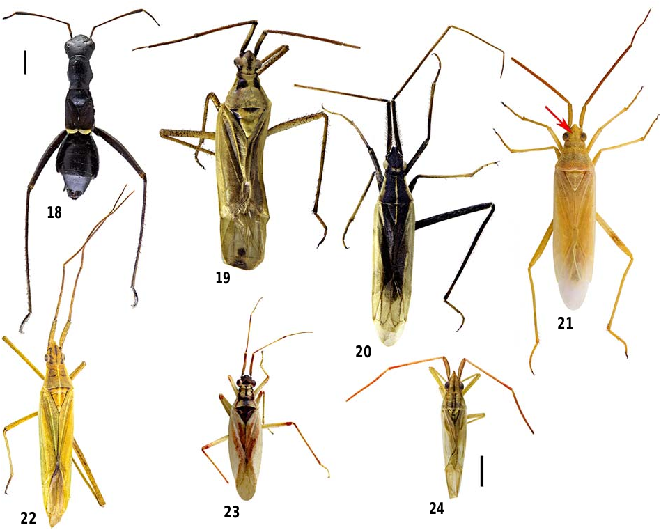
1. Ant like habitus ( Fig. 18 View Figs 18–24 ), collar completely obsolete, brachypterous (if macropterous, medially constricted), anterior segments of bulbous abdomen strongly constricted, lateral margins of abdomen abruptly upturned (tribe Herdoniini ) ............................................. Camponotidea
— Body not as above, collar distinct or posterior margin medially obsolete, macropterous, anterior segments of abdomen not constricted, lateral margins of abdomen not as above ......................................................................... 2
2. Body long and slender, head porrect, collar indistinct and medially not demarcated, antennal fossa stylate, lateral margins of pronotum carinate, 1 st segment of metatarsus longer than other segments (tribe Stenodemini ) ......... 3
— Body oval or elongate–oval, head hypognathous, collar distinct, antennal fossa not stylate, lateral margins of pronotum not carinate (except Pantilius ), 1 st segment of metatarsus shorter than others or subequal to 2 nd segment (except Stenotus ) (tribe Mirini ) ................................... 8
3. Vertex with a longitudinal median sulcus ( Fig. 21 View Figs 18–24 ), eyes almost touching anterior margin of pronotum ............ 4
— Vertex with indistinct longitudinal median sulcus, eyes removed from pronotum ............. Leptopterna ( Fig. 19 View Figs 18–24 )
4. Pronotum, scutellum and sometimes hemelytra distinctly and deeply punctate, vesica without spiculum............... ........................................................ Stenodema ( Fig. 21 View Figs 18–24 )
— Pronotum,scutellum and hemelytra impunctate or smoothly rugose, vesica with spiculum ................................... 5
5. Body large (6–8.5 ♂; 8– 9 ♀ mm), 1 st antennal segment much longer than head width across eyes .................... 6
— Body small (<5.5 ♂; 5– 6 ♀ mm), 1 st antennal segment almost as long as or slightly longer than head width across eyes ................................................................................ 7
6. Clypeus visible from dorsal view, apex of frons smooth, metatibia with short adpressed setae, vesica with two spiculae ( Fig. 30 View Figs 25–33 ), sclerotized rings ovate and relatively small, dorsal structure small or obsolete ( Figs 27–28 View Figs 25–33 ) .... ................................................. Megaloceroea ( Fig. 22 View Figs 18–24 )
— Clypeus not visible from dorsal view,apex of frons notched, metatibia with long erect setae, vesica with single spiculum ( Fig. 26 View Figs 25–33 ); sclerotized rings almost subrectangular and large, dorsal structure large ( Fig. 25 View Figs 25–33 ) ......... Notostria ( Fig. 20 View Figs 18–24 )
7. Clypeus strongly projected, mandibular plates visible from dorsal view, apex of frons almost acute, eyes small, not globular; 1 st antennal segment proximally as thick as its apical part; vesica with single spiculum ( Fig. 32 View Figs 25–33 ); sclerotized rings small, dorsal labiate plate small and not developed ( Fig. 29 View Figs 25–33 ) ..................... Trigonotylus ( Fig. 24 View Figs 18–24 )
— Clypeus blunt, mandibular plate not visible from dorsal view, apex of frons rounded, eyes large and globular; 1 st antennal segment proximally thicker than apically, vesica with 2 or 3 spiculae ( Fig. 33 View Figs 25–33 ); sclerotized rings obsolete, dorsal labiate plate well developed and strongly spinose ( Fig. 31 View Figs 25–33 ) ........................................ Teratocoris ( Fig. 23 View Figs 18–24 )
8. Dorsal surface punctuate or weakly punctate .............. 9
— Dorsal surface impunctate .......................................... 19
9. Vertex carinate ............................................................ 10
— Vertex not carinate ...................................................... 16
10. Tibia with golden spines ............................................. 11 — Tibia with dark spines ................................................. 13
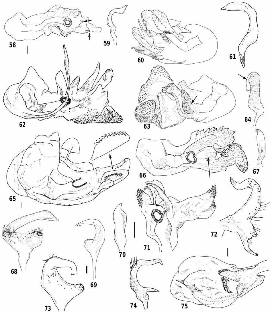
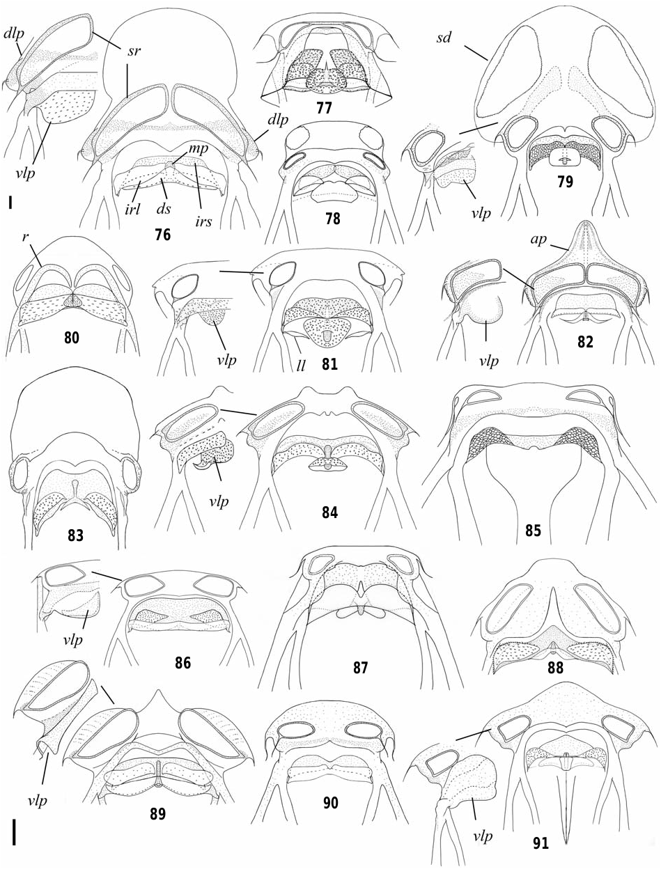
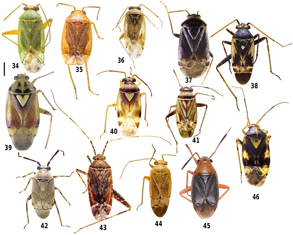
11. Dorsum covered with a mixture of whitish and blackish setae, sensory lobe of left paramere dentate, basally with a small projection and slender process ( Fig. 72 View Figs 58–75 ), vesica without spiculum, sclerotized rings of posterior wall large ( Fig. 88 View Figs 76–91 ) ...................................... Taylorilygus ( Fig. 34 View Figs 34–46 )
— Dorsum covered only with whitish setae, sensory lobe of left paramere smooth, basally without any projection or process, vesica with spiculum, sclerotized rings of posterior wall small ............................................................. 12
12. Body robust and oval, buccula broad, labium reaching mesocoxa (rarely metacoxa), 2 nd antennal segment 2.7– 3.5x as long as first, equals to or shorter than head width, left paramere with almost straight apophysis, distinctly elongated ( Fig. 68 View Figs 58–75 ); posterior wall with large, medially fused interramal lobes ( Fig. 87 View Figs 76–91 ) ................... ........................................................ Agnocoris ( Fig. 35 View Figs 34–46 ) — Body elongate–oval, buccula narrow, labium reaching or surpassing metacoxa, 2 nd antennal segment 4.0–4.3x as long as first, much longer than head width, left paramere with curved apophysis; posterior wall with small, separated interramal lobes ( Fig. 78 View Figs 76–91 ) ............ Pinalitus ( Fig. 36 View Figs 34–46 )
13. Vestiture composed of pale and black setae, vesica with sclerotized appendages; posterior wall without dorsal structure ...................................................................... 14
— Vestiture uniformly whitish, vesica without sclerotized appendages; posterior wall with dorsal structure ...... 15
14. Pronotum coarsely punctuate,cuneus reddish to dark brown, basally with a black marking at inner part, apophysis of left paramere apically expanded ( Fig. 59 View Figs 58–75 ), vesica with two appendages ( Fig. 58 View Figs 58–75 ); outer margin of sclerotized rings rounded ( Fig. 90 View Figs 76–91 ) ...................... Charagochilus ( Fig. 37 View Figs 34–46 )
— Pronotum finely punctuate, cuneus medially red (in species of the subgenus Poeciloscytus present in Iran), apophysis of left paramere apically not expanded ( Fig. 61 View Figs 58–75 ), vesica with more than two appendages ( Fig. 60 View Figs 58–75 ); outer margin of sclerotized rings acute ( Fig. 85 View Figs 76–91 ) ..... Polymerus ( Fig. 38 View Figs 34–46 )
15. Body small ( 3.5–5.2 mm) and elongate–oval, collar narrow ( 0.06 mm), sensory lobe of left paramere with small tubercles ( Fig. 69 View Figs 58–75 ); lateral portion of dorsal structure narrow and reaching lateral edge of interramal sclerites, median process indistinct or absent ( Fig. 86 View Figs 76–91 ) ........ Orthops ( Fig. 40 View Figs 34–46 )
— Body large (5.0– 6.8 mm) and oval, collar broad ( 0.09– 0.11mm), sensory lobe of left paramere dentate ( Fig. 73 View Figs 58–75 ); lateral portion of dorsal structure broad and not reaching lateral edge of interramal sclerites, median process distinct and small ( Fig. 91 View Figs 76–91 ) ........................ Lygus ( Fig. 39 View Figs 34–46 )
16. Eyes very large, vertex not visible in lateral view, spines of hind tibia thick and distinctly longer than width of tibia, interocular width/width of eye < 1♂... Lygidolon ( Fig. 41 View Figs 34–46 )
— Eyes smaller, vertex visible in lateral view, spines of hind tibia narrow and shorter or as long as width of tibia, interocular width/width of eye> 1♂......................... 17
17. Pronotum and hemelytra uniformly black, 2 nd antennal segment clavate, sensory lobe of left paramere edentate, spiculum of conjunctiva long and narrow ( Fig. 62 View Figs 58–75 ) ...... .............................................................. Capsus ( Fig. 45 View Figs 34–46 )
— Pronotum and hemelytra yellowish and black, 2 nd antennal segment cylindrical, sensory lobe of left paramere dentate, spiculum of vesica short and broad .................... 18
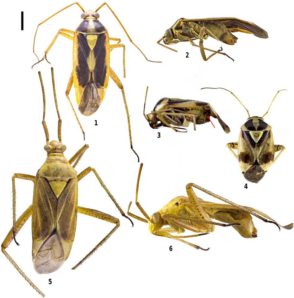
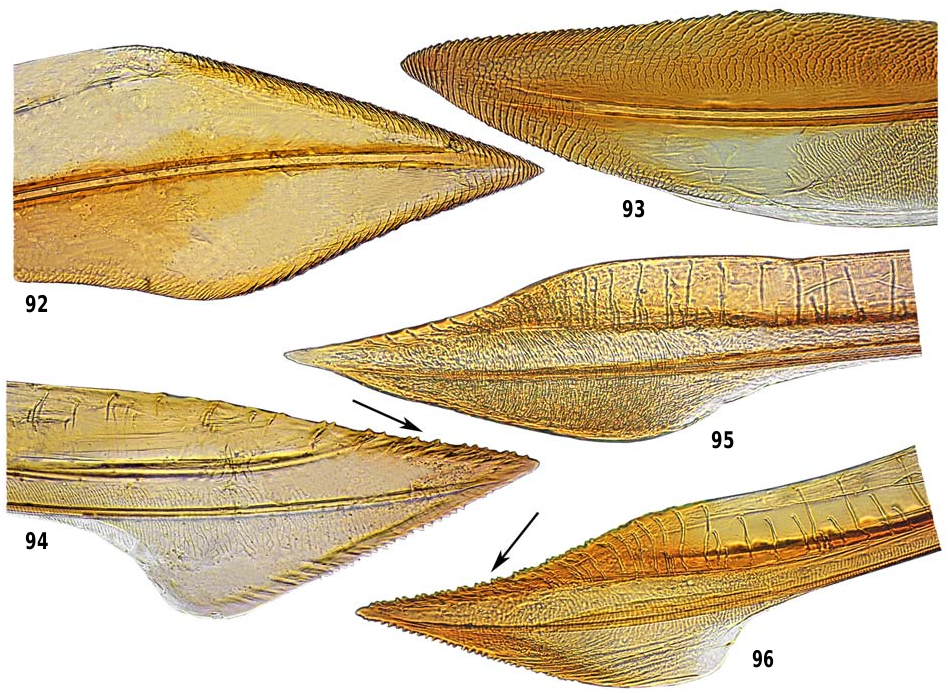
18. Tibia with pale spines, pygophore with large and prominent denticle on the left side ( Fig. 3 View Figs 1–6 ), basal margin of right paramere dentate ( Fig. 9 View Figs 7–17 ); dorsal structure of posterior wall broadly rounded, disk–shaped and spinulate ( Fig. 81 View Figs 76–91 ), dorsal margin of second valvula apically with sparse teeth ( Fig. 94 View Figs 92–96 ) ............................ Cyphodema ( Fig. 3–4 View Figs 1–6 )
— Tibia with dark spines, pygophore without denticle, basal margin of right paramere edentate, dorsal structure of posterior wall not as above; dorsal margin of second valvula with a seri of dense teeth at apex ( Fig. 96 View Figs 92–96 ) ....... ........................................................... Liocoris ( Fig. 41 View Figs 34–46 )
19. Hemelytra greenish, often mottled with small reddish dots, posterior margin of vertex laterally carinate, vesica with short appendage ( Fig. 63 View Figs 58–75 ); margins of sclerotized rings medially form connecting bar, interramal sclerites medially separated ( Fig. 77 View Figs 76–91 ) .... Dichrooscytus ( Fig. 44 View Figs 34–46 )
— Coloration of hemelytra variable, posterior margin of vertex not carinate, vesica without appendage; margins of sclerotized rings not united at middle, interramal sclerites partly or completely fused .......................................... 20
20. First segment of metatarsus distinctly longer than other segments, genital opening oriented posteriorly, apophysis of left paramere with flattened tip ( Fig. 15 View Figs 7–17 ); ventral and dorsal margins of second valvula without teeth ( Fig. 95 View Figs 92–96 ) ......................................................... Stenotus ( Figs 2–3 View Figs 1–6 )
— First segment of metatarsus shorter than other segments, genital opening oriented dorsally, apophysis of left paramere not as above, ventral and dorsal margins of second valvula with a few teeth (except Brachycoleus in its dorsal margin) ........................................................ 21
21. Hemelytra with regular, dense, orange to red or irregular dark mottling, rarely uniformly pale, 1 st antennal segment longer than head width, antenna usually longer than (or as long as) body length, metafemora long, usually reaching beyond apex of abdomen ............... Phytocoris ( Fig. 43 View Figs 34–46 )
— Hemelytra not as above, 1 st antennal segment as long as or shorter than head width, antenna shorter than body length, metafemora not reaching apex of abdomen ............... 22
22. Vestiture mixed, consisting of short adpressed black and whitish setae, or dorsum covered with long and semierect setae (in Brachycoleus ) ............................................... 23
— Vestiture homogenous, blackish or whitish, if mixed, body uniformly pale greenish to brownish (in Rauniella ) ... 27
23. Body small ( 3–4 mm), remarkably robust and oval, 1 st antennal segment tumid, 2 nd antennal segment clavate .. ......................................................... Eurystylus ( Fig. 42 View Figs 34–46 )
— Body larger (> 5 mm), slender and elongated, 1 st antennal segment cylindrical, 2 nd antennal segment cylindrical or weakly incrassate distally (in Pantilius ) .................... 24
24. Body robust, frons markedly projecting over clypeus 25
— Body delicate, frons slightly convex .......................... 26
25. Body covered with short and adpressed setae, vertex with distinct median longitudinal sulcus, 2 nd antennal segment weakly incrassate distally, lateral margins of pronotum carinate ............................................................. Pantilius
— Body covered with long and semierect setae, vertex without median longitudinal sulcus, 2 nd antennal segment cylindrical, lateral margin of pronotum not carinate ..... .................................................... Brachycoleus ( Fig. 47)
26. Labium reaching middle of mesosternum or mesocoxa, interocular width/width of eye < 1♂, cuneus stramineous to greenish, apophysis of right paramere acute at apex ( Fig. 70 View Figs 58–75 ), process of spiculum short and reduced to basal tooth ( Fig. 71 View Figs 58–75 ), sometimes inconspicuous .......... ....................................................... Reuterista ( Fig. 52)
— Labium reaching metacoxa (rarely mesocoxa), interocular width/width of eye> 1♂, cuneus tinged with dark red basally, apex blackish; apophysis of right paramere blunt at apex ( Fig. 67 View Figs 58–75 ), process of spiculum long ( Fig. 66 View Figs 58–75 ) ... .................................................. Closterotomus ( Fig. 49)
27. Vertex with longitudinal shallow or deep midline sulcus, femora with a regular seri of spines, apophysis of left paramere ending with acute process, dorsal structure of posterior wall small .................................................... 28
— Vertex without midline sulcus, femora apically with a few spines, apophysis of left paramere not as above (except Grypocoris fieberi ); dorsal structure of posterior wall large ............................................................................. 30
28. Vertex with deep sulcus, tibiae with pale spines, pygophore without denticle, vesica without spiculum; dorsal labiate plate with anteromedial projection ( Fig. 82 View Figs 76–91 ) ..... ..................................................... Creontiades ( Fig. 48)
— Vertex with shallow sulcus, tibia with black spines, pygophore with denticle, vesica with spiculum; dorsal labiate plate without antero–medial projection ..................... 29
29. Vestiture adpressed, spiculum comb–shaped ( Fig. 65 View Figs 58–75 ); dorsal labiate plate developed, sclerotized rings almost subcontiguous ( Fig. 76 View Figs 76–91 ) ............. Adelphocoris ( Fig. 53)
— Vestiture semierect, spiculum sickle–shaped; dorsal labiate plate reduced, sclerotized rings distinctly separated ( Fig. 80 View Figs 76–91 ) ...................................... Megacoelum ( Fig. 50)
30. Body covered with erect to semierect setae, eyes small, interocular width/width of eye>2 ..... Horistus ( Fig. 57)
— Body covered with adpressed setae, eyes large, interocular width/width of eye <2 ........................................... 31
31. Secondary gonopore like an inverted saddle ( Fig. 10 View Figs 7–17 ); dorsal labiate plate with antero–medial projection (e.g. Figs 82, 89 View Figs 76–91 ) ................................................................. 32
— Secondary gonopore variable; dorsal labiate plate without antero–medial projection ............................................ 33
32. Pubescence mixed of whitish and black, coloration uniformly greenish to brownish, apophysis of left paramere subapically with a large dentate projection ( Figs 11–12 View Figs 7–17 ) ...................................................... Rauniella ( Figs 5–6 View Figs 1–6 )
— Pubescence whitish, coloration black and yellow, apophysis of left paramere subapically without projection. ....................................................... Grypocoris ( Fig. 51)
33. Pubescence whitish, sensory lobe of left paramere with long acute process ( Fig. 74 View Figs 58–75 ), vesica with long spiculum ( Fig. 75 View Figs 58–75 ) ..................................................... Rhabdomiris
— Pubescence black, sensory lobe of left paramere without process, vesica without spiculum ............................... 34
34. Coloration black and yellow or red, antenna almost as long as body length, medial vein between the radial and cubital veins present on corium .............. Miris ( Fig. 55)
— Coloration yellowish to greenish, antenna shorter than body length, medial vein between the radial and cubital veins absent on corium ............................................... 35
35. Labium reaching mesocoxa, 1 st antennal segment 2.5–3x longer than interocular distance, lateral margin of left paramere with a projection ( Fig. 64 View Figs 58–75 ); dorsal labiate plate membranous, sclerotized rings oval ( Fig. 79 View Figs 76–91 ), first valvula broadly triangular, apically acute ( Fig. 92 View Figs 92–96 ) ................ ............................................... Mermitelocerus ( Fig. 56)
— Labium reaching metacoxa, 1 st antennal segment 1.4– 1.6x longer than interocular distance, lateral margin of left paramere without projection; dorsal labiate plate moderately sclerotized, sclerotized rings elongate–oval ( Fig. 84 View Figs 76–91 ), first valvula blade shaped ( Fig. 93 View Figs 92–96 ) ....................................................................... Calocoris ( Fig. 54)


No known copyright restrictions apply. See Agosti, D., Egloff, W., 2009. Taxonomic information exchange and copyright: the Plazi approach. BMC Research Notes 2009, 2:53 for further explanation.