
Abernessia giga, Waichert & Pitts, 2013
|
publication ID |
https://doi.org/ 10.11646/zootaxa.4801.1.12 |
|
publication LSID |
lsid:zoobank.org:pub:FEDD641B-B0AD-43F1-9BDF-516E002BDCEB |
|
DOI |
https://doi.org/10.5281/zenodo.10564172 |
|
persistent identifier |
https://treatment.plazi.org/id/038D9366-045F-3F13-8B83-8CC39C333D94 |
|
treatment provided by |
Plazi |
|
scientific name |
Abernessia giga |
| status |
|
A bernessia giga Waichert & Pitts, 2013 View in CoL
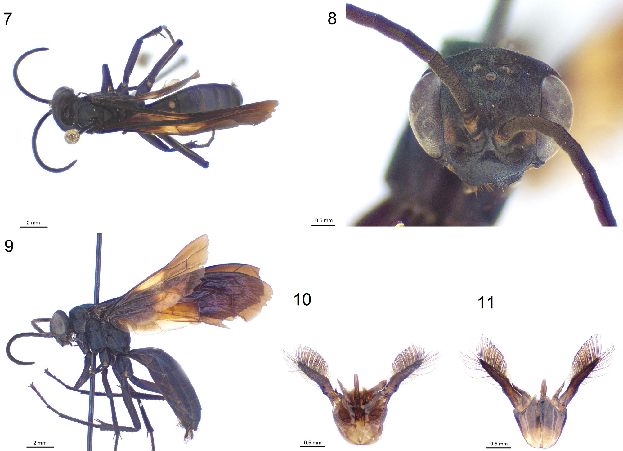
( Figs. 7–11 View FIGURES 7–11 )
Diagnoses. The male of this species can be distinguished from all other species of Abernessia by the following combination of characters: the clypeus is black, the forewings have orange maculations and the hindwings have pale orange to white maculations, the SGP apex broadly truncated, and the paramere is about 2 × as long as the aedeagus. Description. MALE. Body length 13.2 mm, forewing length 13.7 mm.
Color ( Figs 7, 9 View FIGURES 7–11 ). Body black; labrum, most of mandible, setae, legs, and spurs brown; mandible center dark red; dull yellow patches on face along inner eye margin at antenna base level; first tergite segment with two distinct white patches on dorsal face ( Fig. 6 View FIGURES 2–6 ); wings tinted brown, darker at base; forewing darker than hindwing, middle with orange patch; wing veins light brown on hindwing, middle of forewing, dark brown on forewing darker areas; hindwing middle with translucent off-white patch extending to lower margin including anal lobe.
Head ( Fig. 8 View FIGURES 7–11 ). Round, TFD 1.2 × FD, UID 2.9 × A3; clypeus polished, length 1.8 × height, finely punctured, with median indent, rest of body densely punctured; antennae base set in deep sockets, ratio of flagellomeres 1–4 8:3:9:9, antennal segments short, A3 length 2.25 × width; mandible bidentate, malar space inconspicuous; vertex extended above ocelli, with long setae compared to lower face; ocelli in line with top of eye, in obtuse angle; lateral ocelli closer to each other than to compound eye, OOL 1.5 × POL.
Mesosoma . Pronotum trapezoidal, dorsal face about 2 × long as anterior face, posterior margin arcuate; propodeum evenly curved throughout, sparse long setae over posterior half; fore leg tarsal claws symmetrical, bidentate; mid and hind tarsal claws widely dentate, even curvature with small tooth at basal third; ultimate tarsus ventral side with two lateral rows of short spines, up to three per row; pulvillar pad circular with about six long thin bristles; hind tibia with complete brush, posterior side slightly flattened with one row of longitudinal scale-like integumental teeth, longer tibial spur slightly shorter than half of basitarsus; tegula asymmetrical, narrowed posteriorly; hindwing cu-a joining M+CuA much beyond juncture of M with CuA; forewing stigma half of marginal cell, 1m-cu meeting SMC2 slightly beyond half way, 2m-cu meeting SMC3 at midpoint.
Metasoma. S6 with long inward-curved hooked bristles starting transversely at basal third continuing along lateral margins; shorter bristles in center and posterior areas of S6; two tooth-like projections along posterior margin of S6; SGP flat, wide at base, at halfway slight but distinct inward angle to broadly truncated apex; long setae cover ventral face.
Genitalia ( Fig. 10, 11 View FIGURES 7–11 ). Paramere thick, spade-like, basal ridge with stout thick bristles on margin; upper margin with thick long setae, lower margin with thick dense hooked bristles; digitus flat, sides straight, length less than half of paramere.
Material examined. 1 ♂: Brazil: MG , Águas Vermelhas , 15°45’S 11°28’W, alt. 800m, xii.1983, Alavarenga [col.] ( EMUS _816) GoogleMaps .
Distribution. Minas Gerais, Brazil.
Remarks. The association of the male of A. giga with the female is based on similarities in wing venation, wing coloration and distribution. The male specimen of A. giga ( Figs 7–9 View FIGURES 7–11 ) looks similar to A. prima and A. capixaba . It can be distinguished from A. capixaba by having pale markings on the wings, white markings on the metasoma, and the SGP distinctly narrowed midway with apex truncated. It can be distinguished from A. prima by also having pale markings on the wings and the SGP distinctly narrowed midway with apex truncated, in addition to the clypeus being completely black and having more prominent hind tibial teeth with smaller distance between each tooth. Abernessia giga also differs from A. capibaxa by having the SMC3 short, as long as wide, square-shaped; whereas in A. capixaba the SMC3 cell is about 2× as long as wide, rectangular-shaped. The male specimen matches the wing coloration of the previously recorded female of A. giga and was collected in the same ecoregion. Molecular sequencing may prove that these male characters correspond to intraspecific variation, as the A. giga specimen was collected close to the locations of three of the A. prima males listed above ( Fig. 1 View FIGURE 1 ). Additionally, the metasoma white patches greatly vary in A. prima , and one variety does match that of the described A. giga ( Figs 4–6 View FIGURES 2–6 ).


| MG |
Museum of Zoology |
No known copyright restrictions apply. See Agosti, D., Egloff, W., 2009. Taxonomic information exchange and copyright: the Plazi approach. BMC Research Notes 2009, 2:53 for further explanation.
|
Kingdom |
|
|
Phylum |
|
|
Class |
|
|
Order |
|
|
Family |
|
|
Genus |