
Anaptomecus paru, Guala, Mariel E., Labarque, Facundo M. & Rheims, Cristina A., 2012
|
publication ID |
https://doi.org/10.5281/zenodo.280012 |
|
DOI |
https://doi.org/10.5281/zenodo.3507452 |
|
persistent identifier |
https://treatment.plazi.org/id/038DE965-AB76-FFEC-FF73-FDBFFC9DFD21 |
|
treatment provided by |
Plazi |
|
scientific name |
Anaptomecus paru |
| status |
sp. nov. |
Anaptomecus paru View in CoL sp. nov.
Figs 1–30 View FIGURES 1 – 7 View FIGURES 8 – 13 View FIGURES 14 – 18 View FIGURES 19 – 24 View FIGURES 25 – 30 Type material. Holotype: 3 from ECUADOR, Santo Domingo de los Tsáchilas Province, Cantón Santo Domingo, Parroquia Santo Domingo, Jatun Tinalandia lodge, km 85 Road Aloaj, S 0,321033, W 78,951583, 758 m, 8 December 2009, M. Ramírez, C. Grismado, M. Izquierdo & F. Labarque leg. in PBI Oonpidae Expedition (QCAZ; preparation code MEG-00015-17). Paratypes: 1Ƥ, 1 immature, same data as holotype (MACN-Ar 26896, 26897; preparation codes MEG-00018-20, MEG-00021-22); 23, 1Ƥ, 2 immature, same locality data as holotype, C.A. Rheims leg. (IBSP 160967).
Etymology. The specific name means golden in Quechua, which is the language of the native people of Ecuador, and refers to the spots distributed along the opisthosoma; term in apposition.
Diagnosis. Males are distinguished from all other Anaptomecus species by a massive embolus that is twisted anticlockwise in ventral view (left palp). It arises dorsally on the tegulum and is prolaterally bent, with a short lamina widest medially similar to A. longiventris ( Figs 1–4 View FIGURES 1 – 7 , 11–13 View FIGURES 8 – 13 ). Females have a pair of copulatory openings situated laterally, appearing as large circles that resemble those of A. longiventris ; but differ from it by the relative positions of the spermathecae, which are situated laterally rather than in the middle line. They are distinguished from the rest of the species by the posterior epigynal margin with two small invaginations and a transversal median septum in ventral view ( Figs 5, 7 View FIGURES 1 – 7 , 17–18 View FIGURES 14 – 18 ).
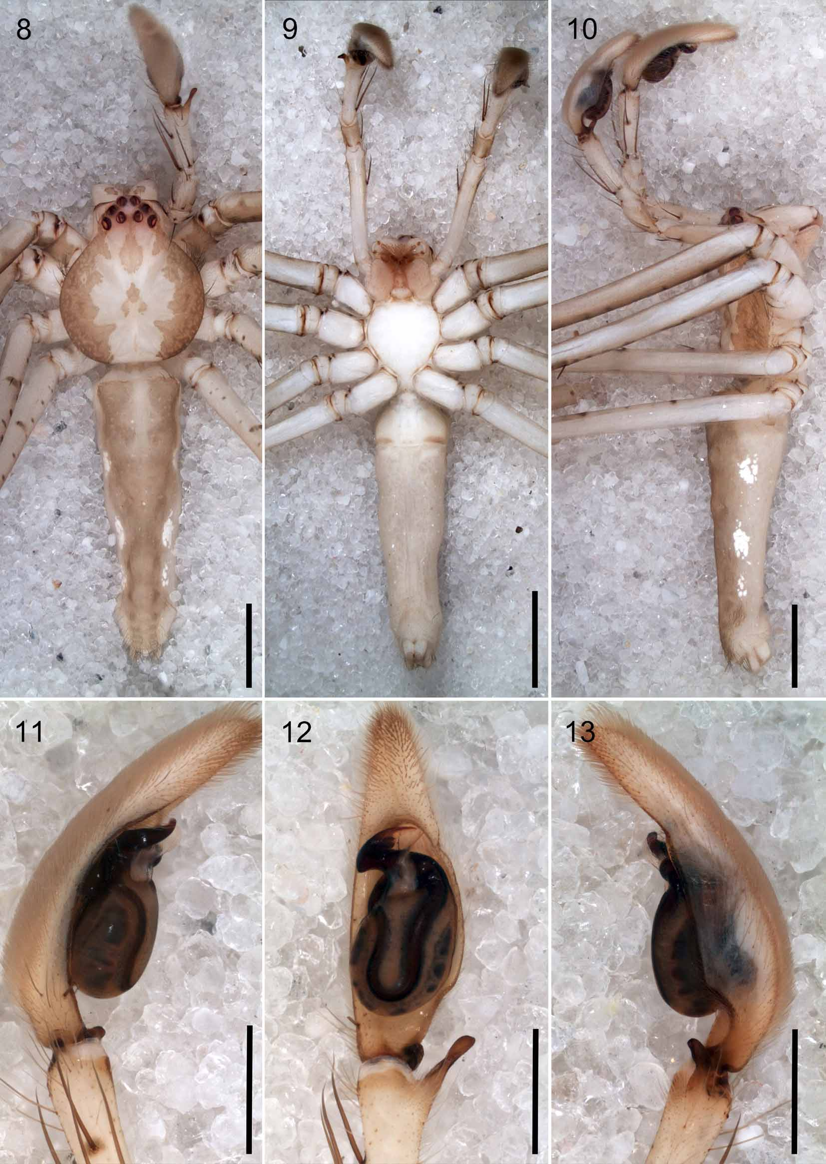
Description. Male ( holotype): Total length 11.0. Prosoma: 4.1 long, 3.4 wide. Opisthosoma: 6.6 long, 2.1 wide. Eye diameters: 0.22, 0.22, 0.18, 0.20; interdistances: 0.16, 0.08, 0.26, 0.24, 0.24, 0.26. Legs (leg formula: 1243): I: 44.7 (11.1, 2.2, 12.9, 14.8, 3.7); II: 43.9 (10.9, 2.2, 12.7, 14.5, 3.6); III: 28.5 (7.7, 1.8, 8.3, 8.5, 2.2); IV: 33.3 (9.1, 1.7, 8.9, 10.8, 2.8). Spination: femur I–II: p1-1-1; d0-1-1; r1-1-1; femur III: p1-1-1; d0-0-1; r1-1-1; femur IV: p1-1-1; d0-0-1; r0-0-1; tibia I–II: p1-0-1; d0-1-1; r1-0-1; v2-2 -0; tibia III–IV: p1-0-1; d0-1-0; r1-0-1; v2-2 -0; metatarsus I–II: p1-0-0; r1-0-0; v2-2 -0; metatarsus III: p1-1-0; r1-0-0; v2-2 -0; metatarsus IV: p1-1-1; r1-1-1; v2-2 - 1. Palp as in diagnosis. Small teeth behind the lamina of the embolus. Hyaline conductor well developed, leafshaped ( Figs 1–4 View FIGURES 1 – 7 , 11–13 View FIGURES 8 – 13 ). Cymbium with two prolatero-ventral spines, and a basal retrolateral pit with a sclerotized projecting spur ( Figs 1–2 View FIGURES 1 – 7 , 11–12 View FIGURES 8 – 13 ). Coloration: General coloration pale yellow. Dorsal shield of prosoma laterally with brown thick bands with lobed inner margins, one anterior patch in cephalic region extending between PME, three central small patches next to fovea and one patch behind fovea. Chelicerae frontally with a brown dot. Pedipalps and legs with brown spots distributed randomly and one spot per spine base encircling it. Sternum, endites and labium pale yellow. Opisthosoma light brown with dorsal rhomboidal cream colored marks, three pairs of lateral bright guanine crystal spots and four pairs of muscle sigilla ( Figs 8–10 View FIGURES 8 – 13 ).
Female (MACN-Ar 26896, paratype): Total length 12.6. Prosoma: 4.8 long, 4.1 wide. Opisthosoma: 7.8 long, 2.5 wide. Eye diameters: 0.20, 0.22, 0.18, 0.20; interdistances: 0.20, 0.08, 0.32, 0.26, 0.32, 0.32. Legs (leg formula: 1243): I: 31.9 (8.6, 2.3, 9.4, 9.2, 2.4); II: 31.5 (8.6, 2.46, 9.3, 9.0, 2.3); III: 22.5 (6.3, 2.0, 6.5, 6.0, 1.6); IV: 25.0 (7.4, 1.9, 6.6, 7.0, 2.0). Spination as in male except femur II r1-1-1-1; tibiae I–II d0-1-0 and metatarsus IV v2- 2 -0. Epigyne as in diagnosis. Fertilization ducts short, twisted and terminating medially; spermathecae with glandular projection pointing dorsally ( Figs 5, 7 View FIGURES 1 – 7 , 17–18 View FIGURES 14 – 18 ); schematic course of internal duct system shown in Fig. 6 View FIGURES 1 – 7 . Coloration as in male but lighter. Opisthosoma with a dorsal cream colored arrow-shaped mark, bright guanine crystals dots distributed randomly and four pairs muscle sigilla with dark hairs ( Figs 14–16 View FIGURES 14 – 18 ).
Immature (MACN-Ar 26897): For permanent scanning electron preparations we have dissected a juvenile instar instead of an adult specimen of Anaptomecus , because there are few of them in world´s collections. Here we describe characters of the chelicerae and legs in high magnification. Chelicerae longer than wide ( Figs 19, 21, 23 View FIGURES 19 – 24 ), with anterior median tooth larger than the others and posterior teeth gradually decreasing in size from cheliceral fang base to paturon margin. ( Figs 20, 22, 24 View FIGURES 19 – 24 ). Cheliceral boss at the ectal base ( Fig. 21 View FIGURES 19 – 24 ). Cheliceral furrow with 14 denticles in a distinct patch ( Fig. 22 View FIGURES 19 – 24 ). Tarsi I–IV with pair of pectinate claws bearing 12–20 teeth and a dense claw tuft ( Figs. 25–26 View FIGURES 25 – 30 ). Median lobe of trilobate membrane acuminate longer than laterals, may be bent ( Figs 27–28 View FIGURES 25 – 30 ). Tarsal organ capsulated, with oval opening and five sensilla, located dorsally at the distal end of tarsi ( Fig. 29 View FIGURES 25 – 30 ). Trichobothrium with basal crescent plate projecting over a plate with one distal groove ( Fig. 30 View FIGURES 25 – 30 ).
Variation. Males (n=3): total length 10.4–11.1; prosoma length 4.0–4.1; femur I length 10.2–11.4. Females (n=2): total length 12.6–13.3; prosoma length 4.4–4.8; femur I length 7.9–8.6.
Natural history. A female was collected together with a spherical egg-sac under a tree leaf. Egg-sac was attached to the leaf, containing 115 spiderlings.
Distribution. Ecuador (known only from the type locality).


No known copyright restrictions apply. See Agosti, D., Egloff, W., 2009. Taxonomic information exchange and copyright: the Plazi approach. BMC Research Notes 2009, 2:53 for further explanation.
|
Kingdom |
|
|
Phylum |
|
|
Class |
|
|
Order |
|
|
Family |
|
|
Genus |