
Asceua thrippalurense, Sankaran, 2023
|
publication ID |
https://doi.org/ 10.11646/zootaxa.5296.3.4 |
|
publication LSID |
lsid:zoobank.org:pub:B60263A8-DD7F-4D3C-93B5-8C280D6C55B1 |
|
DOI |
https://doi.org/10.5281/zenodo.7983985 |
|
persistent identifier |
https://treatment.plazi.org/id/038E87AC-FFB2-0421-FF26-FC090159AA4C |
|
treatment provided by |
Plazi |
|
scientific name |
Asceua thrippalurense |
| status |
sp. nov. |
Asceua thrippalurense sp. nov.
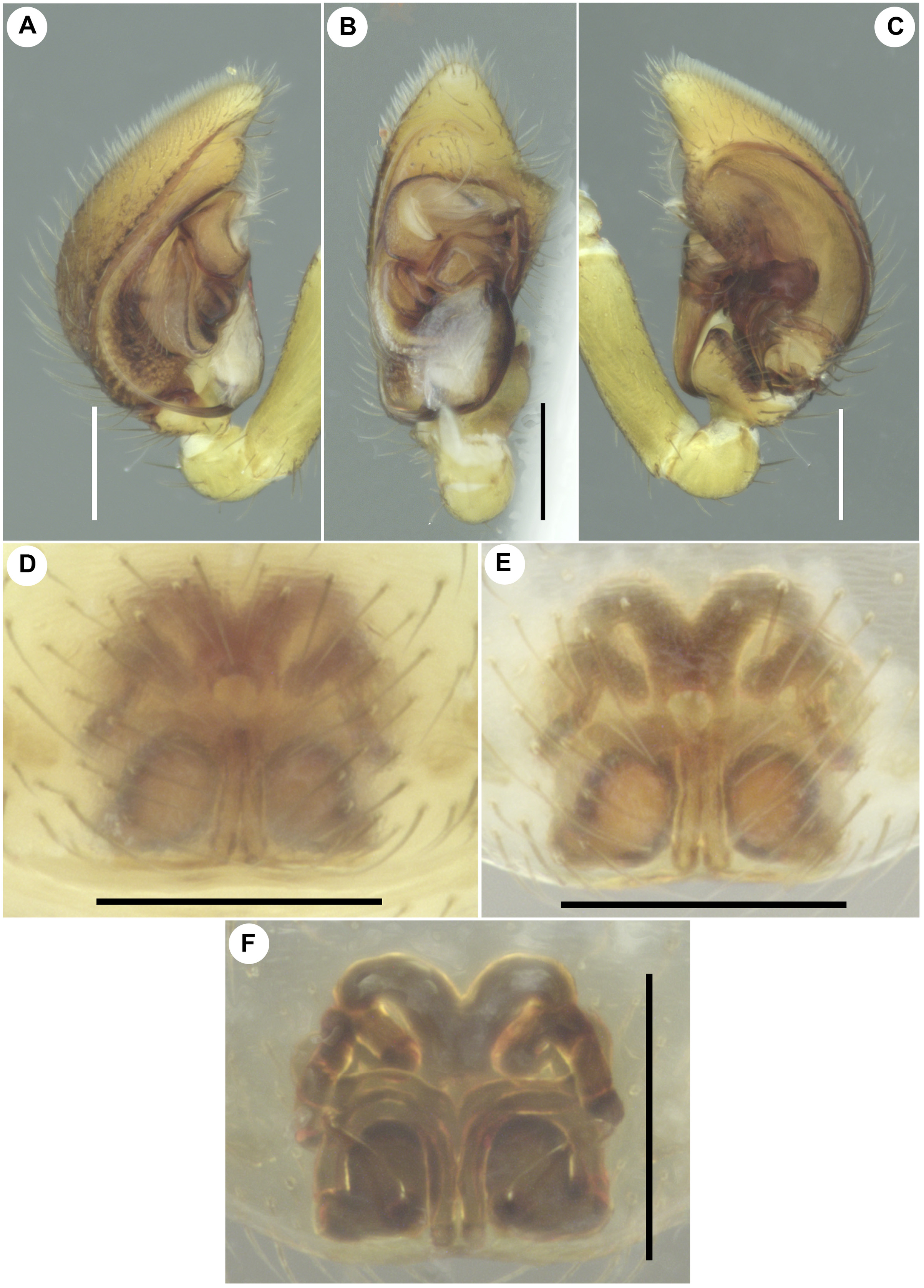
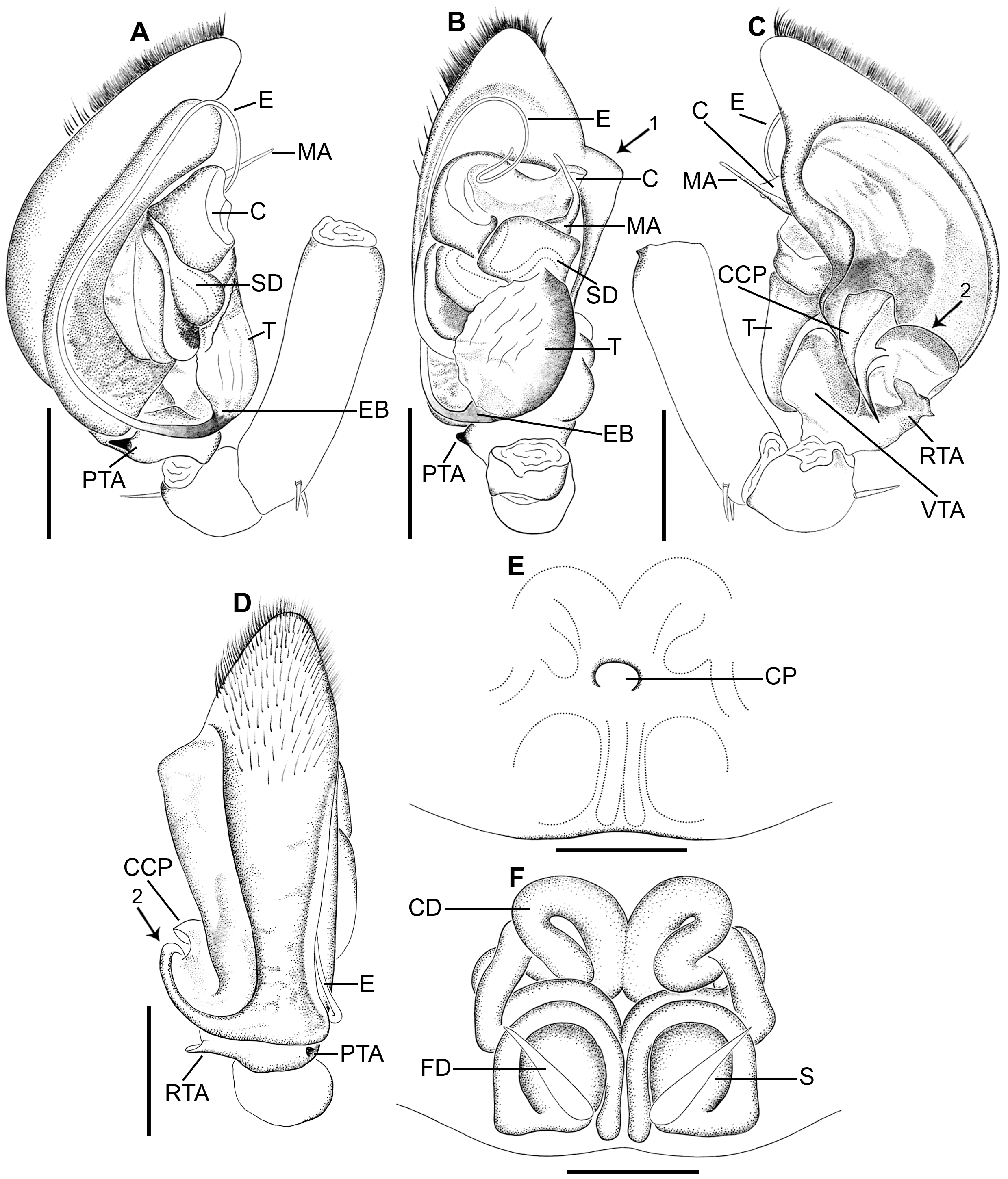
Figs 7–10 View FIGURE 7 View FIGURE 8 View FIGURE 9 View FIGURE 10 , 17 View FIGURE 17
Type material. Holotype ♁ ( ADSH196 View Materials ) from INDIA: Kerala: Palakkad, Thrippalur, Pullodu , 10°38’16.58’’N 76°33’52.87’’E, 70 m a.s.l., 24 January 2016, on the ground, by hand, leg. M.S. Pradeep. GoogleMaps Paratype: 2 ♀♀ ( ADSH197 View Materials ), same data as for the holotype GoogleMaps .
Etymology. The specific epithet is an adjective and referring to Thrippalur, where the type locality of the new species is located.
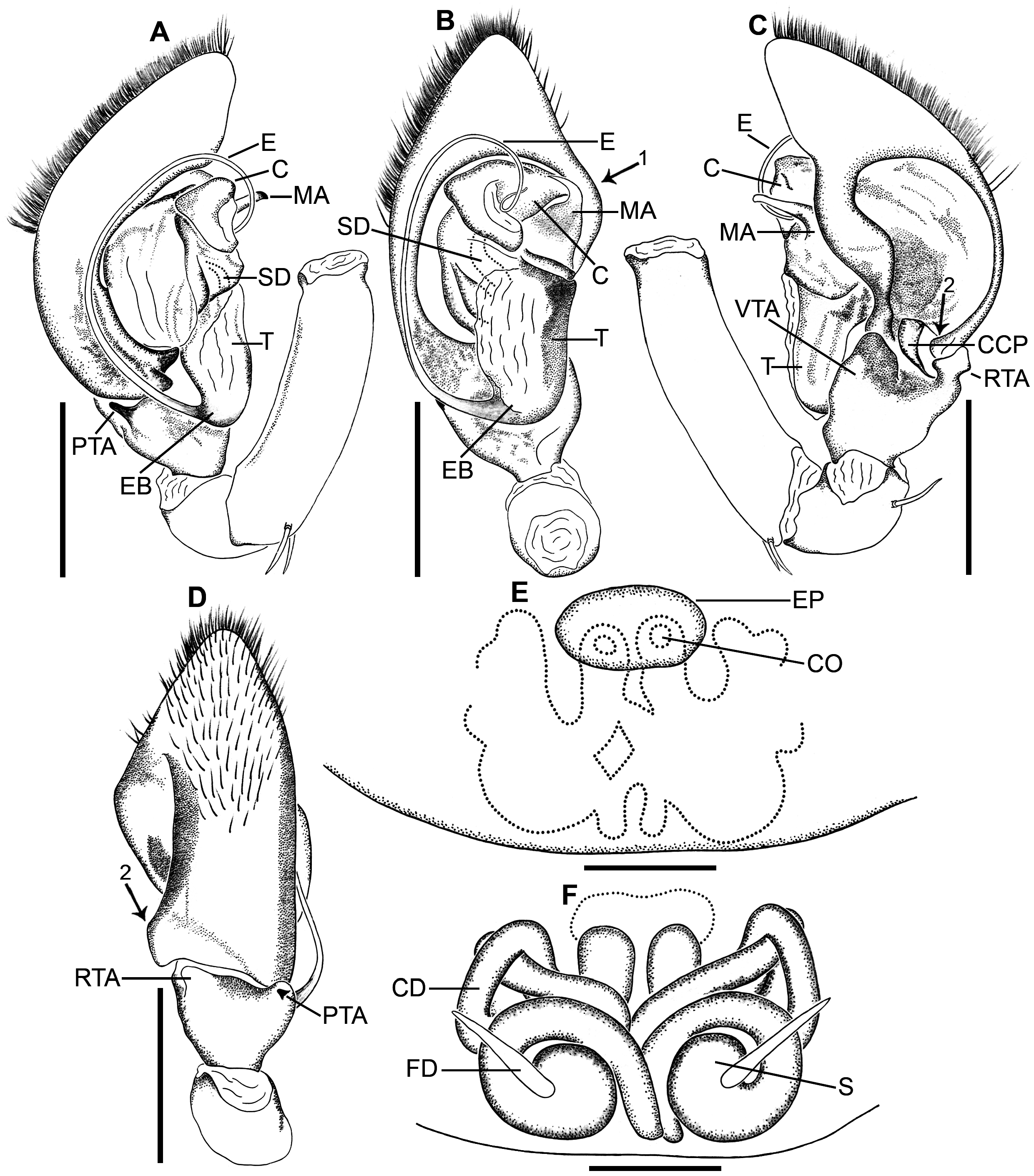
Diagnosis. Males are similar to A. cingulata , but can be distinguished from it as stated above (compare Figs 9A–C View FIGURE 9 , 10A–D View FIGURE 10 with Figs 4A–C View FIGURE 4 , 5A–D View FIGURE 5 ). Females are closely related to the females of Asceua wallacei Bosmans & Hillyard, 1990 as both share spherical spermathecae and copulatory ducts with prominent windings near copulatory openings, but can be separated from the latter species by epigyne with anteromedian pore (vs. absent in A. wallacei ), and without anteromedian process (vs. present in A. wallacei ) (compare Figs 9D–F View FIGURE 9 , 10E–F View FIGURE 10 with Bosmans & Hillyard 1990: figs 54–55). Females of A. thrippalurense sp. nov. can easily be distinguished from the females of A. cingulata by the presence of longitudinal striae retrolaterally on femur I ( Fig. 8H View FIGURE 8 ) which is absent in A. cingulata .
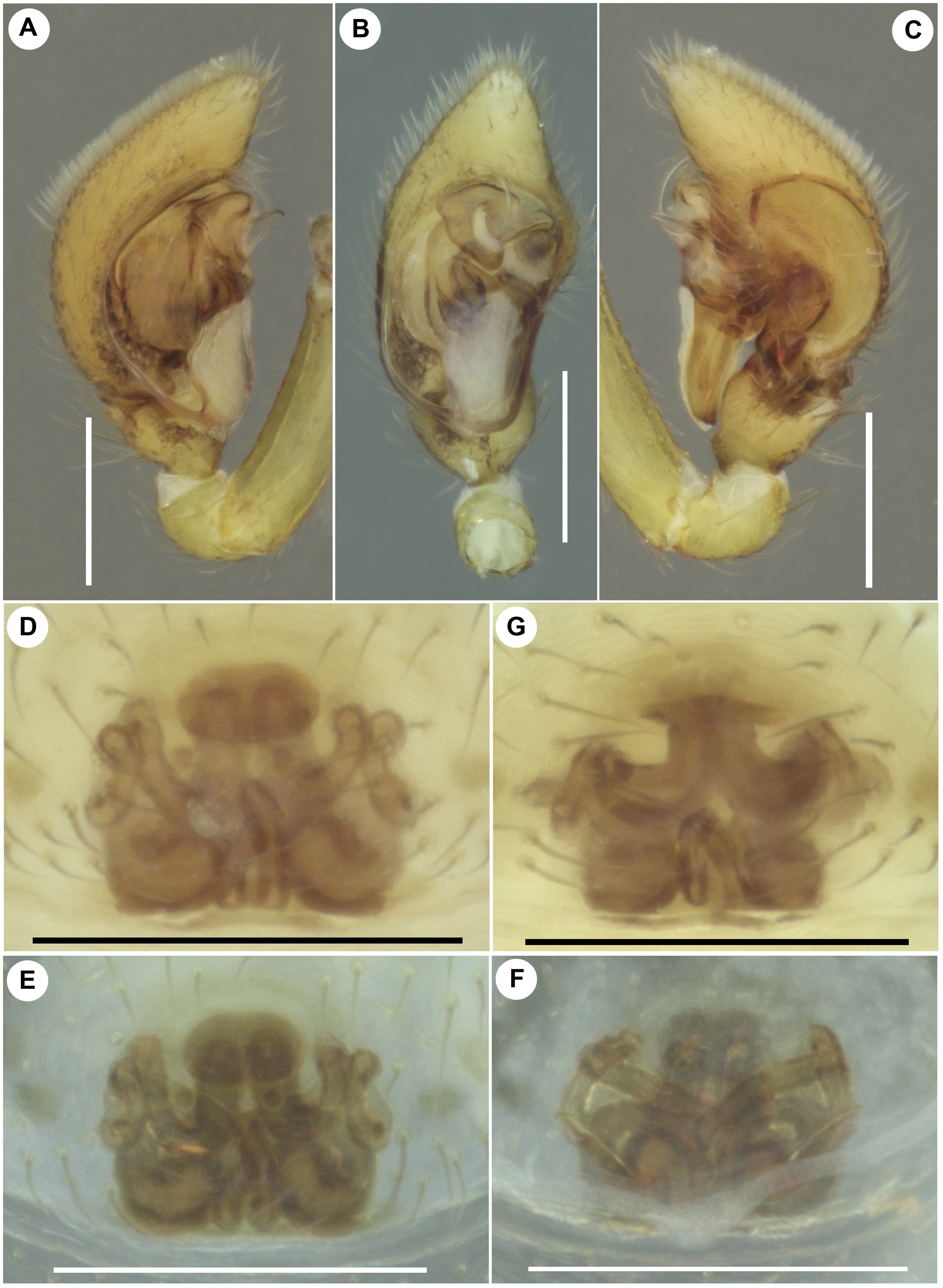
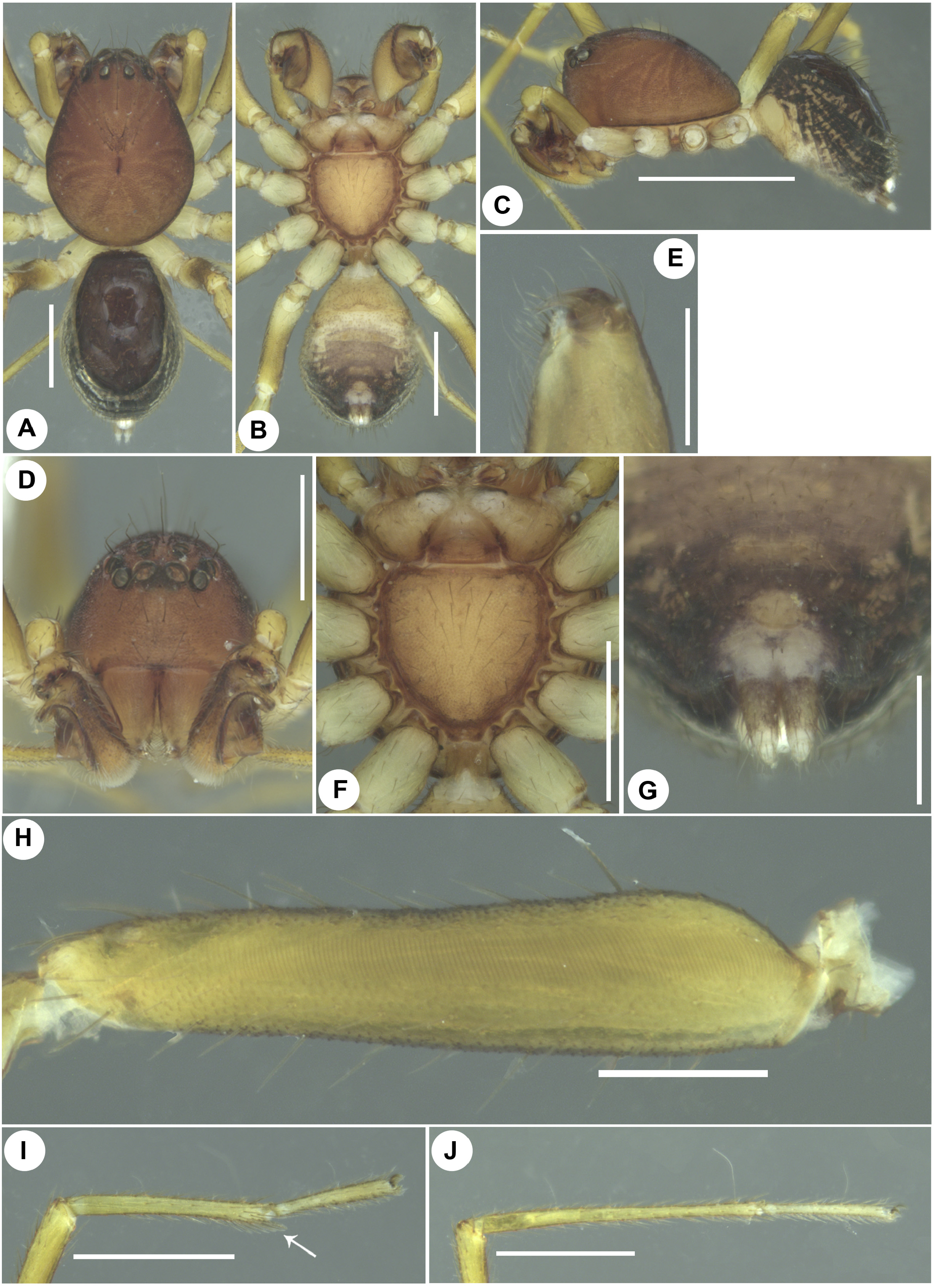
Description. Male in alcohol (holotype, ADSH196) ( Figs 7A–J View FIGURE 7 ). Body length 2.06. Carapace 1.09 long, 0.88 wide. Opisthosoma 0.97 long, 0.73 wide. Carapace, eye region, clypeus dark brown; chilum, chelicerae, endites, labium, sternum brown; leg and palp segments yellowish brown to creamy-white; dorsal and lateral opisthosoma black with creamy-white streaks on laterals, dorsal scutum shiny, brownish black ( Fig. 7A View FIGURE 7 ), venter creamy-white with yellowish brown and greyish patches; spinnerets creamy-white with black shades ( Fig. 7G View FIGURE 7 ). Carapace finely rugose; cephalic part provided with scattered few long black setae. Fovea short, longitudinal, straight, dark. Clypeus high ( Fig. 7D View FIGURE 7 ). Chilum inverted triangular, unipartite ( Fig. 7D View FIGURE 7 ). Cheliceral promargin with two tiny teeth, retromargin without tooth visible ( Fig. 7E View FIGURE 7 ). Sternum rebordered, rugose, shield-shaped with posterior end truncated, provided with scattered greyish black setae, with coxal and intercoxal extensions ( Fig. 7F View FIGURE 7 ). Opisthosoma oval, scutum covering 2/3 rd of dorsum area ( Fig. 7A View FIGURE 7 ), covered with long black setae; rear and lateral opisthosoma with corrugations. Colulus and anal tubercle prominent ( Figs 7A, G View FIGURE 7 ). Femur I retrolaterally provided with longitudinal striae almost along its entire length ( Fig. 7H View FIGURE 7 ); metatarsi II–III with distal preening brush ( Fig. 7I View FIGURE 7 ); all metatarsi and tarsi without scopula ( Fig. 7J View FIGURE 7 ); all tarsi with reduced claw tuft ( Figs 7I–J View FIGURE 7 ). Eye diameters and interdistances: ALE 0.08, AME 0.10, PLE 0.07, PME 0.05; AME–ALE 0.02, AME–AME 0.03, AME–PME 0.07, ALE–PLE 0.03, PME–PLE 0.08, PME–PME 0.09. Clypeus height at AMEs 0.32, at ALEs 0.30. Chelicerae 0.28 long. Sternum 0.54 long, 0.52 wide. Measurements of palp and legs: palp 1.36 [0.42, 0.17, 0.13, 0.64], I 3.22 [0.87, 0.27, 0.79, 0.82, 0.47], II 2.55 [0.73, 0.28, 0.54, 0.64, 0.36], III 2.46 [0.68, 0.26, 0.52, 0.67, 0.33], IV 3.54 [0.93, 0.28, 0.81, 1.08, 0.44]. Leg formula: 4123. Spination of palp: femur rld 1, patella do 1, tibia spineless, tarsus/cymbium spineless; legs: femora I–IV do 1; patellae I–IV spineless; tibiae I–IV spineless; metatarsus I spineless, II–IV plv 1 rlv 1; tarsi I–IV spineless. Palp ( Figs 9A–C View FIGURE 9 , 10A–D View FIGURE 10 ). Tibia with short retrolateral, broad ventral and narrow, finger-like prolateral apophyses ( Figs 9A, C View FIGURE 9 , 10A–D View FIGURE 10 ); RTA with prolaterally oriented distal part ( Figs 9C View FIGURE 9 , 10C View FIGURE 10 ); RTA and VTA enclose large concavity in which fits basomedial projection of cymbium ( Figs 9C View FIGURE 9 , 10C View FIGURE 10 ). Cymbium with large retrolateral fold having prominent anterolateral margin and highly modified basoretrolateral extremity ( Figs 9B–C View FIGURE 9 , 10B–C View FIGURE 10 ), with basomedial conical process extending between RTA and VTA ( Figs 9C View FIGURE 9 , 10C View FIGURE 10 ). Tegulum broad, with basomedian extension ( Figs 9B View FIGURE 9 , 10B View FIGURE 10 ). Median apophysis with short proximal and long, slender, prolaterally oriented distal parts ( Figs 9B–C View FIGURE 9 , 10A– C View FIGURE 10 ), lying adjacent to retrolateral process of conductor ( Figs 9B View FIGURE 9 , 10B View FIGURE 10 ). Conductor broad, sclerotized, with retrolateral and mediolateral, blunt processes ( Figs 9B View FIGURE 9 , 10B View FIGURE 10 ); retrolateral process anteriorly oriented ( Figs 9B View FIGURE 9 , 10B View FIGURE 10 ). Embolus long, filiform, with short embolar base originated basally to tegulum, at first running anteriad along prolateral side of cymbium, then turning retrolaterad, then to posterolaterad, then to anteroretrolaterad without forming distal loop, with blunt tip directed at 1-o’ clock ventrally ( Figs 9A–B View FIGURE 9 , 10A–B View FIGURE 10 ).
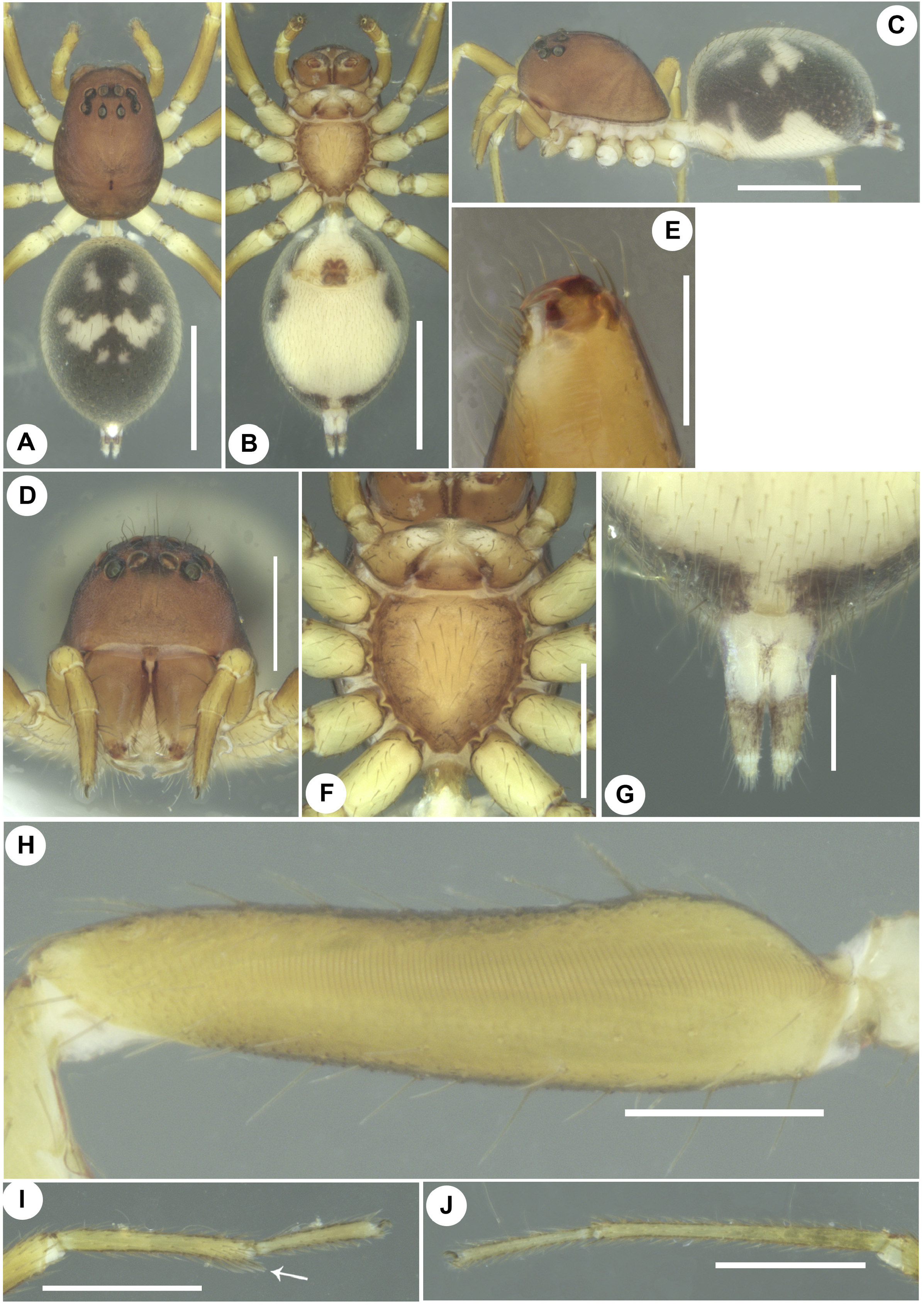
Female in alcohol (paratype, ADSH197) ( Figs 8A–J View FIGURE 8 ). Body length 2.46. Carapace 1.04 long, 0.88 wide. Opisthosoma 1.42 long, 1.00 wide. Habitus and details like male except for the following: leg and palp segments yellowish brown to light brown. Opisthosoma without scutum and corrugations ( Fig. 8A View FIGURE 8 ); dorsum with three pairs of creamy-white patches, anterior C-shaped, median V-shaped and posterior dot-like ( Fig. 8A View FIGURE 8 ); laterals black and creamy-white without streaks; venter uniformly creamy-white with black at the rear end. Eye diameters and interdistances: ALE 0.07. AME 0.08. PLE 0.04. PME 0.05; AME–ALE 0.02. AME–AME 0.04. AME–PME 0.09. ALE–PLE 0.05. PME–PLE 0.10. PME–PME 0.09. Clypeus height at AMEs 0.33, at ALEs 0.28. Chelicerae 0.42 long. Sternum 0.58 long, 0.55 wide. Measurements of palp and legs: palp 1.04 [0.34, 0.20, 0.21, 0.29], I 2.97 [0.82, 0.30, 0.67, 0.72, 0.46], II 2.44 [0.66, 0.28, 0.52, 0.62, 0.36], III 2.51 [0.69, 0.28, 0.52, 0.67, 0.35], IV 3.42 [0.91, 0.30, 0.78, 1.01, 0.42]. Leg formula: 4132. Spination of palp: tibia pld 1; tarsus pl 1 pld 1 v 2; legs: metatarsi I–III spineless. Genitalia ( Figs 9D–F View FIGURE 9 , 10E–F View FIGURE 10 ). Epigyne with W-shaped posterior margin, with a central pore leading to copulatory openings ( Figs 9D–E View FIGURE 9 , 10E View FIGURE 10 ). Copulatory openings contiguous, situated inside the central pore. Copulatory ducts long, slender, with multiple windings ( Figs 9F View FIGURE 9 , 10F View FIGURE 10 ). Spermathecae large, globular, lying adjacent to each other, situated posteriorly ( Figs 9F View FIGURE 9 , 10F View FIGURE 10 ). Fertilization ducts long, narrow, diverging ( Figs 9F View FIGURE 9 , 10F View FIGURE 10 ).
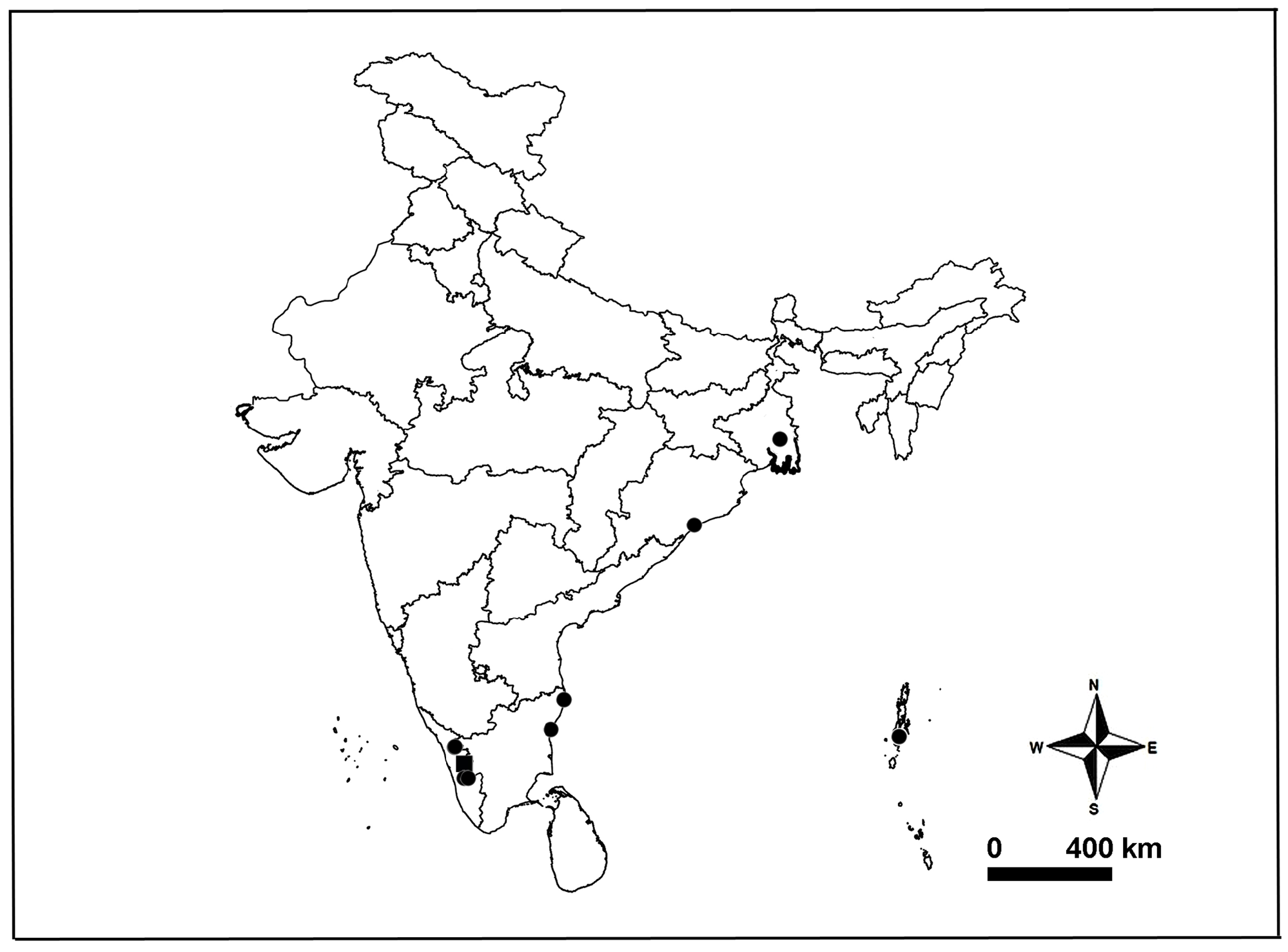
Distribution. Known only from the type locality ( Fig. 17 View FIGURE 17 ).
No known copyright restrictions apply. See Agosti, D., Egloff, W., 2009. Taxonomic information exchange and copyright: the Plazi approach. BMC Research Notes 2009, 2:53 for further explanation.
|
Kingdom |
|
|
Phylum |
|
|
Class |
|
|
Order |
|
|
Family |
|
|
Genus |