
Latinopsis anisitsi ( Daday, 1905 ) Daday, 1905
|
publication ID |
https://doi.org/ 10.5281/zenodo.190875 |
|
DOI |
https://doi.org/10.5281/zenodo.6213445 |
|
persistent identifier |
https://treatment.plazi.org/id/038E87ED-3961-960B-08B1-2690BD2FFCE1 |
|
treatment provided by |
Plazi |
|
scientific name |
Latinopsis anisitsi ( Daday, 1905 ) |
| status |
comb. nov. |
Latinopsis anisitsi ( Daday, 1905) comb. nov.
( Figures 4 View FIGURE 4 , 5 View FIGURE 5 )
Synonymy
Candonopsis anisitsi n. sp. – Daday, 1905: 256, Plate 16, Figs 16-26. Candonopsis anisitsi Daday – Klie, 1930: 226.
Candonopsis anisitsi Daday – Tresseler, 1956: 334, Fig. 6 View FIGURE 6 . [Not]. Cubacandona anisitsi ( Daday, 1905) comb. nov. – Karanovic, 2004: 106.
Material examined: Three slides were available from the collection of the HMHN, all containing dissected specimens of Candonopsis anisitsi , as marked on the slides. Since Daday (1905) didn’t mark the holotype, and there is no doubt that the specimens deposited in the HNHM are types (therefore syntypes), we designate here a lectotype and paralectotypes. The lectotype is a male dissected on one slide (IV/P-35) together with one male paralectotype which is half dissected (A1, A2, Md, Mxl, T1 not separated one from another but separated from the T2, T3, CR and hemipenis). Slide IV/P-34 contains one paralectotype, male; slide IV/P-33 contains two paralectotype males and one paralectotype female. The slide with the lectotype is in good condition, while the other two slides contain specimens poorly mounted. Carapaces were not present on the slides, and thus not observed by the present authors; description of the carapace is based on Daday (1905) illustrations.
Type locality: Daday (1905) mentioned three localities in Paraguay for Candonopsis anisitsi: Aregua, Acuncion , and Villa Rica. All three were puddles or small freshwater lagoons. The type locality is not given in Daday (1905), nor marked on the slides, nor mentioned in Forró et al. (1987), containing a list of ostracod taxa described by Daday and deposited in the HMNH.
Diagnosis: Dorsal margin in lateral outline straight. Lobe “b” of the hemipenis small and rounded, lobe “a” narrow and triangular.
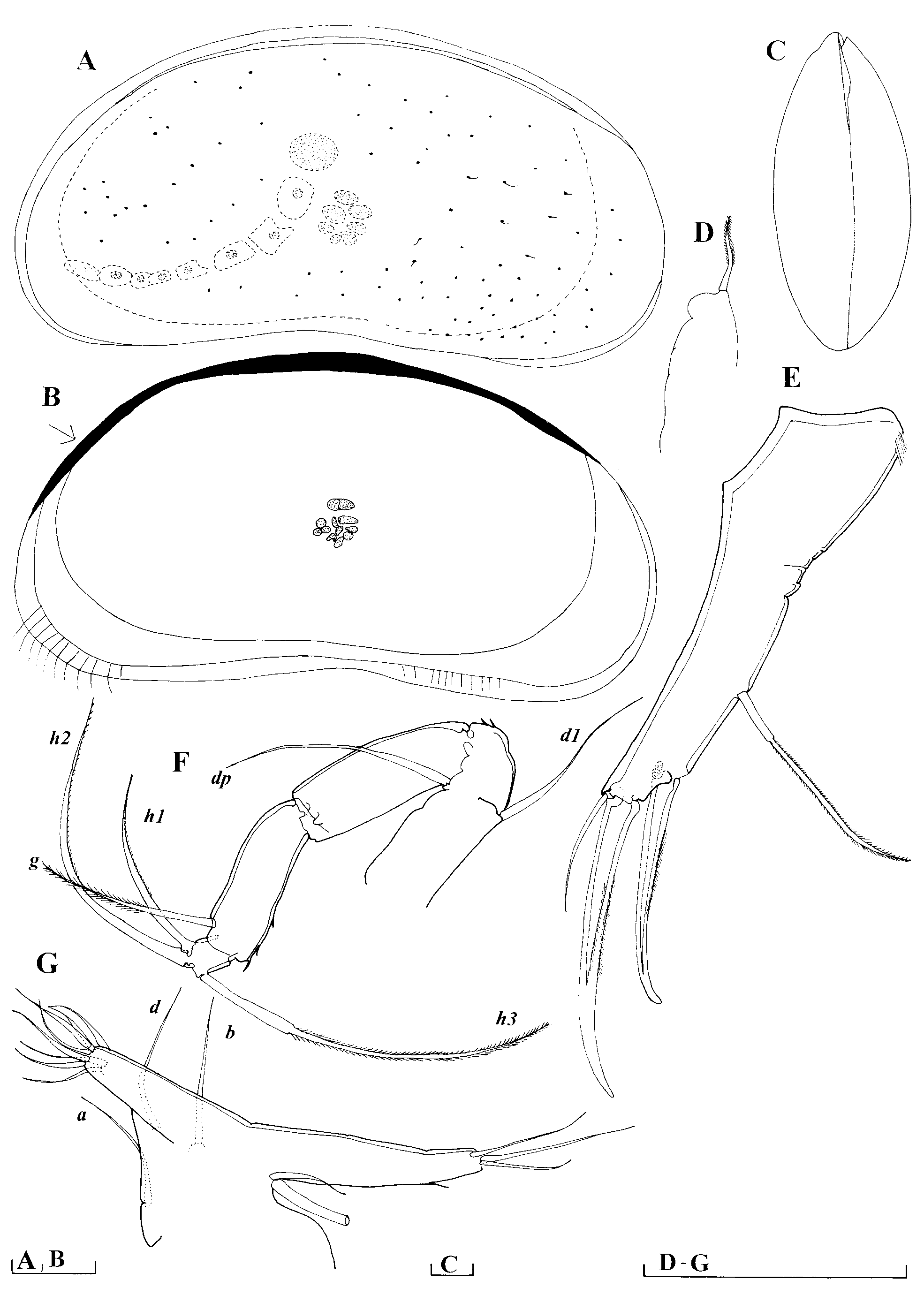
Description. Male: In lateral outline dorsal margin is straight line. Anterior and posterior margins (in same outline) evenly rounded and equally wide. Ventral margin slightly concave. Inner calcified lamella narrow: anteriorly 9 percent, posteriorly 5 percent of total L. Marginal pore canals straight, short and more dense anteriorly. Surface of carapace smooth, covered with sparse and short setae.
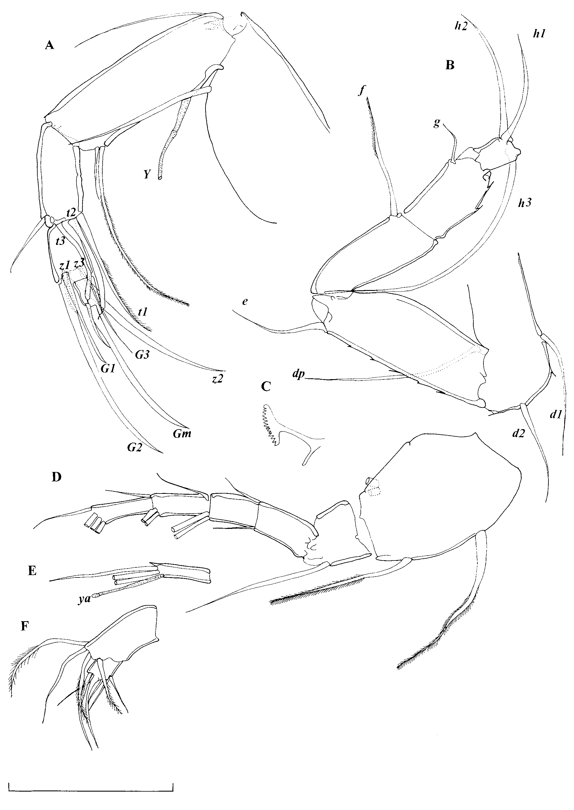
A1 ( Figure 4 View FIGURE 4 D, E): 7-segmented. First segment with four setae. Second segment with one long anterior seta, reaching distal end of fourth segment. Third segment with one anterior and one posterior seta, both being short. Fourth and fifth segments with two anterior and one posterior seta each. Penultimate segment with two posterior and two anterior setae. Alpha seta not observed. Terminal segment ( Figure 4 View FIGURE 4 E) with three setae and aesthetasc ya, which is 1.4 times longer than terminal segment. L ratios of endopodal segments 1.2: 1.4: 1: 1: 1.2: 1.
A2 ( Figure 4 View FIGURE 4 A): 6-segmented (first one not drawn). Penultimate segment subdivided with two t-setae transformed into male sexual bristles. Exopod with one long and two short setae. Seta t1 very long and plumed (well exceeding distal end of terminal segment). Claw G2 as long as first endopodal segment. Claw G1 reduced and half as long as G2, G3 seta-like and as long as G1. Seta z1 short and only slightly exceeding distal end of terminal segment. Seta z2 transformed into claw, as long as G2; z3 seta not reaching distal end of terminal segment. Terminal segment with long Gm claw, and short GM claw. Aesthetasc Y half as long as first endopodal segment. L ratios between endopodal segments 7.2: 3.4: 2.3: 1.
Md ( Figure 5 View FIGURE 5 D): palp 4-segmented. First segment with two short, and two long and plumed setae. Second segment with one exterior seta, reaching distal end of penultimate segment. Inner edge of same segment with a group of 3+2 setae. Penultimate segment three setae extero-distally, three setae intero-distally and two setae distally. Terminal segment short, only about 1.5 times longer than wide, with one central claw, one exterior claw and two interior setae.
Rake-like organ ( Figure 4 View FIGURE 4 C): with about 14 unequal small teeth.
Mxl ( Figure 4 View FIGURE 4 F): Palp 2-segmented. First segment with three plumed and one smooth seta. Second segment with two prominent claws and four seta/claws.
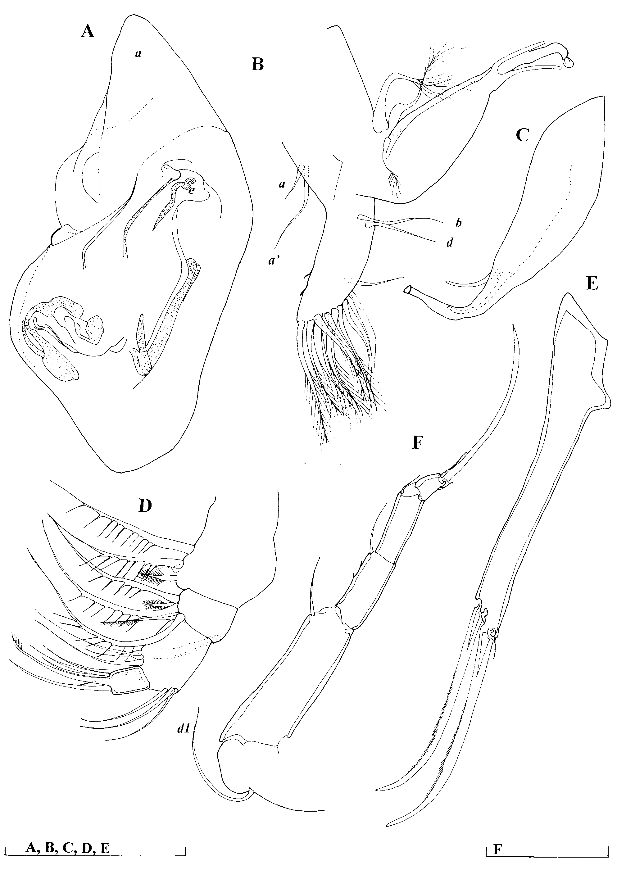
T1 and prehensile palps ( Figure 5 View FIGURE 5 B, C): left and right limbs equally developed, with elongated finger and prominent subterminal setae. Both a and a’ setae present.
T2 ( Figure 5 View FIGURE 5 F): 5-segmented. Basal segment with one seta, first and second endopodal segments with one seta each. Penultimate segment with one seta observed. Terminal claw 0.9 times as long as three terminal segments combined.
T3 ( Figure 4 View FIGURE 4 B): 5-segmented. Basal segment with three setae; first, second and third segments with one seta each, seta f considerably longer than g. Terminal segment with three setae, h1 being the shortest, but only slightly shorter than h2, which is 1/3 times as long as h3.
CR ( Figure 5 View FIGURE 5 E): without posterior seta. L ratios between anterior margin, anterior and posterior claw 1.2: 1: 1.
Hemipenis ( Figure 5 View FIGURE 5 A): Lobe “a” triangular; middle part “g” short, ejaculatory process “e” small. No second lobe “a”. Margins of lobe “b” hard to distinguish of the slide, but lobe “b” being rounded.
Female: Slide in a poor condition. A2 observed and with G2 as long as G1, CR similar to that of the male. Daday’s (1905) descriptions of female should be followed.
Remarks and affinities. The species can be easily separated from other representatives of the genus by its appearance in the lateral view: it has a flat dorsal margin, which inclines towards frontal and caudal ends. The drawings of the hemipenis of C. anisitsi , presented by Daday (1905), are somewhat confusing, and it is not clear if there is a second lobe “a”. The examination of the type material has revealed that there is only one lobe “a” and that the proximal part, which could be mistaken for the “a1” lobe, is an unidentified parasite, with many more present on the same slide, the animals having probably been infected prior to dissection. Unfortunately, the later record of this species from Paraguay ( Klie 1930) did not clarify the appearance of the carapace, and carapaces are not kept in the collection of the Zoological Museum in Hamburg, where three slides (collection numbers 324a, 324b, and 324c) containing only soft parts are deposited. Tressler (1956) reported Candonopsis anisitsi from Jamaica, but the shape of the carapace presented in the paper does not correspond with that of C. anisitsi at all, as it is more rounded than in Daday’s drawings ( Daday, 1905: Plate 16), so the identity of that species remains uncertain.
No known copyright restrictions apply. See Agosti, D., Egloff, W., 2009. Taxonomic information exchange and copyright: the Plazi approach. BMC Research Notes 2009, 2:53 for further explanation.
|
Kingdom |
|
|
Phylum |
|
|
Class |
|
|
Order |
|
|
Family |
|
|
SubFamily |
Candoninae |
|
Genus |
Latinopsis anisitsi ( Daday, 1905 )
| Karanovic, Ivana & Datry, Thibault 2009 |
Candonopsis anisitsi
| Karanovic 2004: 106 |
Candonopsis anisitsi
| Klie 1930: 226 |
| Daday 1905: 256 |