
Abelocephala albula, Ishikawa, Tadashi, Cai, Wanzhi & Tomokuni, Masaaki, 2015
|
publication ID |
https://doi.org/ 10.11646/zootaxa.3936.2.1 |
|
publication LSID |
lsid:zoobank.org:pub:157EDA4A-00A3-469F-8234-7E7175BF13E7 |
|
DOI |
https://doi.org/10.5281/zenodo.6112386 |
|
persistent identifier |
https://treatment.plazi.org/id/038E87FC-FFD4-1369-FF43-BBE7FAFDA500 |
|
treatment provided by |
Plazi |
|
scientific name |
Abelocephala albula |
| status |
sp. nov. |
Abelocephala albula View in CoL sp. nov.
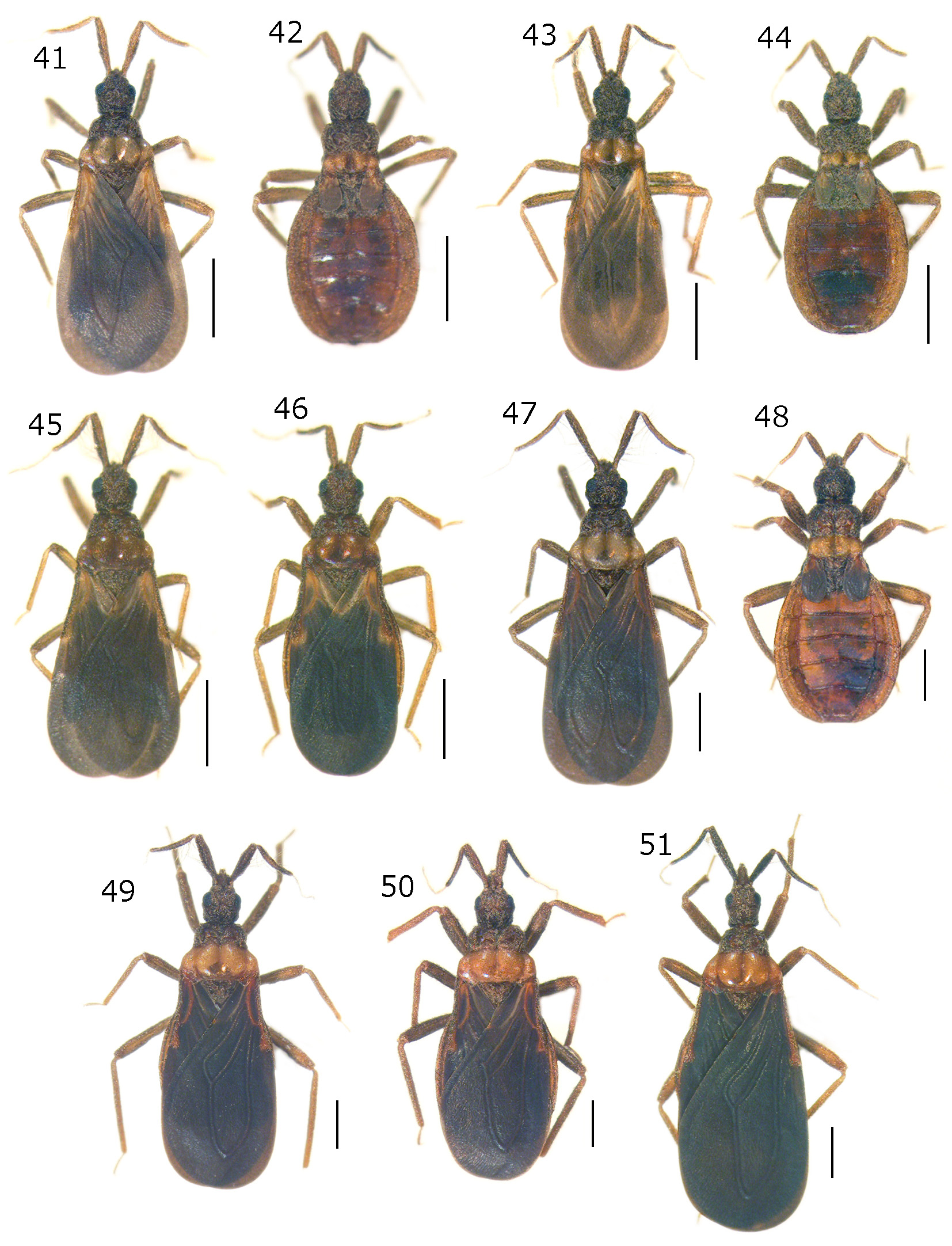
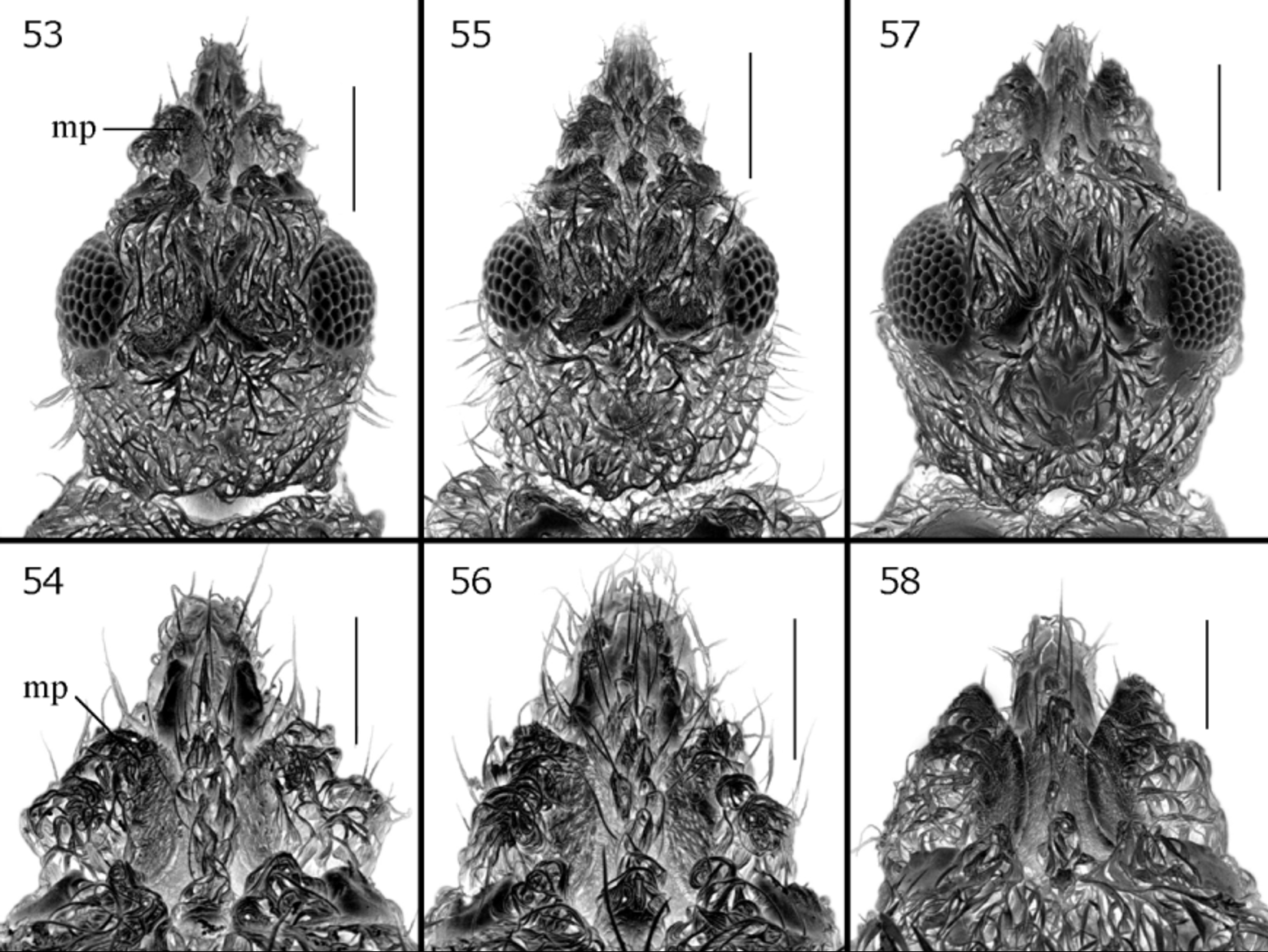
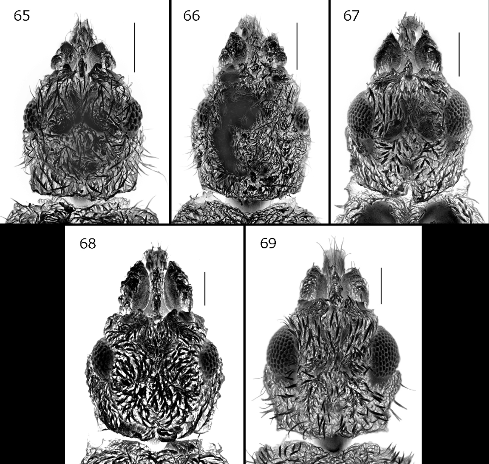
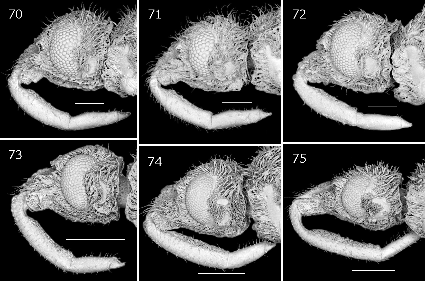
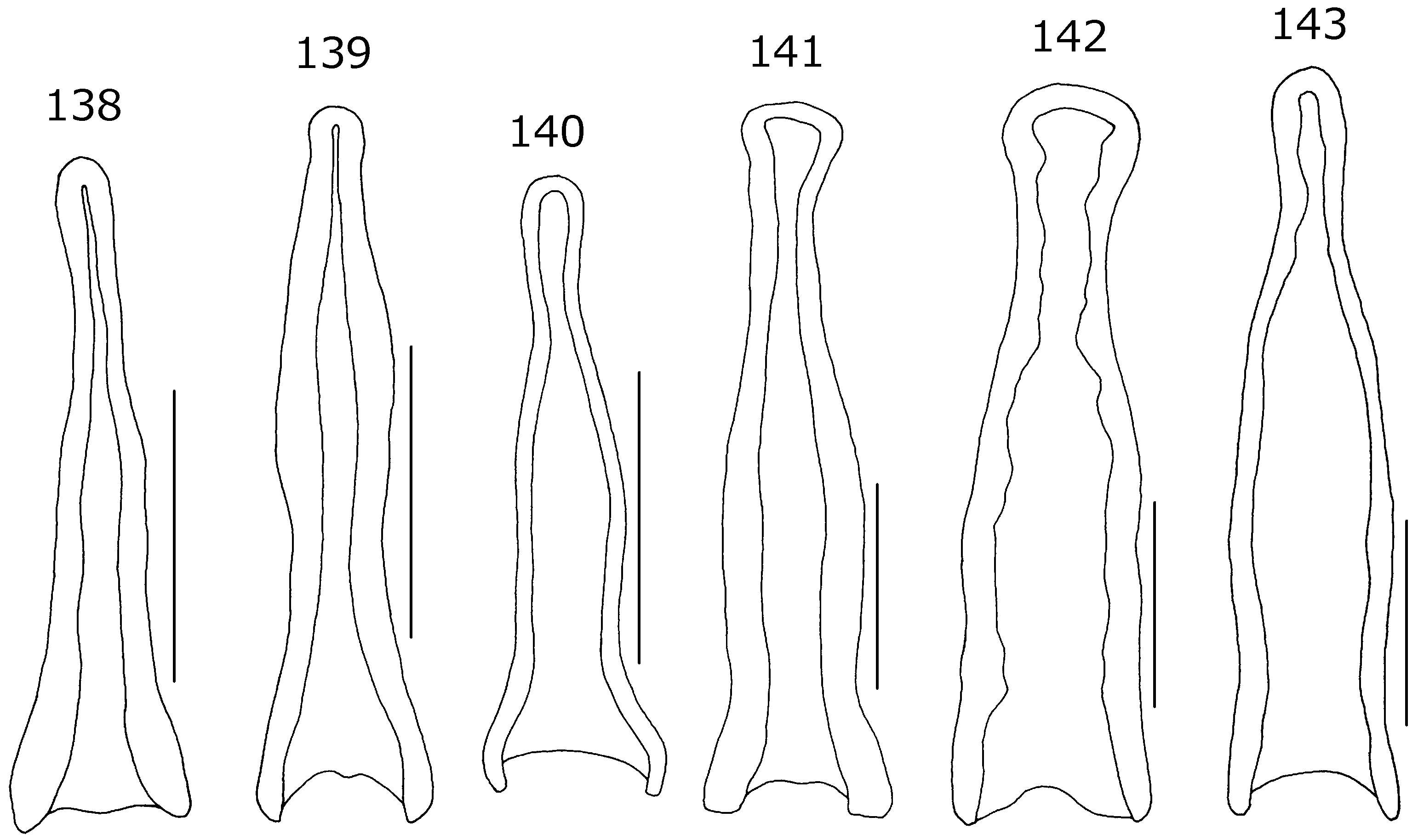
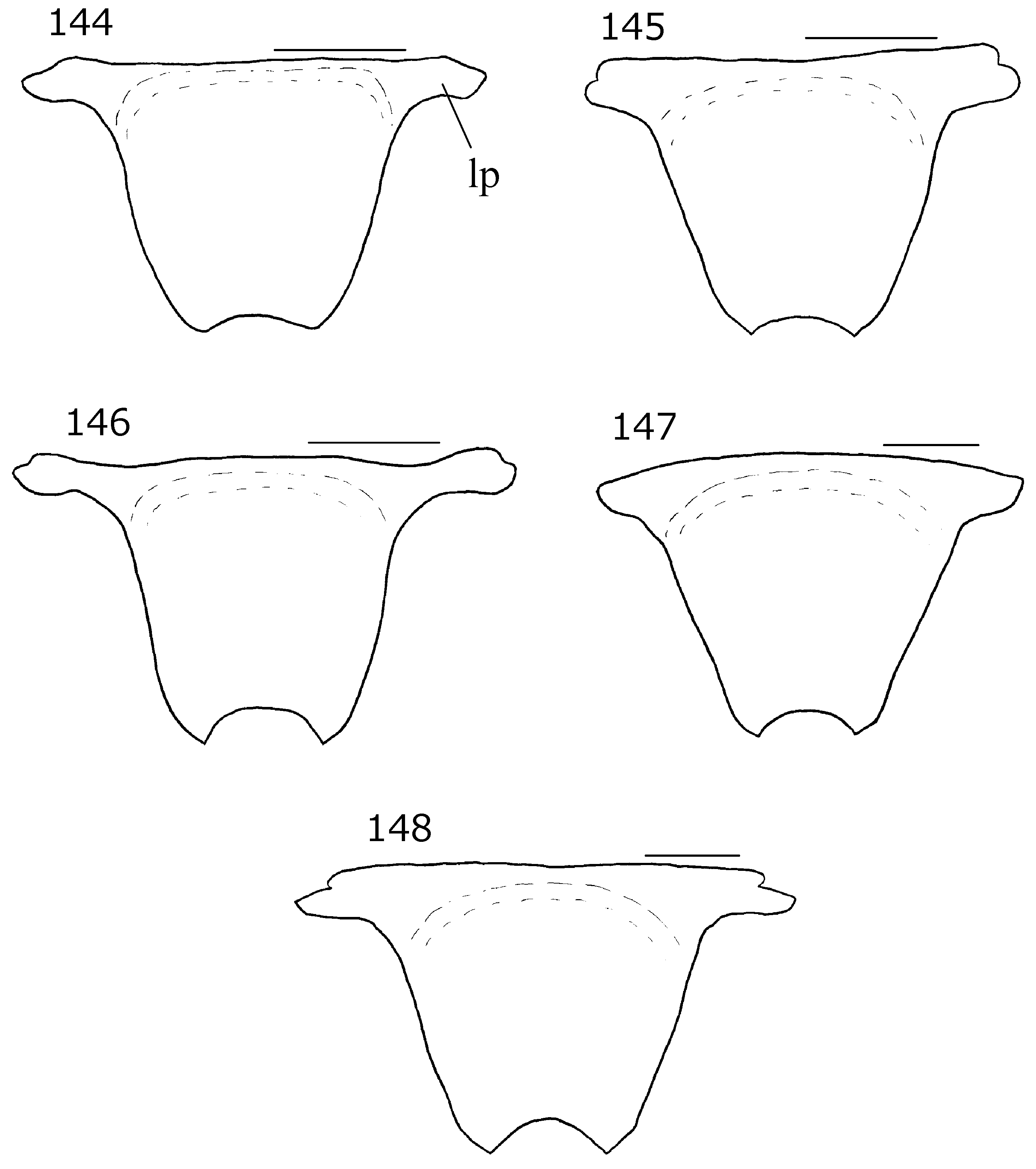
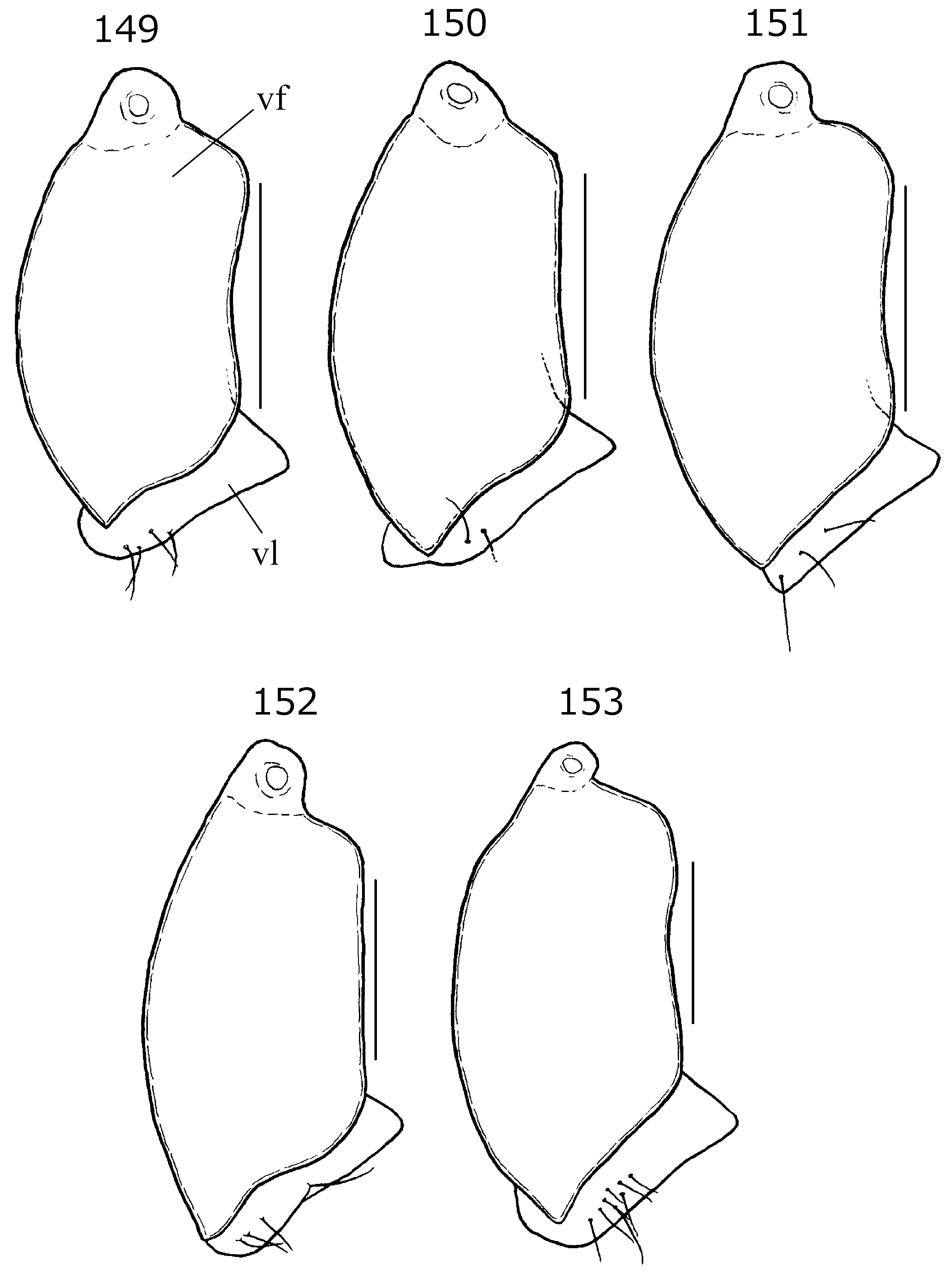
( Figs. 41, 42 View FIGURES 41 – 51 , 53, 54 View FIGURES 53 – 58 , 65 View FIGURES 65 – 69 , 70 View FIGURES 70 – 75 , 76 View FIGURES 76 – 80 , 81–86 View FIGURES 81 – 92 , 114 View FIGURES 114 – 119 , 120 View FIGURES 120 – 125 , 126, 132, 138, 144, 149)
Diagnosis. This species is recognized by the following combination of character states: body approximately 3 mm long; head 1.4 to 1.5 times longer than width across eyes; mandibular plate nearly right-angled at anterior corner and weakly projected anteriorly in dorsal view; posterior pronotal lobe whitish to pale yellow; outer (larger) cell of hemelytral membrane acutely angled apically; and posterior process of pygophore wide with straight apical margin in dorsal view.
Description. Male (macropterous). Body ( Fig. 41 View FIGURES 41 – 51 ) mostly brownish. Antennae, rostrum, and legs brownish yellow. Antennal flagella pale yellow. Posterior pronotal lobe whitish to pale yellow except for median longitudinal sulcus and posterior marginal area brownish. Hemelytra brown, with basal part pale yellow to pale brown; corial cell more or less fuscous ( Fig. 41 View FIGURES 41 – 51 ). Abdomen yellowish brown to brown.
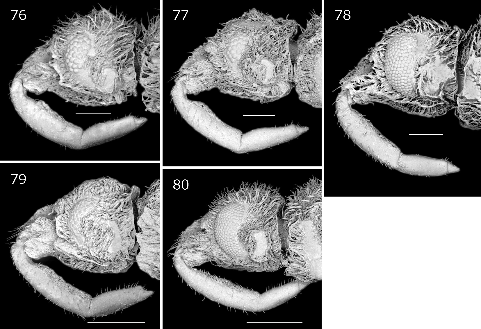
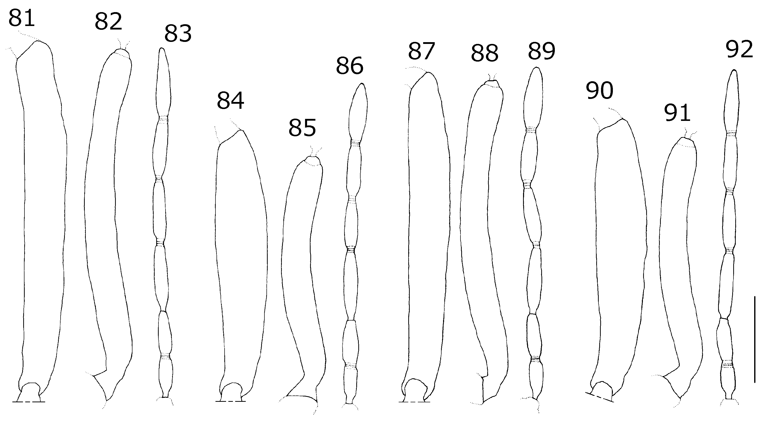
Head ( Figs. 41 View FIGURES 41 – 51 , 53, 54 View FIGURES 53 – 58 , 70 View FIGURES 70 – 75 ) approximately 1.4 times longer than width across eyes, as long as pronotum; anteoculus 1.1 times longer than postoculus; mandibular plate ( Fig. 54 View FIGURES 53 – 58 ) nearly right-angled at anterior corner and weakly projected anteriorly in dorsal view. Eye ( Figs. 53 View FIGURES 53 – 58 , 70 View FIGURES 70 – 75 ) approximately 0.35 times as wide as interocular space in dorsal view. Antennal segment I slender, approximately 8.5 times longer than its maximum width, as long as segment II ( Figs. 81, 82 View FIGURES 81 – 92 ); flagellum as long as segment I ( Fig. 83 View FIGURES 81 – 92 ). Rostral segment I stout, 1.5 times longer than segment II ( Fig. 70 View FIGURES 70 – 75 ).
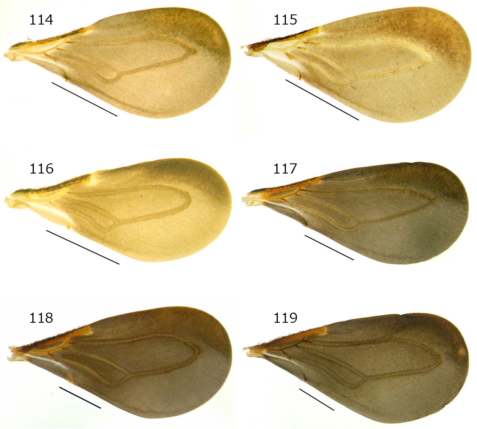
Pronotum ( Fig. 41 View FIGURES 41 – 51 ) approximately 0.7 times as long as humeral width; anterior lobe 0.6 times as long as posterior lobe along midline, 0.7 times as wide as humeral width. Hemelytron ( Figs. 41 View FIGURES 41 – 51 , 114 View FIGURES 114 – 119 ) wide, twice as long as its maximum width, exceeding apex of abdomen by approximately 0.35 times of its length; outer (larger) cell of membrane ( Fig. 114 View FIGURES 114 – 119 ) acutely angled apically.
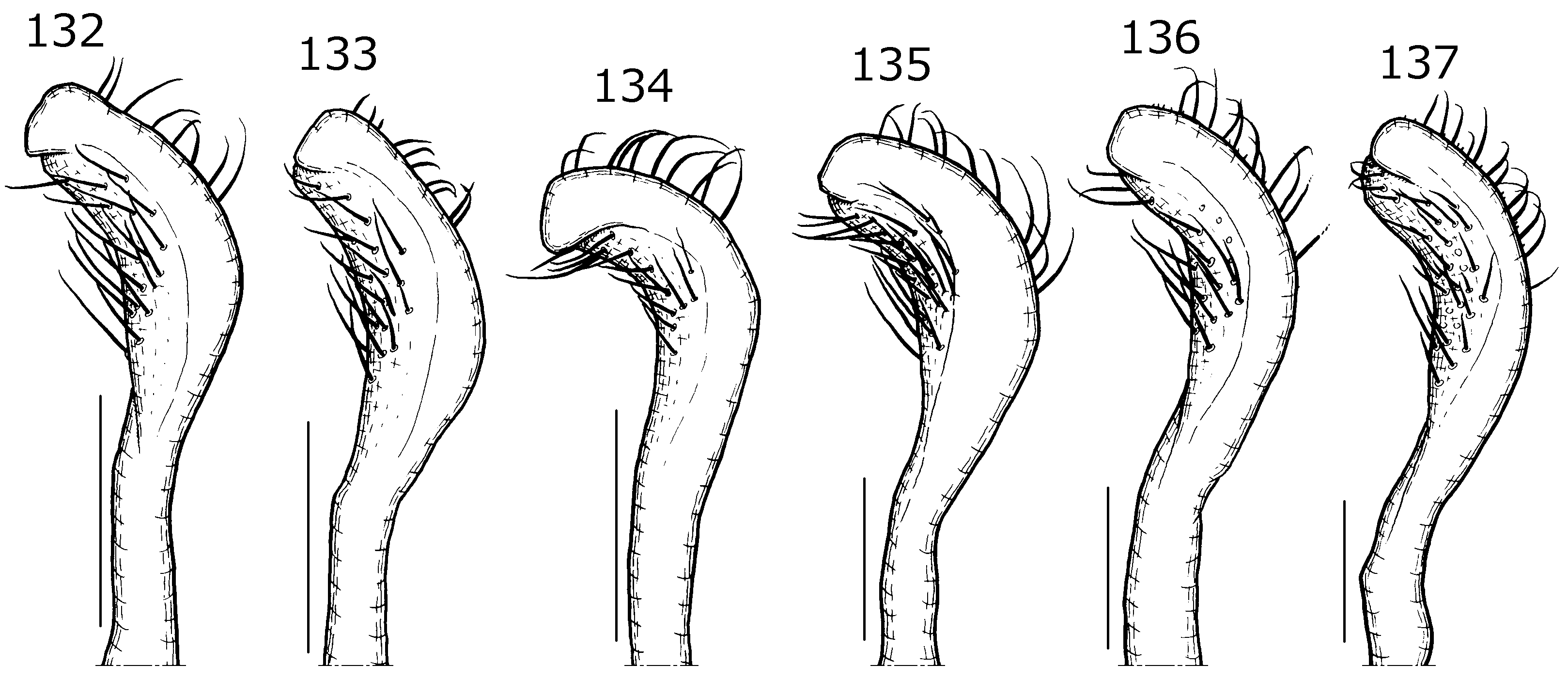
Pygophore ( Fig. 120 View FIGURES 120 – 125 ) somewhat compressed dorsoventrally; posterior process (Fig. 126) wide, with straight apical margin in dorsal view. Parameres ( Fig. 132 View FIGURES 132 – 137 ) weakly curved in apical two-thirds, with obtuse, inwardly projected apex in dorsal view. Struts of phallus ( Fig. 138 View FIGURES 138 – 143 ) tapering apicad, obtuse at apex, and with lateral walls thickened at its base in dorsal view.
Female (micropterous). At first sight, obviously differing from male due to micropterous condition ( Fig. 42 View FIGURES 41 – 51 ). Head ( Figs. 42 View FIGURES 41 – 51 , 65 View FIGURES 65 – 69 , 76 View FIGURES 76 – 80 ) approximately 1.5 times longer than width across eyes, approximately 1.2 times longer than pronotum; anteoculus 0.9 times as long as postoculus. Eye ( Figs. 65 View FIGURES 65 – 69 , 76 View FIGURES 76 – 80 ) small, approximately 0.2 times as wide as interocular space in dorsal view. Antennal segment I stouter and much shorter than that of male, approximately 5.5 times longer than its maximum width, a little longer than segment II ( Figs. 84, 85 View FIGURES 81 – 92 ); flagellum approximately 1.2 times longer than segment I ( Fig. 86 View FIGURES 81 – 92 ). Rostral segment I stout, 1.4 times longer than segment II ( Fig. 76 View FIGURES 76 – 80 ). Pronotum ( Fig. 42 View FIGURES 41 – 51 ) approximately 0.85 times as long as humeral width; anterior lobe 1.1 times longer than posterior lobe along midline, as wide as humeral width. Hemelytra ( Fig. 42 View FIGURES 41 – 51 ) small, pad-like, reaching to posterior margin of abdominal tergite II; venation inconspicuous. Abdominal tergite IX ( Fig. 144 View FIGURES 144 – 148 ) with lateral projection at each basal angle; lateral projection short, tapering in apical part, obtuse at apex. Valvifer I ( Fig. 149 View FIGURES 149 – 153 ) oblong; valvula I ( Fig. 149 View FIGURES 149 – 153 ) with approximately 4 setae.
Measurements [in mm, ♂ (n=51) /♀ (n=8), holotype in parentheses]. Body length 2.76–3.17/2.91–3.17 (3.00). Head length 0.66–0.74/0.71–0.74 (0.72), width across eyes 0.53–0.57/0.47–0.49 (0.54). Lengths of antennal segments I and II 0.72–0.80/0.55–0.56 (0.77) and 0.71–0.77/0.49–0.53 (0.74), respectively. Lengths of rostral segments I and II 0.49–0.55/0.52–0.56 (0.49) and 0.31–0.35/0.30–0.35 (0.34), respectively. Pronotum length 0.66–0.67/0.53–0.60 (0.67), width across humeri 0.92–1.01/0.63–0.68 (0.99). Hemelytron length 2.74–2.82/ 0.37–0.46 (2.74). Lengths of femur and tibia of fore leg 0.86–0.98/0.78–0.87 (0.95) and 0.91–1.00/0.80–0.85 (0.98); of mid leg 0.84–0.91/0.71–0.83 (0.88) and 0.87–0.95/0.79–0.81 (0.88); of hind leg 1.08–1.21/0.98–1.05 (1.19) and 1.15–1.31/1.01–1.09 (1.26). Abdomen length 1.44–1.67/1.57–1.66 (1.56), maximum width 1.22–1.44/ 1.40–1.59 (1.34).
Holotype. ♂ ( Fig. 41 View FIGURES 41 – 51 ), “[ JAPAN] Komi, Iriomote-jima Is., the Ryûkyûs, 23–27.IV.2004, FIT, T. Ishikawa et al.” ( LETUA IC 2014-00069) ( TUA).
Paratypes (50 ♂, 8 ♀). JAPAN [Ishigaki Is.] Shiramizu: 1 ♂, 1 ♀ ( Fig. 42 View FIGURES 41 – 51 ), 15.vi.2003, H. Mizushima ( LETUA IC 2014-00070–00071) ( TUA), 1 ♂, 3–6.v.2004, FIT-M, T. Ishikawa ( LETUA IC 2014-00072) ( TUA). Mt. Omoto-dake: 2 ♀, 16.vi.2003, H. Mizushima ( LETUA IC 2014-00073–00074) ( TUA). [Iriomote Is.] Haemidahama: 1 ♀, 30.v.1999, K. Takahashi ( LETUA IC 2014-00075) ( TUA). Ôtomi-rindô: 2 ♂ (one shown in Fig. 114 View FIGURES 114 – 119 ), 7–11.vi.2002, Malaise trap, T. Tsuru ( LETUA IC 2014-00076–00077) ( TUA), 1 ♀, 23.iv.2004, T. Ishikawa ( LETUA IC 2014-00078) ( TUA), 1 ♂, 26.iv.2004, T. Ishikawa ( LETUA IC 2014-00079) ( TUA). Fusatoruba, Ôhara: 1 ♀ ( Figs. 144 View FIGURES 144 – 148 , 149 View FIGURES 149 – 153 ), 29.v.2000, H. Mizushima ( LETUA IC 2014-00080) ( TUA). Komi: 1 ♂, 2.iii.2002, T. Ishikawa ( LETUA IC 2014-00081) ( TUA), 2 ♀ (one shown in Figs. 65 View FIGURES 65 – 69 , 76 View FIGURES 76 – 80 , 84–86 View FIGURES 81 – 92 ), 2.v.2002, S. Nagashima ( LETUA IC 2014-00082–00083) ( TUA), 1 ♂, 1.iv.2003, S. Nagashima ( LETUA IC 2014-00084) ( TUA), 28 ♂ (each one shown in Figs. 53, 54 View FIGURES 53 – 58 , 70 View FIGURES 70 – 75 , 81–83 View FIGURES 81 – 92 , 132 View FIGURES 132 – 137 , and Figs. 120 View FIGURES 120 – 125 , 126, 138), same data as holotype ( LETUA IC 2014-00085–00112) ( TUA, CAU, NSMT). Near Aira-gawa: 1 ♂, 28.iv.2003, T. Kurihara ( LETUA IC 2014-00113) ( TUA). Funaura: 3 ♂, 6–11.iv.2005, FIT-M, J. Kantoh ( LETUA IC 2014-00114–00116) ( TUA). Shirahama: 11 ♂, 23–27.iv.2004, FIT-M, T. Ishikawa et al. ( LETUA IC 2014-00117–00127) ( TUA).
Distribution. Japan: Ryukyu Islands (Ishigaki Is., Iriomote Is.).
Etymology. From the Latin albula , referring to the whitish to pale yellow pronotum; an adjective.
Remarks. Macropterous males and micropterous females of this species are known. In general habitus, this new species resembles Abelocephala thai , which is the type species of the genus, known from Thailand ( Maldonado Capriles 1996). The male of A. albula sp. nov. can be distinguished from the male of A. thai by the longer head [approximately 1.4 times longer than the width across the eyes ( Fig. 53 View FIGURES 53 – 58 ) vs. approximately 1.1 times longer than width across the eyes], shorter antennal flagellum [as long as antennal segment I ( Figs. 81, 83 View FIGURES 81 – 92 ) vs. approximately 1.2 times longer than antennal segment I], shorter rostral segment I [1.4 times longer than segment II ( Fig. 70 View FIGURES 70 – 75 ) vs. approximately 3 times longer than segment II], and acutely angled outer (larger) cell of the hemelytral membrane at apex ( Fig. 114 View FIGURES 114 – 119 ) (vs. rounded angled outer (larger) cell of the hemelytral membrane at apex). We were unable to compare females between these two species because no female specimens of A. thai were available.
Many male specimens (all with macropterous wings) examined in the present study were collected from FIT- Ms or a Malaise trap placed in humid forests. This finding suggests that the males fly actively near the forest floor. All the females (with micropterous wings) and a few males were collected directly from forest leaf litter.

No known copyright restrictions apply. See Agosti, D., Egloff, W., 2009. Taxonomic information exchange and copyright: the Plazi approach. BMC Research Notes 2009, 2:53 for further explanation.