
Megaperlodes tiunovi Te
|
publication ID |
https://doi.org/ 10.11646/zootaxa.3904.4.4 |
|
publication LSID |
lsid:zoobank.org:pub:D8D15BFD-4954-4976-B17C-31DDDE20BE66 |
|
DOI |
https://doi.org/10.5281/zenodo.6105919 |
|
persistent identifier |
https://treatment.plazi.org/id/038F6B41-512B-FFB5-48DA-FB67FCD6FE51 |
|
treatment provided by |
Plazi |
|
scientific name |
Megaperlodes tiunovi Te |
| status |
|
Megaperlodes tiunovi Te View in CoL sl e nk o s p. n.
( Figs. 1–19 View FIGURES 1 – 3 View FIGURES 4 – 10 View FIGURES 11 – 16 View FIGURES 17 – 19 )
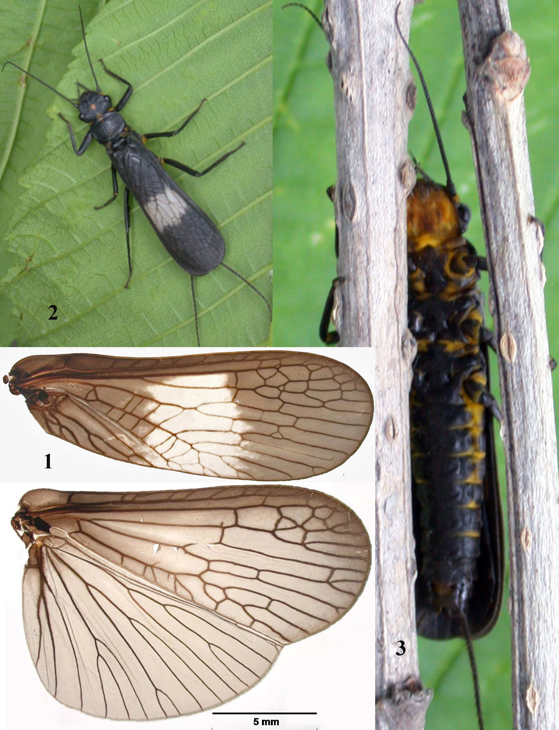
Description. Adult habitus. Sexually dimorphic, female larger than male. Mean body length from tip of the head to the apex of abdomen, males, 17.0–17.3 mm; females 22.0–26.2 mm. Wings slightly shortened. Forewing length, males, 15.2–16.0 mm; females, 19.8–21.8 mm; wingspan, males, 32.3–34.1 mm; females 42.8–46.8 mm. The front wing of male and female ( Fig. 1 View FIGURES 1 – 3 ) black, fumose, veins dark, in the middle with a contrastive white frosted spot, occupying about ⅓ of the wing length. The spot extends from R1 to the posterior wing edge, lateral margins of the spot are indistinct, and veins in the spot paler. Wing venation includes an irregular net near the apex sometimes consisting of four rows of cells. Seven to ten cross veins between C and Sc; five apical veins between Sc and R1 ( Fig. 1 View FIGURES 1 – 3 ). Rs with five apical branches. Nine to thirteen veins between М and Cu2; four anal veins branching into six. Hindwing black fumose, slightly paler in the cord area, in males especially. Anal area enlarged and pleated, A2–A5 forked; A5 with dark small spot at base.
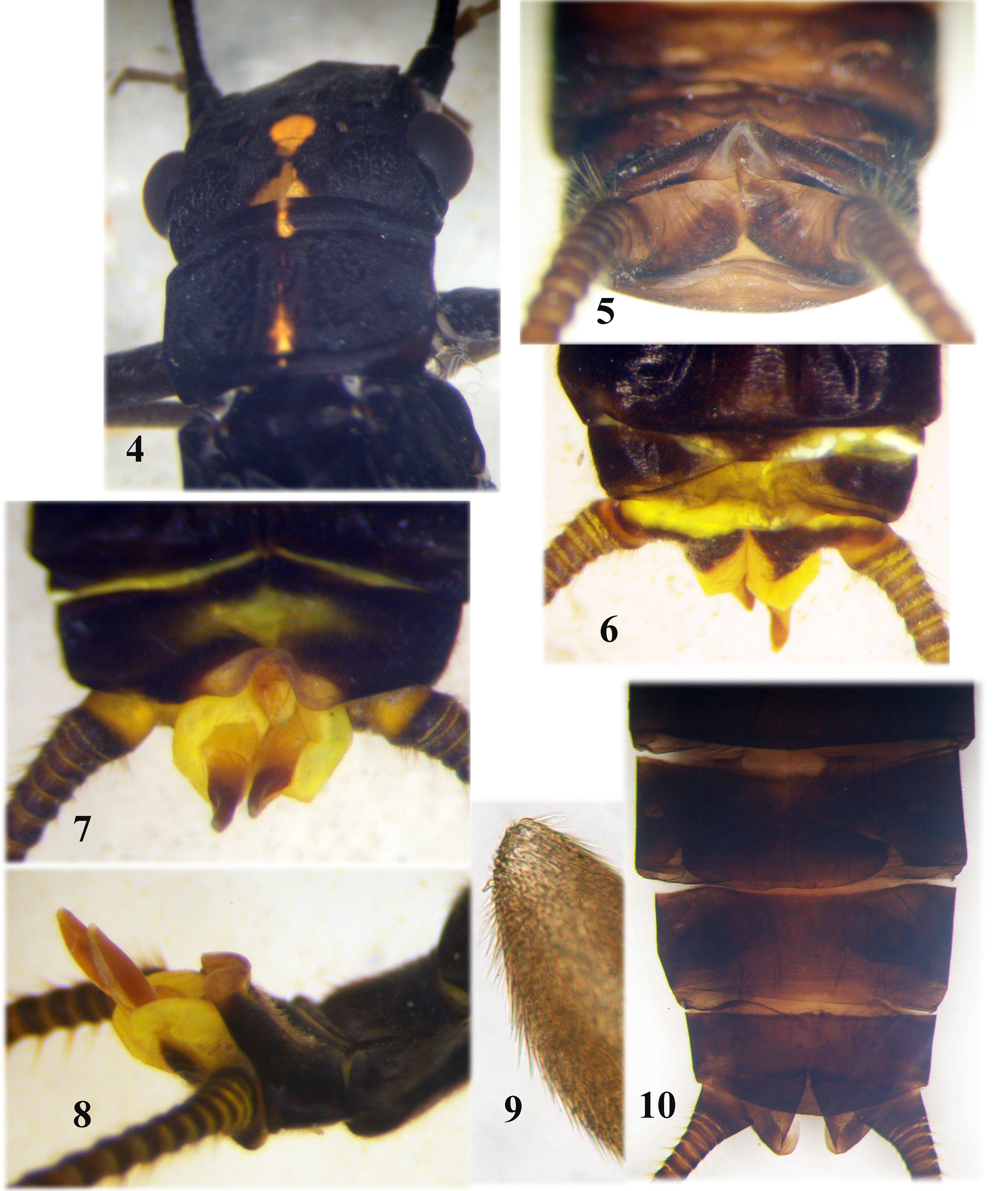
General body colour black with yellow spots; membranous ventral portions also yellow ( Figs. 2, 3 View FIGURES 1 – 3 ). Head and pronotum matt black, with sharply delimited bright yellow marks. Head with compound eyes wider than the pronotum; M-line smooth and distinct ( Fig. 4 View FIGURES 4 – 10 ). Interocellar area with a small tear-shaped bright-yellow spot, widest anteriorly. A small yellow triangle extends along the epicranial stem on the occiput ( Fig. 4 View FIGURES 4 – 10 ). A posterolateral reticulate callosity behind each compound eye. Antennae shorter than cerci, black; palpi dark brown. Head ventrally yellow brown ( Fig. 3 View FIGURES 1 – 3 ). Submentum almost square, with slightly rounded lateral margins, the submental gills very small to almost absent in both sexes. The pronotum rectangular, same width as the head width under the compound eyes, with narrow longitudinal thread-like median yellow band widened triangularly at the anterior pronotal margin as well as before the posterior pronotal furrow ( Fig. 4 View FIGURES 4 – 10 ). Pronotal rugosites smooth, the lateral fields matt. The arms of the mesosternal ridge meet the posterior corners of the furcal pits; suture between basisternum and furcasternum indistinct ( Fig. 3 View FIGURES 1 – 3 ).
Abdominal tergites black, strongly sclerotized and covered by short colourless clothing hairs, only tergum 1 with X-shaped white spot medially, with a rounded inside black spot. Abdominal segments 1–5 divided laterally by yellow pleural folds, more distinct in females; posterior segments ring-shaped, entire, fully sclerotized ( Fig. 3 View FIGURES 1 – 3 ). Legs black except bright yellow joints ( Fig. 2 View FIGURES 1 – 3 ).
Male. Abdominal tergum 8 black, slightly impressed dorsomedially; two submedial rounded swellings covered by short colourless hairs close to the posterior margin, especially apparent in lateral view ( Figs. 5, 8 View FIGURES 4 – 10 ). Posterior margin of tergum 9 with a medial triangular notch, which extends ½ of the length of tergum 9, two posterolateral rounded swellings covered by short fine colourless hairs ( Fig. 7 View FIGURES 4 – 10 ); in repose tergum 9 hidden by tergum 8 and noticeable as narrow band with a triangular posteromedial notch ( Fig. 5 View FIGURES 4 – 10 ). Sternum 9 bears a scoopshaped subgenital plate extending backward and curved upward ( Figs. 5, 6 View FIGURES 4 – 10 ). Sternum 10 narrow, pale medially with two black lateral bands hidden by the subgenital plate in ventral view ( Fig. 6 View FIGURES 4 – 10 ). Tergum 10 entire, black, with median diamond-shaped yellow spot, covered by short fine colourless hairs ( Fig. 7 View FIGURES 4 – 10 ). In repose, tergum10 tapers backward and caudally triangular, the paraprocts touch in contact dorsomedially, pressed against the lower face of tergum10. In everted condition, a yellow membranous strip with rounded posterolateral edges protrudes from under the edge of tergum10 and supported by a small medial conical peg located between the paraproct lobes and concealed by them in repose ( Figs. 7, 8 View FIGURES 4 – 10 ). The paraproct sclerite wide, heavily sclerotized basally, narrowed and bluntly rounded distally ( Figs. 5, 6, 8 View FIGURES 4 – 10 ). Eversible paraproct with lobe (EPL) arises from the base of the paraproct sclerite ( Fig. 8 View FIGURES 4 – 10 ). EPL large, inflated, ball-shaped basally, yellow and bears a long fingerlike projection, extended at the base and pointed towards the top ( Figs. 6–8 View FIGURES 4 – 10 ); ventral face of the projection membranous, the dorsal face covered by long thin brown-red sclerotized setae, more densely in the distal ⅓ of the projection ( Figs. 8, 9 View FIGURES 4 – 10 ). Both projections closely appressed, directed obliquely backward and upward ( Figs. 6–8 View FIGURES 4 – 10 ).
Female. Sternum 8 black with a small pale spot anteromedially, a short transversal subgenital plate extending slightly beyond the edge of sternum 8 ( Fig. 10 View FIGURES 4 – 10 ), posterior margin of the plate with a weak medial notch separating two lateral lobes, slightly rounded posteriorly, covered by small setae ( Fig. 10 View FIGURES 4 – 10 ). Lateral margins of subgenital plate rounded. Abdominal sternum 9 black with pale spot medially, a pair of dark posterolateral sclerites looks like oblique bands directed to anterior margin. Abdominal sternum 10 and the blunt paraprocts unmodified and back.
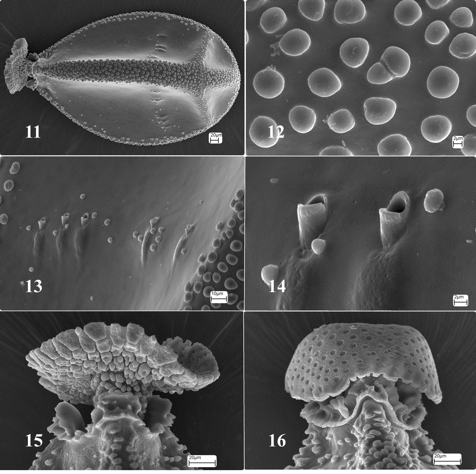
Egg. Trilateral ( Fig. 11 View FIGURES 11 – 16 ), 614 X 345 µm with narrow anterior pole. The longitudinal ridges (maximal width 65 Μm) delimiting the three faces of the egg, densely covered by globular bodies ( Figs. 11, 12 View FIGURES 11 – 16 ). Each face additionally with a transverse ridge close to the posterior pole, also covered by globular bodies smaller and less dense, than on the longitudinal ridges ( Fig. 11 View FIGURES 11 – 16 ). A row of 3–7 micropyles near transverse ridge ( Figs. 11, 13 View FIGURES 11 – 16 ) on each of the three faces. Micropyle openings elevated above the chorion surface, rostral with rounded margins ( Figs. 13, 14 View FIGURES 11 – 16 ). Collar short, formed by medially projecting extensions of the three longitudinal ridges, each top of extension flaky, and petal-shaped, smooth on inner surface ( Fig. 15 View FIGURES 11 – 16 ). Collar delimited by rim on outer side basally ( Figs. 15, 16 View FIGURES 11 – 16 ). Anchor mushroom-shaped with short pedicel ( Fig. 16 View FIGURES 11 – 16 ); external surface pleated with globular bodies ( Fig. 16 View FIGURES 11 – 16 ). Internal surface of anchor as Fig. 15 View FIGURES 11 – 16 . The margin of the anchor covers the pedicel, not reaching the collar’s rim ( Fig. 16 View FIGURES 11 – 16 ). The chorion surface smooth ( Fig. 14 View FIGURES 11 – 16 ).
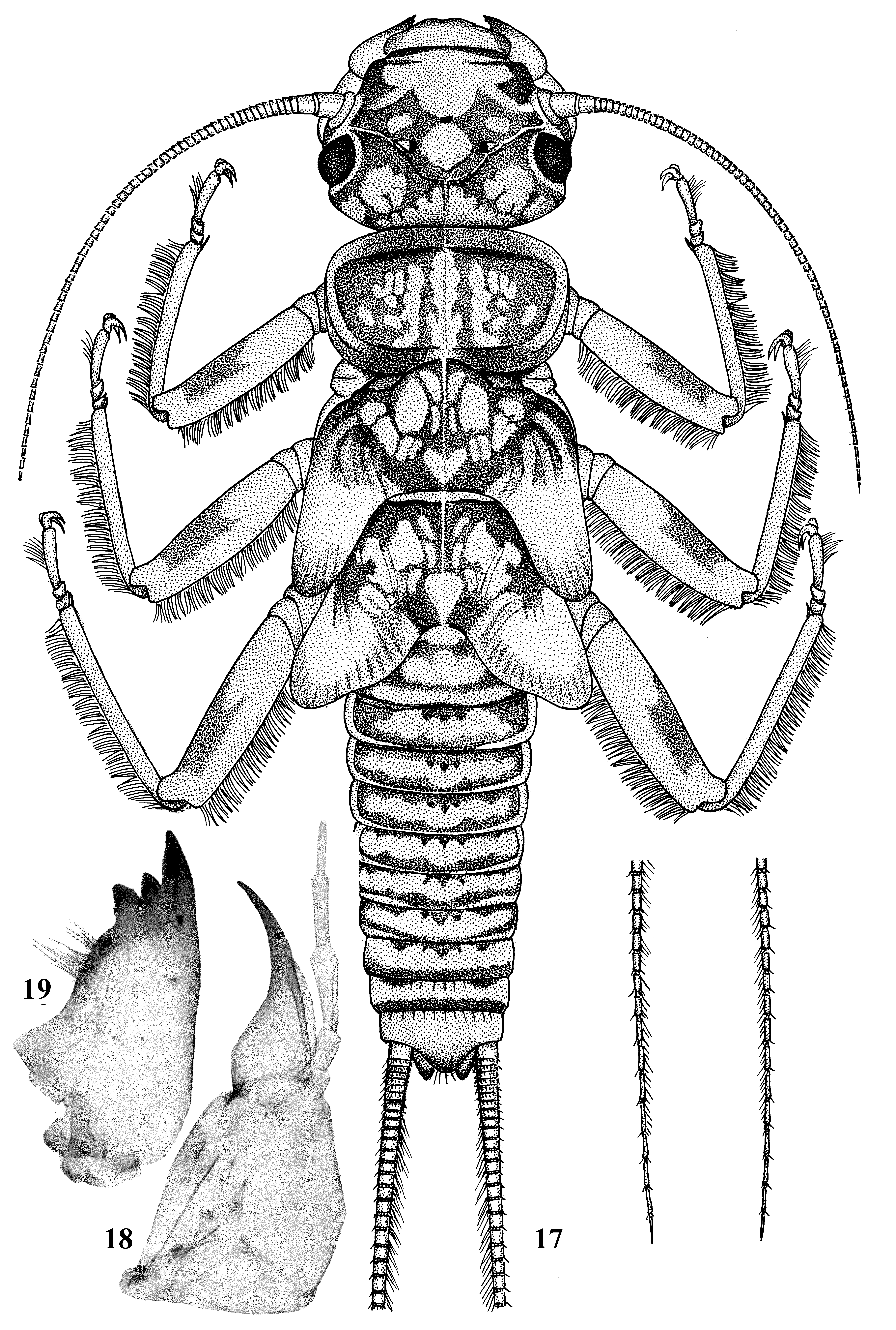
Larvae. Mature female larvae 20.5–21.5 mm long (n=2). General colour grey-brown with a pale pattern. Body covered by short brown clothing hairs. Antennae, cerci and ventral body side pale grey. Head wider than the pronotum with a large, triangular, pale spot in front of the anterior ocellus extending onto the clypeus, labrum pale ( Fig. 17 View FIGURES 17 – 19 ). M-line pale, distinct; interocellar area brown with tear-shaped central pale spot ( Fig. 17 View FIGURES 17 – 19 ). A small triangular pale spot laterally from each posterior ocellus and the tentorial callosities pale. Occiput with a large triangular pale spot around the epicranial stem, two pairs of pale spots bordered by sinuate brownish rims behind each eye. No postocular spinule row. Lacinia unidentate ( Fig. 18 View FIGURES 17 – 19 ), narrow apically, pointed and well sclerotized, basal ½ expanded. Submarginal setae absent. Galea reaches more than ½ of the lacinia length. Mandible ( Fig. 19 View FIGURES 17 – 19 ) simple, not deeply cleft, with medial setae; mandibular teeth without serrations; a small patch of acanthae basally from the last tooth. Submentum square with almost straight lateral margins; submental gills very short or almost absent. Pronotum trapeziform, transverse with rounded angles; anterior margin wider than posterior one; median, dorsal stripe pale, narrow, slightly wider posteriorly, a pale reticulate stripe presents on each lateral field of pronotum; pronotal margins without fringe of bristles ( Fig. 17 View FIGURES 17 – 19 ). Legs pale-grey, femur ventrally in distal ½ with elongate brownish mark. Outer margins of femur, tibia and tarsus with a fringe of colourless silky hairs.
Abdominal segments 1–5 divided by hairless pleural membranes. Terga with a few colourless long hairs posteriomedially, more pronounced on the last terga; the bristles at posterior tergal margins not detected. Terga brown with irregular yellow median band, the brown edge in front of it with three prominent dark spots; the distinct caudal setae present on terga 8 and 9; the pattern on terga 9 and 10 diffuse ( Fig. 17 View FIGURES 17 – 19 ). All abdominal terga covered by brown clothing hairs. Paraprocts normal length, apex rounded. Cerci pale greyish with dorsal fringe of fine, silky, colourless hairs ( Fig. 17 View FIGURES 17 – 19 ) increasing in length towards apical cercal segments, each cercal segment with an apical whorl of short brown setae.
Material examined. Holotype, male. Russian Far East, Primorsky Region, the Brus’ya River under road bridge, near Bamburovo Settlement, 42.56.480'N 131.19.120'E, 12.05.2014, coll. Teslenko V. Paratypes: 1 male, 6 females, 7 exuviae, the same locality and data as holotype, coll. Teslenko V.; 2 males (dissected in vial), 10 females (one dissected in vial).
Other material. 5 egg masses, 7 exuviae, the same locality, 18.05.2014, coll. V. Teslenko; 2 nymphs, the same locality, 28.04.2013, coll. I. Tiunov.
Etymology. The species is named in honor of the ornithologist Ivan M. Tiunov, who also made significant contributions to the knowledge of the stonefly and mayfly fauna of the Russian Far East.
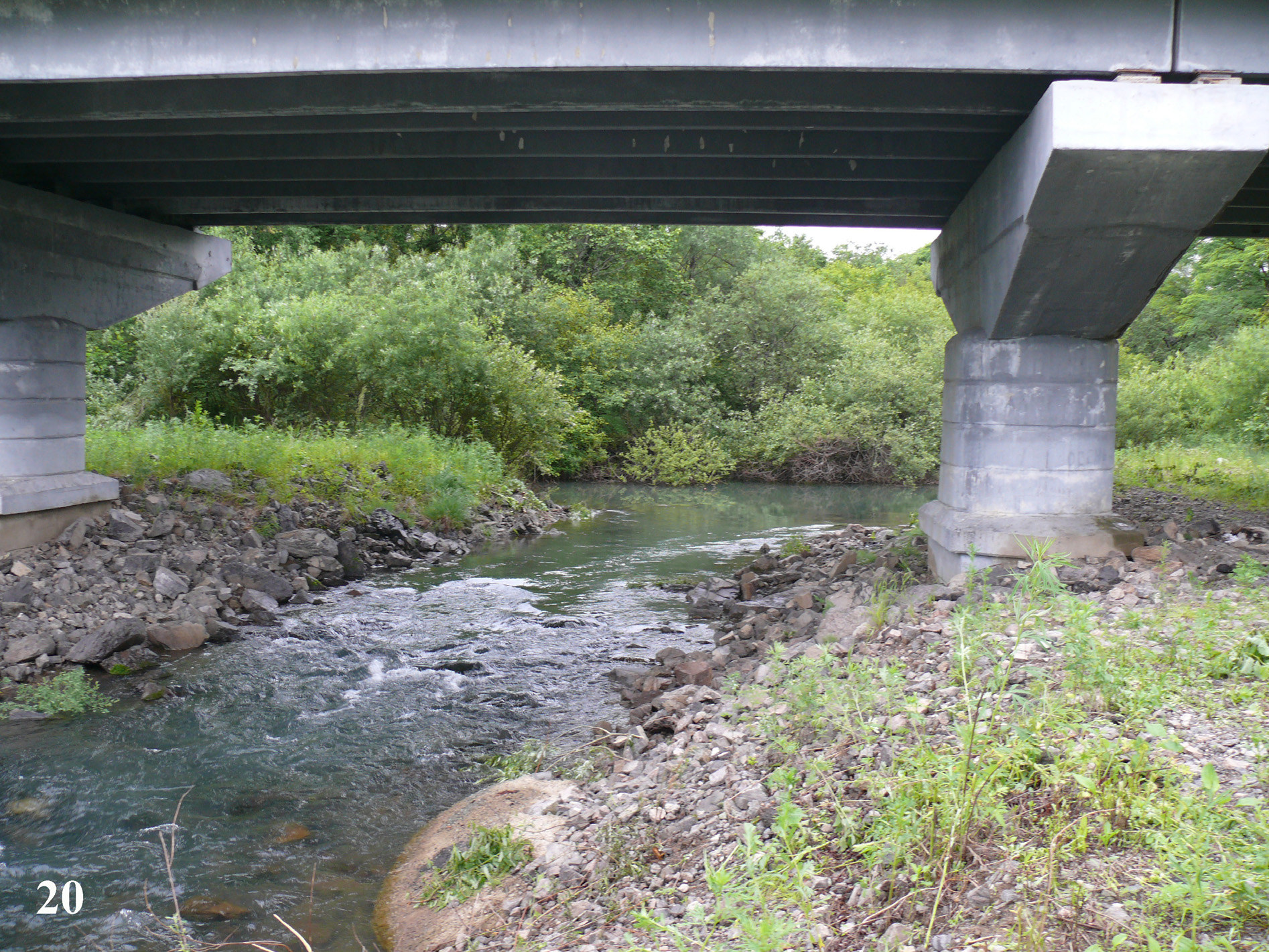
Distribution. The new species is known from the small foothill salmonid stream, the Brus’ya River. This stream is located in the South Russian Far East in the Primorsky Region near the border with China and Korea. The Brus’ya River flows out from the East Manchurian Mountains to the Peter the Great Bay of the Sea of Japan. The type locality is situated about 5 km from the mouth ( Fig. 20 View FIGURE 20 ). Water temperature at the time of collection (12 of May 2014 at 12 am) was 3.8ºC.
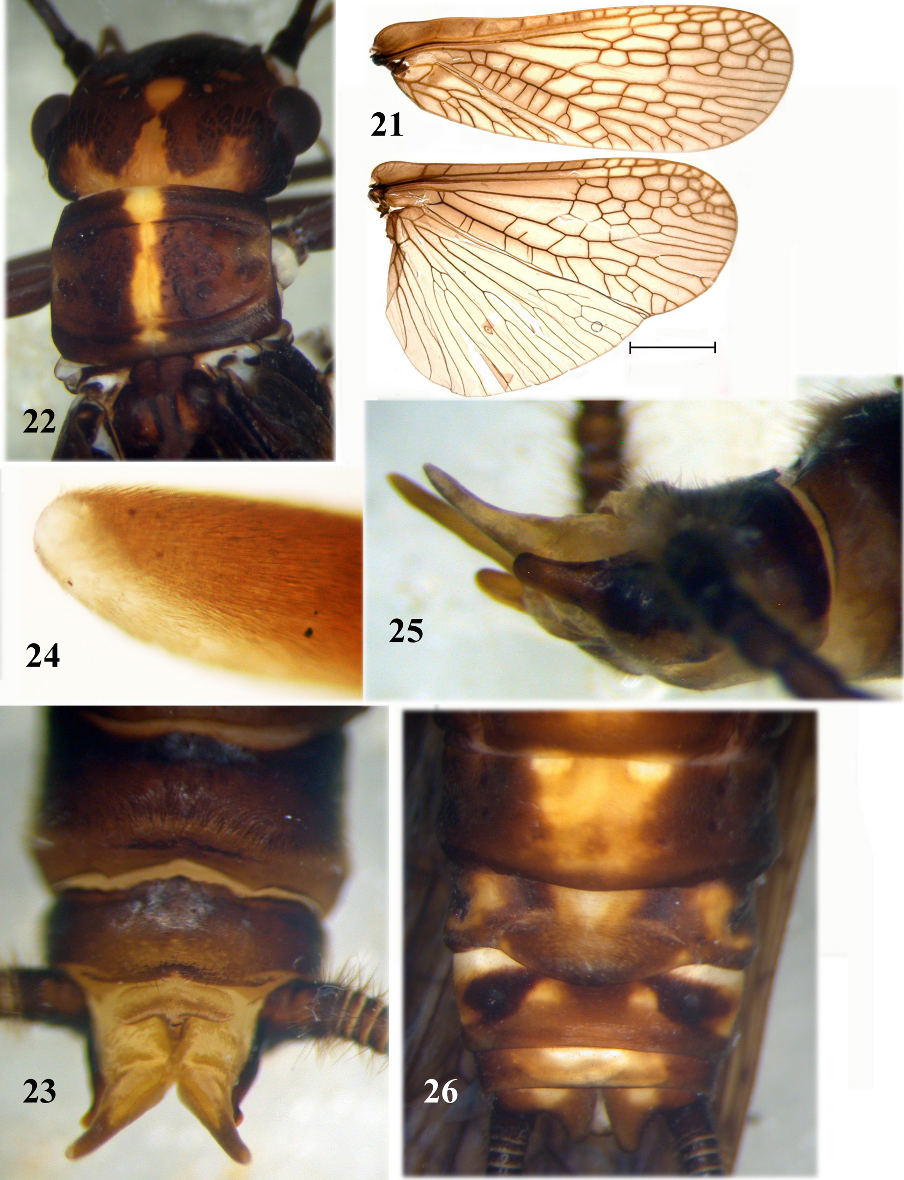
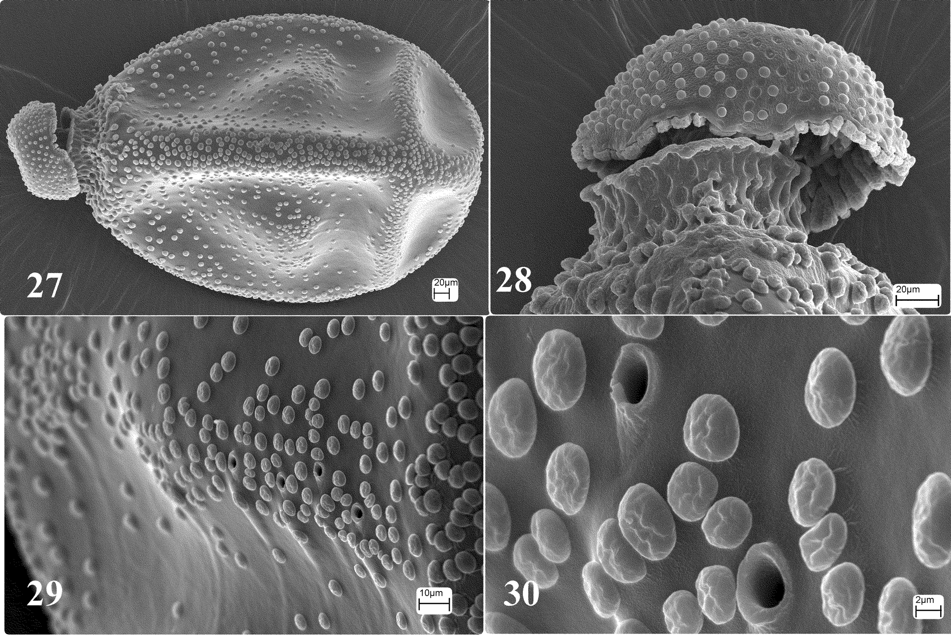
Remarks. Megaperlodes tiunovi is smaller in size than M. niger and differs in number of the abdominal segments divided laterally by yellow pleural folds, segments 1–5 are divided in M. tiunovi , whereas segments 1–6 are divided laterally by pleural folds in M. niger . The front wing M. tiunovi is black with a contrasting white frosted spot medially ( Fig. 1 View FIGURES 1 – 3 ), the front wing M. niger ( Fig. 21 View FIGURES 21 – 26 ) lacks a spot. The male of M. niger lacks a median yellow spot on tergum 10. In everted condition, the posterior margin of tergum 10 of M. niger appears like a long transversal slightly sclerotized sclerite with rounded lateral margins ( Fig. 23 View FIGURES 21 – 26 ). The paraproct sclerite of M. niger is larger than in M. tiunovi and completely heavily sclerotized, with the base hemispherical, the sclerite long, medially narrowed, and then widened and distally bluntly rounded ( Figs. 23, 25 View FIGURES 21 – 26 ). The rectangular base of the EPL of M. niger has a long fingerlike projection, narrowed from base to the pointed tip ( Figs. 23, 25 View FIGURES 21 – 26 ), and densely covered by short setae ( Fig. 24 View FIGURES 21 – 26 ). The female subgenital plate of M. niger is caudally rounded and covered by short setae ( Fig. 26 View FIGURES 21 – 26 ). The egg of M. niger has a wider anterior pole with an entire collar ( Figs. 27, 28 View FIGURES 27 – 30 ). Each egg face has medially an additional hemispherical swelling ( Fig. 27 View FIGURES 27 – 30 ); the micropyles are shorter than in M. tiunovi , slightly elevated above the chorion surface, openings with rounded margins ( Figs. 29, 30 View FIGURES 27 – 30 ). The larvae differ in color, shape of submentum and pronotum ( Fig. 3 View FIGURES 1 – 3 E in Inada et al. 1998).
No known copyright restrictions apply. See Agosti, D., Egloff, W., 2009. Taxonomic information exchange and copyright: the Plazi approach. BMC Research Notes 2009, 2:53 for further explanation.