
Phallusia fragilis, Bonnet, Nadia Y. K. & Rocha, Rosana M., 2011
|
publication ID |
https://doi.org/ 10.5281/zenodo.277398 |
|
DOI |
https://doi.org/10.5281/zenodo.6186581 |
|
persistent identifier |
https://treatment.plazi.org/id/038F878E-FFBB-FFEB-0BB5-FF48FDB94B9A |
|
treatment provided by |
Plazi |
|
scientific name |
Phallusia fragilis |
| status |
sp. nov. |
Phallusia fragilis sp. nov.
( Figs. 16–17 View FIGURE 16 View FIGURE 17 )
Material examined. Holotype: MZUSP 0 0 0 12 —1 ind.; Pastores island; 1.0 m, coral reef; 10/viii/2003.
Paratypes: DZUP PHA 23—1 ind.; Casa Blanca; coral reef; 05/vi/2009. DZUP PHA 19—3 ind.; Garden; 7.0– 9.0 m, in dead coral reef; 10/viii/2008; DZUP PHA 20—1 ind.; 6.0 m, coral reef; 23/xii/2008. MZUSP 0 0 0 13 —1 ind.; South Solarte; 06/viii/2003. DZUP PHA 17—1 ind.; 1.0 m, Garden, coral reef; 10/viii/2003; DZUP PHA 18— 2 ind.; Garden, coral reef; 17/viii/2006; DZUP PHA 21—1 ind.; Garden, coral reef; 25/ii/2009. DZUP PHA 22—1 ind.; Big Bight; coral reef; 05/vi/2009.
Etymology. The specific epithet is due to the delicate tunic and body wall.
Diagnosis. Long cylindrical body; no musculature on the oral siphon and on the left side; musculature on the right side formed by short parallel fibers on the dorsal and ventral margins; oral tentacles project from a delicate membrane joining them by their bases; dorsal tubercle absent; accessory apertures concentrated near to the neural gland; 58–78 longitudinal vessels on the right side and 47–64 on the left side of the pharynx; 3–5 stigmata per mesh; filiform primary papillae on the first anterior transverse vessel; elongated or sac-like dilation at the rectum; ovary inside the primary intestinal loop, ramified on the intestine wall; gonads of both sexes are not found at the same time.
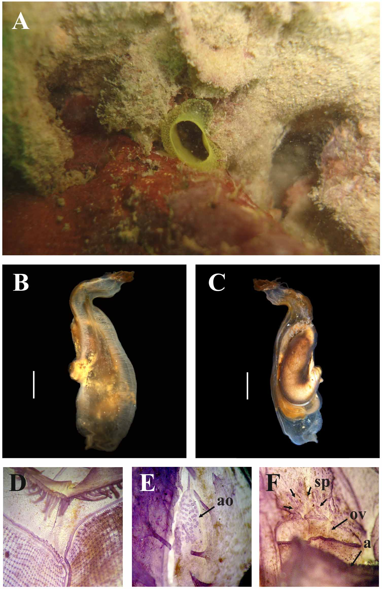
This species is usually found buried in coral rubble, between 2.0–10.0 m deep, with only the oral siphon visible above the surface. In the field the oral siphon projects 1.0–2.0 cm from the bottom and is easily recognized by the pale yellow color and by its shape, with the aperture diameter smaller than the siphon itself (about 1.0 cm). Numerous yellow papillae project from tunic around the siphons. The animal is attached to coral fragments and shells by the left side which is very delicate and easily broken when attempting to remove the animal from the substrate. The tunic is yellow and transparent; on the right side it is thicker (2.0 mm) and gelatinous, with some protuberances. Blood vessels are very ramified and have swollen blind ends which appear as small yellow papillae on the surface of the tunic.
The body is elongate, up to 7.5 cm total length (include siphons). Without the tunic the distance between the oral tentacles and the posterior margin is 3.5–6.4 cm, and 1.2–1.7 cm wide. The body wall is usually colorless, but some animals fixed in formalin are brownish on the right side. The oral siphon is apical with the atrial siphon somewhat posteriorly displaced 1.8–3.9 cm from the circle of oral tentacles. The oral siphon is from 5.0–35.0 mm long and the atrial siphon is shorter at 2.0–5.0 mm. Both siphons have a plain rim or eight small rounded transparent lobes. The neural ganglion is at the level of the tip of the intestinal loop, midway between the oral tentacles and the atrial siphon.
On the right side, the body wall musculature comprises short and thick fibers perpendicular to the dorsal and ventral margins. The posterior margin has no fiber and the anterior margin has a band of well organized circular fibers forming a sphincter at the level of the oral tentacles. Posterior to this sphincter the musculature forms a net of fibers on both ventral and dorsal margins. Some very slender fibers run longitudinally to the posterior end over the transverse fibers. No muscle fibers are on the left side nor on the oral siphon. The atrial siphon has a conspicuous sphincter of circular fibers.
The 46–70 simple oral tentacles are of three sizes, the largest are 1.4–2.5 mm long and the tentacles extend from a wide muscular membrane. Oral tentacles are wide and flat along most of their length narrowing at the extremity. The double prepharyngeal groove has no projections and the anterior membrane may be wider; the width of the area between the line of tentacles and the prepharyngeal groove may be 2.5 mm in which are many papillae. The peritubercular area is V-shaped, sometimes very deep and lacking a dorsal tubercle. The fusiform neural gland is displaced posteriorly alongside the neural ganglion and has 24–92 small sessile apertures to the atrial cavity ( Fig. 16 View FIGURE 16 E). The dorsal lamina is double anteriorly and wide posteriorly with finger like projections formed by the ends of the transverse vessels without any intermediate projections; there are no projections close to the esophageal aperture. The dorsal lamina narrows alongside the esophageal aperture on its left and then widens towards the end of the pharynx (7–18 mm beyond the stomach). The area between the dorsal lamina and the first right longitudinal vessel is wide and often is perforated by large openings, just prior to the esophageal aperture. On the right side of the esophageal aperture, the transverse vessels form long languets. The pharynx has 58–78 longitudinal vessels on the right side, 47–64 on the left side and 192–317 transverse vessels, is not pleated and with few stigmata in the meshes (3–5). Primary papillae are bilobed, and only those on the first anterior transverse vessel have a filiform lobe and longer; intermediate papillae are very small, button-like or completely lacking. Parastigmatic vessels were not seen.
The alimentary canal is large, occupying half or more of the left side of the body. The stomach is oval, short, with four internal folds. The intestine forms two loops and the descending part is dilated forming an elongate or sac-like pouch. The anus with a plain rim is located 23.2–37.8 mm from the oral tentacles. The atrial epithelium covering the alimentary canal has diminutive papillae, except on the stomach and ascending intestine walls where they are larger. Long filiform papillae were seen in some animals. Renal vesicles are 0.1–0.25 mm in diameter and they cover the entire atrial side of the alimentary canal.
The species is apparently sequentially hermaphrodite: only the ovary was developed in individuals collected in June and August, while only the testis was visible in those collected in December and February. The ovary is mostly located inside the primary intestinal loop. Outside the loop it may branch on the intestinal wall at the top of the primary loop. From the inside, there is a portion inside the primary loop and also a portion on the rectum at the extremity of the secondary loop, which do not show ramifications, instead it is more cauliflower like. Oocytes are 0.1 mm in diameter. The oviduct adheres to the body wall instead to the wall of the rectum and its aperture is very large and anterior to the anus. The testis has long and little ramified follicles covering the wall of the ascending intestine and of the intestinal dilation, both externally and on the atrial cavity side. The sperm duct has many small apertures spread on the body wall anterior to the oviducal aperture ( Fig. 16 View FIGURE 16 F).
Remarks. In the Caribbean sea, there are two known Phallusia species: P. nigra and P. caguayensis ( Millar & Goodbody, 1974) . Both are very different from Phallusia fragilis sp. nov.: P. nigra has a rigid and dark tunic (black or gray), the body musculature formed by a net of fibers on the right side of the body and it has a dorsal tubercle; P. caguayensis has thin and light gray tunic, musculature restrict to the dorsal margin of the body, about 120 oral tentacles, up to 200 accessory openings, only primary papillae and 8–10 stigmata per mesh ( Millar & Goodbody 1974). No other Phallusia registered in the world is similar to Phallusia fragilis sp. nov.
Ascidia muricata Heller, 1874 , described from the Mediterranean and Azores is similar to P. fragilis sp. nov. in having a yellow tunic (not all individuals) with papillae, elongate body, similar number of oral tentacles (40–60) projecting from a muscular membrane, lack of the dorsal tubercle in some individuals, and presence of small intermediate papillae ( Monniot 1974). However, A. muricata differs from Phallusia fragilis sp. nov. by the absence of secondary openings of the neural gland, different musculature pattern on the right side of its body, fewer number of longitudinal vessels, presence of digitiform and long primary pharyngeal papillae, dorsal lamina not surpassing the esophageal aperture, smaller alimentary canal and lack of rectum dilation, simultaneous presence of both gonads, ovary not visible from the outside.

No known copyright restrictions apply. See Agosti, D., Egloff, W., 2009. Taxonomic information exchange and copyright: the Plazi approach. BMC Research Notes 2009, 2:53 for further explanation.