
Diamphidaxona
|
publication ID |
https://doi.org/ 10.5281/zenodo.173379 |
|
DOI |
https://doi.org/10.5281/zenodo.6259058 |
|
persistent identifier |
https://treatment.plazi.org/id/038F8A52-FFE0-FFB1-8341-FA4C5C76AF18 |
|
treatment provided by |
Plazi |
|
scientific name |
Diamphidaxona |
| status |
|
Key for adults of north american species of Diamphidaxona
1a. Gnathosoma with rostrum short and not extending beyond insertion of pedipalps ( Figs. 7 View FIGURES 1 – 7 , arrow, 22, 53); pedipalps with segments relatively long and slender ( Figs. 5 View FIGURES 1 – 7 , 20 View FIGURES 16 – 22 , 51 View FIGURES 47 – 53 ) ............................................................ Diamphidaxona (s. s.) 2
1b. Gnathosoma with rostrum long and extending well beyond insertion of pedipalps ( Figs. 68 View FIGURES 62 – 68 , arrow, 72, 78, 84, 94) and pedipalps with segments relatively short and stocky ( Figs. 66 View FIGURES 62 – 68 , 71 View FIGURES 69 – 74 , 77 View FIGURES 75 – 80 , 83 View FIGURES 81 – 86 , 91 View FIGURES 87 – 94 ) ............................................................ ............................................................. Diamphidaxona (Diamphidaxonella) 10
2a (1a). Suture lines between third and fourth coxal plates with lateral loops dropshaped and closed anteriorly ( Figs. 3, 6 View FIGURES 1 – 7 )......................................... D. cramerae sp. nov.
2b. Suture lines between third and fourth coxal plates with lateral loops Ushaped
and open anteriorly ( Figs. 10, 15 View FIGURES 8 – 15 ) ....................................................................... 3 3a (2a). Dorsal portion of camerostome strongly acutely angled and projecting beyond anterior end of idiosoma ( Figs. 10, 15 View FIGURES 8 – 15 ; arrows) .................... D. imamurai Cook
3b. Dorsal portion of camerostome weakly acutely angled or rounded and not extending beyond anterior end of idiosoma ( Figs. 18, 21 View FIGURES 16 – 22 , 56, 59 View FIGURES 54 – 61 )...................... 4
4a (3b). Ventral shield with projections covering insertions of fourth pair of legs relatively short and extending laterally to edges of shield ( Figs. 18, 21 View FIGURES 16 – 22 ) ................... ........................................................................................... D. chihuahua sp. nov.,
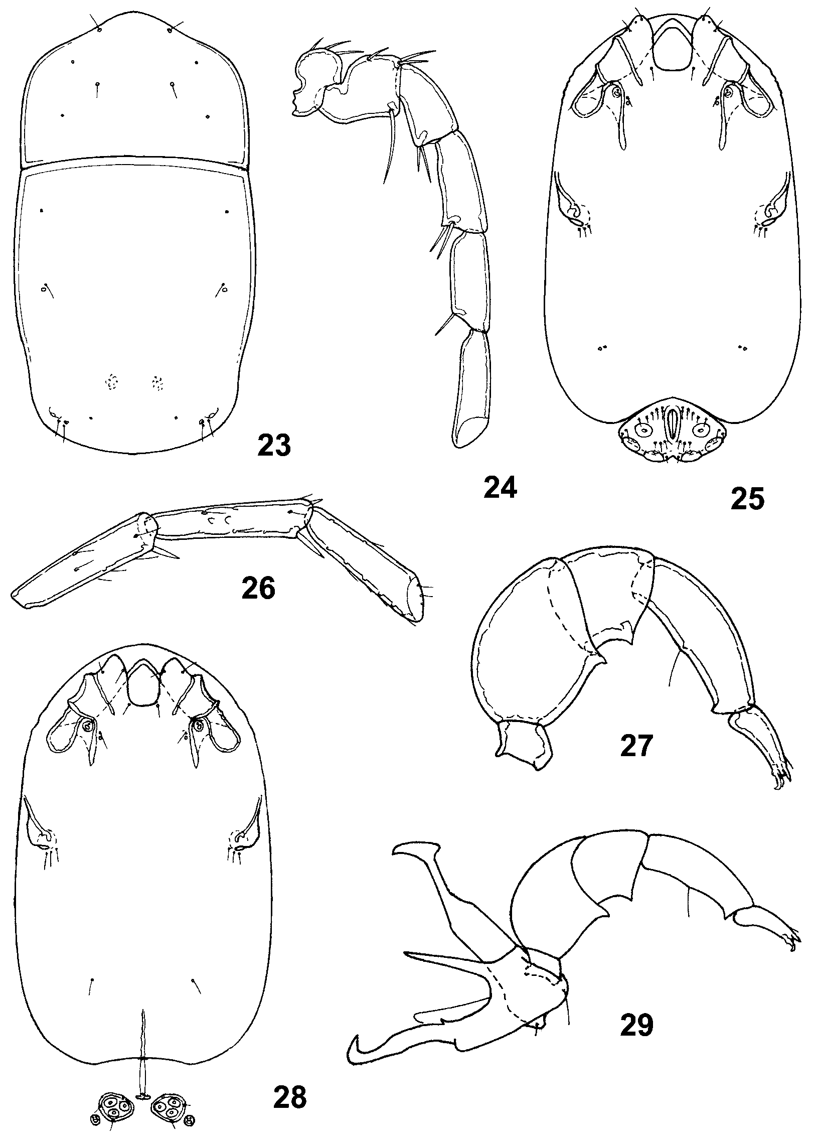
4b. Ventral shield with projections covering insertions of fourth pair of legs relatively long and extending anteriorly parallel to edges of shield ( Figs. 25, 28 View FIGURES 23 – 29 , 56, 59 View FIGURES 54 – 61 )........................................................................................................................ 5
5a (4b). Pedipalps with ventral spinelike setae on tarsus lacking denticles ( Fig. 27 View FIGURES 23 – 29 ) ....... ............................................................................................ D. dolichosoma Cook
5b. Pedipalps with ventral spinelike setae on tarsus bearing ventral denticles ( Figs. 32 View FIGURES 30 – 34 , 39 View FIGURES 35 – 41 , 51 View FIGURES 47 – 53 , 57 View FIGURES 54 – 61 ) .................................................................................................... 6
6a (5b). Pedipalps with tarsus relatively short and stocky, with ventral spinelike seta extending only slightly beyond end of segment and bearing two relatively large and strongly recurved ventral denticles ( Fig. 32 View FIGURES 30 – 34 , arrow)....................................... ......................................................................................... D. sabinalensis sp. nov.
6b. Pedipalps with tarsus relatively much longer and more slender, with ventral spinelike seta extending well beyond end of segment and bearing relatively small, straight or slightly curved ventral denticles ( Figs. 39 View FIGURES 35 – 41 , 51 View FIGURES 47 – 53 , 57 View FIGURES 54 – 61 ) ................. 7
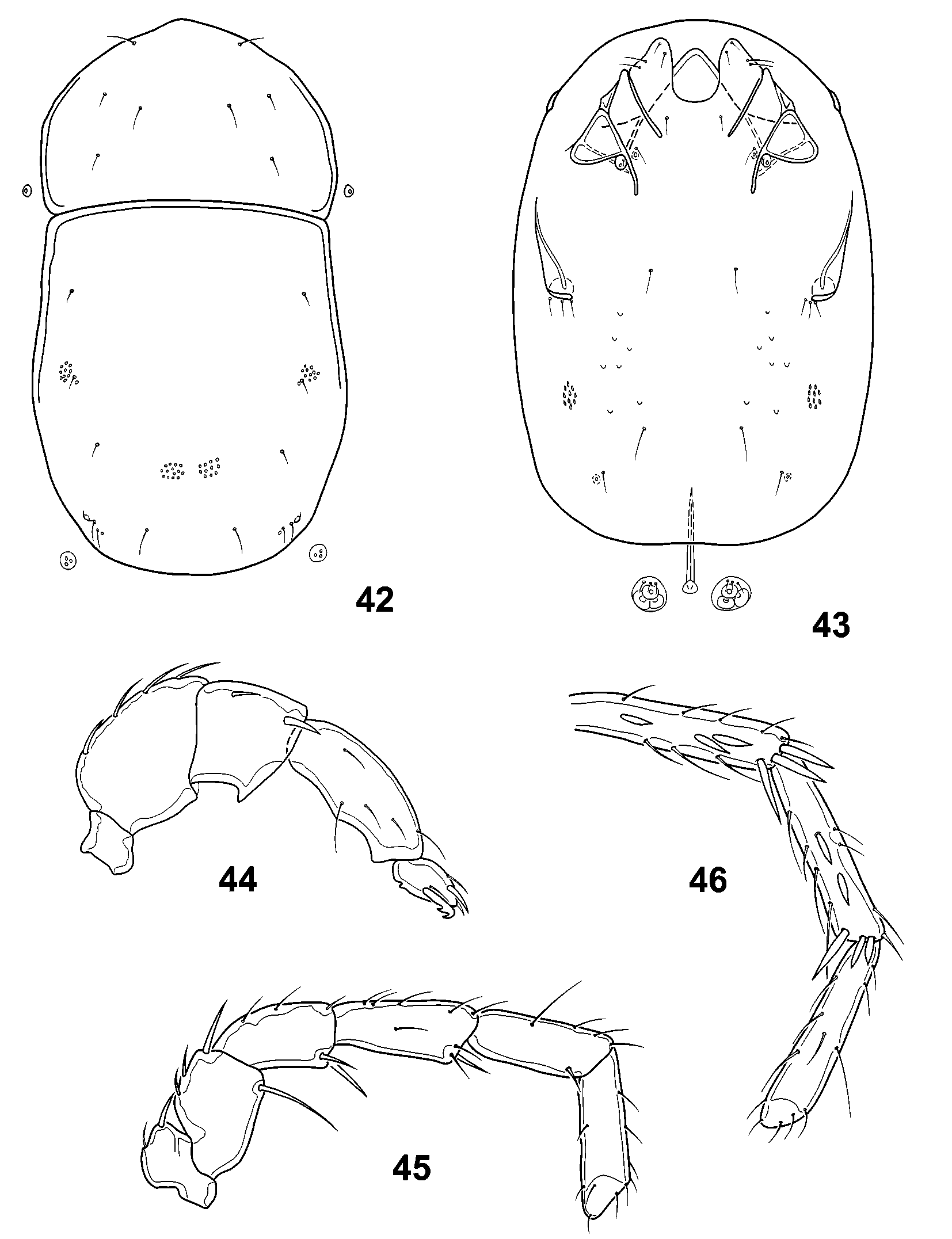
7a (6b). Suture lines between third and fourth coxal plates with medial loops bearing glands of fourth coxal plates relatively shallow ( Figs. 37, 40 View FIGURES 35 – 41 , 43 View FIGURES 42 – 46 ) ................... 8
7b. Suture lines between third and fourth coxal plates with medial loops bearing glands of fourth coxal plates relatively deeply arched ( Figs. 49, 52 View FIGURES 47 – 53 , 56, 59 View FIGURES 54 – 61 ) ..... 9
8a (7a). Idiosoma and appendages relatively large in size (ventral shield of female more than 490 μm in length); posterior dorsal plate slightly narrowed near midlength ( Fig. 35 View FIGURES 35 – 41 ); leg segments relatively long and slender ( Figs. 36, 38 View FIGURES 35 – 41 ) ........................ ........................................................................................... D. chiricahua sp. nov.
8b. Idiosoma and appendages relatively small in size (ventral shield of female less than 450 μm in length); posterior dorsal plate slightly widened near midlength ( Fig. 42 View FIGURES 42 – 46 ); leg segments relatively short and stocky ( Figs. 45, 46 View FIGURES 42 – 46 ) ........................ ..................................................................................... D. cavecreekensis sp. nov.
9a (7b). Dorsal shield relatively long and slender ( Fig. 47 View FIGURES 47 – 53 ); male with genital acetabula relatively small and first pair widely separated from second and third pairs ( Fig. 49 View FIGURES 47 – 53 ) ................................................................................ D. parvacetabula sp. nov.
9b. Dorsal shield relatively short and wide ( Fig. 54 View FIGURES 54 – 61 ); male with genital acetabula relatively large and first pair closer to second and third pairs ( Fig. 56 View FIGURES 54 – 61 ) ................... ..................................................................................................... D. pallida Cook
10a (1b). Pedipalps with segments very short and stocky and tibia bearing 2 ventral setae ( Fig. 66 View FIGURES 62 – 68 ) ......................................................................... D. neomexicana sp. nov.
10b. Pedipalps with segments relatively long and slender and tibia bearing 1 ventral seta ( Figs. 71 View FIGURES 69 – 74 , 77 View FIGURES 75 – 80 , 83 View FIGURES 81 – 86 , 91 View FIGURES 87 – 94 ) ................................................................................. 11
11a (10b). Fourth pair of legs with tarsi short and stocky ( Fig. 74 View FIGURES 69 – 74 ) ..................................... ............................................................................................ D. brevitarsa sp. nov.
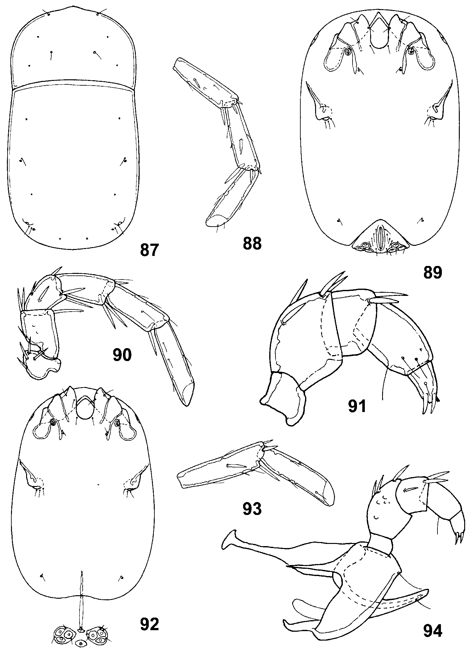
11b. Fourth pair of legs with tarsi relatively long ( Figs. 80 View FIGURES 75 – 80 , 86 View FIGURES 81 – 86 , 88 View FIGURES 87 – 94 )......................... 12
12a (11b). Dorsal shield relatively wide and with maximum width in anterior region of anterior plate ( Fig. 75 View FIGURES 75 – 80 ) ........................................................ D. platysoma sp. nov.
12b. Dorsal shield relatively narrow and with same width throughout or maximum width in posterior region of posterior plate ( Figs. 81 View FIGURES 81 – 86 , 87 View FIGURES 87 – 94 ) ................................ 13
13a (12b). Dorsal shield with anterior plate relatively long and strongly pointed anteriorly ( Fig. 81 View FIGURES 81 – 86 ) ............................................................................ D. californica sp. nov.
13b. Dorsal shield with anterior plate relatively short and weakly pointed anteriorly ( Fig. 87 View FIGURES 87 – 94 ) ............................................................................... D. arizonica sp. nov.


No known copyright restrictions apply. See Agosti, D., Egloff, W., 2009. Taxonomic information exchange and copyright: the Plazi approach. BMC Research Notes 2009, 2:53 for further explanation.