
Diamphidaxona Cook, 1963
|
publication ID |
https://doi.org/ 10.5281/zenodo.173379 |
|
DOI |
https://doi.org/10.5281/zenodo.6259023 |
|
persistent identifier |
https://treatment.plazi.org/id/038F8A52-FFF4-FFAD-8341-FA045830AE90 |
|
treatment provided by |
Plazi |
|
scientific name |
Diamphidaxona Cook, 1963 |
| status |
|
Genus Diamphidaxona Cook, 1963
Diamphidaxona Cook, 1963 . American Midland Naturalist, 70, 111.
Diamphidaxona Cook, 1974 . Memoirs of the American Entomological Institute, 21, 347–348. Diamphidaxona Cook, 1980 . Memoirs of the American Entomological Institute, 31, 181–182.
Type species. Diamphidaxona pallida Cook. Original designation.
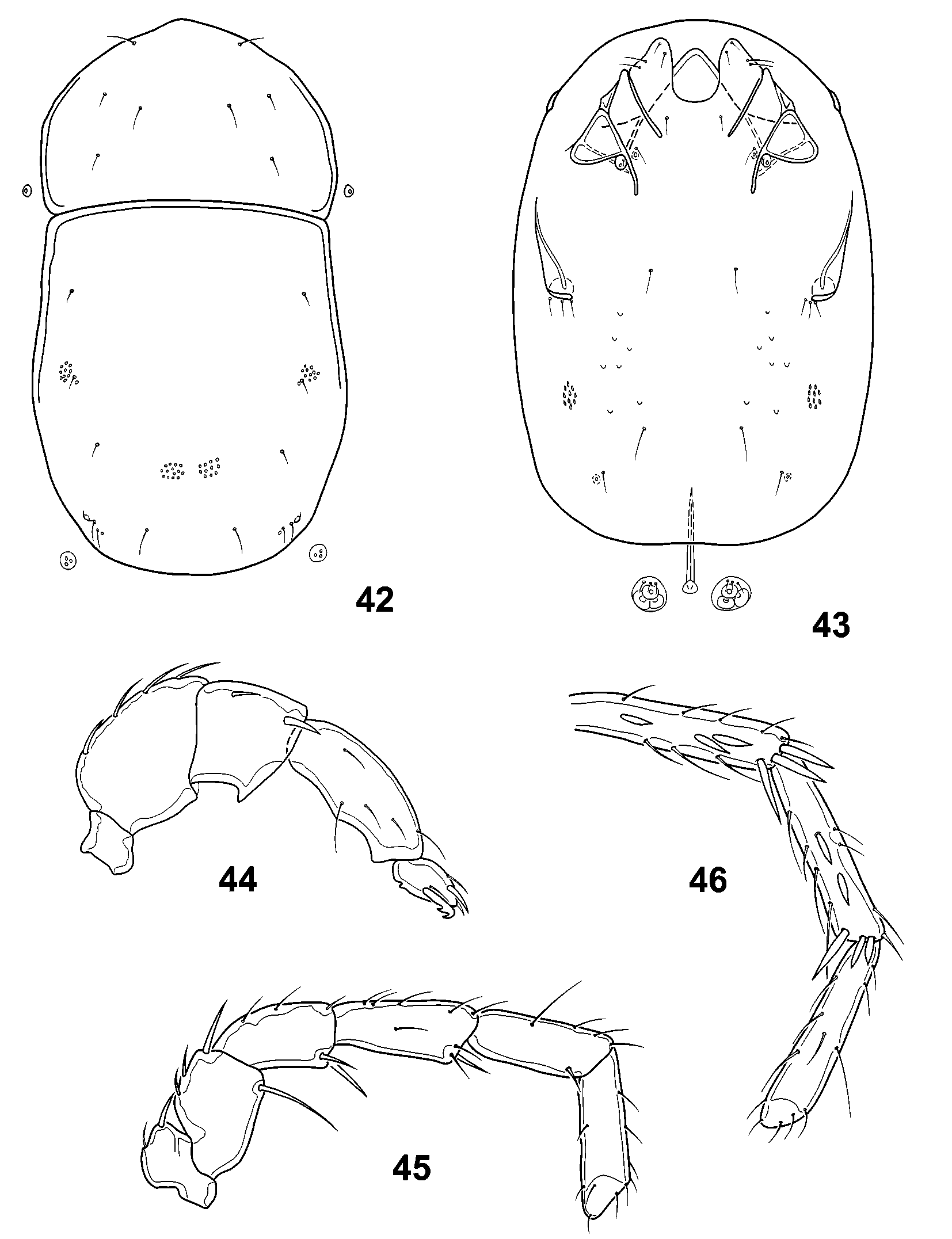
Diagnosis. Hygrobatid water mites (see Cook 1974, 1980) with following combination of character states: Gnathosoma with rostrum short and inconspicuous or long and extending well beyond insertions of pedipalps, with capitular apodemes (anchoral process) very long. Dorsal edge of camerostome smoothly arched, acutely rounded or sharply pointed anteriorly. Pedipalps with segments variable in shape from short and stocky to long and slender; tibia with small ventral setiferous tubercle near midlength; tarsus with large, spinelike seta inserted ventromedially that may be smooth or bear conspicuous denticles on ventral surface ( Figs. 98–100 View FIGURES 95 – 100 ). Idiosoma lacking eyes and bearing well developed dorsal and ventral shields ( Figs. 95 View FIGURES 95 – 100 , 101, 102 View FIGURES 101 – 106 . Dorsal shield divided into relatively small anterior plate and larger posterior plate ( Fig. 95 View FIGURES 95 – 100 ); anterior plate ( Fig. 96 View FIGURES 95 – 100 ) much wider than long, rounded or acutely pointed anteriorly, bearing preocular setae anteriorly, postocular setae medially near midlength and two pairs of setae laterally representing reduced glandularia with gland portion absent ( Figs. 42 View FIGURES 42 – 46 , 96, 97 View FIGURES 95 – 100 ); posterior plate much longer than wide, bearing one pair of glandularia laterally near midlength, two pairs of dissimilar glandularia grouped together posterolaterally ( Fig. 106 View FIGURES 101 – 106 ) and three pairs of setae representing reduced glandularia with the gland portion absent ( Fig. 42 View FIGURES 42 – 46 ). Ventral shield ( Figs. 101, 102 View FIGURES 101 – 106 ) with anterior three pairs of coxal plates grouped together anteriorly and separated by suture lines that are distinct laterally but obliterated by fusion medially; coxoglandularia I located anterior to medial end of partial suture line between second and third coxal plates; suture lines between third and fourth coxal plates conspicuously undulating, with medial loop arched and open posteriorly to accommodate glandularia of fourth coxal plates and lateral loop Ushaped and open anteriorly or dropshaped with anterior opening constricted ( Figs. 103, 104 View FIGURES 101 – 106 ); fourth coxal plates with edges obliterated by fusion, with insertions of fourth pair of legs covered by conspicuous projections with anterior extensions which are short and directed laterally to edges of ventral shield, moderately long, sinuous and directed anterolaterally or long and directed anteriorly parallel to edges of ventral shield. Coxoglandularia II located near posterior end of ventral shield. Ventral shield ( Figs. 101, 102 View FIGURES 101 – 106 ) with three pairs of medial setae in regions of first, third and fourth coxal plates, respectively, and small groups of setae near anterior edges of first coxal plates and posterior to insertions of fourth pair of legs ( Fig. 43 View FIGURES 42 – 46 ). Genital field bearing three pairs of acetabula on acetabular plates flanking gonopore. Genital field of males with acetabular plates fused with one another to surround gonopore ( Fig. 105 View FIGURES 101 – 106 ) and either fused with or separate from ventral shield. Genital field of females with acetabular plates flanking gonopore and separate from ventral shield. Legs not exhibiting sexual dimorphism and lacking swimming setae.
Habitat. Interstitial gravels in streams.
Distribution. North, Central and South America.
Remarks. Cook (1963, 1974) originally provisionally placed Diamphidaxona in the family Aturidae because adults possess well developed dorsal and ventral shields, but later transferred the genus to Hygrobatidae ( Cook 1980) . Diamphidaxona belongs to a small and highly distinctive clade of hygrobatid genera, along with Hopkinsobates Cook, 1983 from New Zealand and Camposea Schwoerbel, 1986 from Chile, in which adults have the dorsal shield divided into anterior and posterior plates and the suture lines between the third and fourth coxal plates conspicuously looped to accommodate the glandularia of the fourth coxal plates. Adults of Diamphidaxona can be distinguished from those of other North American genera using the key by Smith et al. (2001). Larvae of Diamphidaxona are unknown. Absence of the gland portion of several dorsal glandularia exhibited by adult Diamphidaxona illustrates a tendency for these organs to be reduced in many unrelated taxa adapted for living in interstitial hyporheic habitats. The setae associated with these glandularia continue to be expressed in Diamphidaxona species, but are very slender and transparent and are usually only visible either under high magnification using differential interference contrast with light microscopy or with scanning electron microscopy (as in Fig. 97 View FIGURES 95 – 100 ).


No known copyright restrictions apply. See Agosti, D., Egloff, W., 2009. Taxonomic information exchange and copyright: the Plazi approach. BMC Research Notes 2009, 2:53 for further explanation.
|
Kingdom |
|
|
Phylum |
|
|
Class |
|
|
Order |
|
|
Family |
Diamphidaxona Cook, 1963
| Smith, Ian M. & Cook, David R. 2006 |
Diamphidaxona
| Cook 1980 |
Diamphidaxona
| Cook 1974 |
Diamphidaxona
| Cook 1963 |