
Truttaedacnitis clitellarius (Ward & Magath, 1917) Petter, 1974
|
publication ID |
https://doi.org/10.11646/zootaxa.4185.1.1 |
|
publication LSID |
lsid:zoobank.org:pub:0D054EDD-9CDC-4D16-A8B2-F1EBBDAD6E09 |
|
DOI |
https://doi.org/10.5281/zenodo.5626960 |
|
persistent identifier |
https://treatment.plazi.org/id/038FB248-FF26-FF2E-89B9-C595211E9933 |
|
treatment provided by |
Plazi |
|
scientific name |
Truttaedacnitis clitellarius (Ward & Magath, 1917) Petter, 1974 |
| status |
|
Truttaedacnitis clitellarius (Ward & Magath, 1917) Petter, 1974
Synonym: Cucullanus clitellarius Ward & Magath, 1917
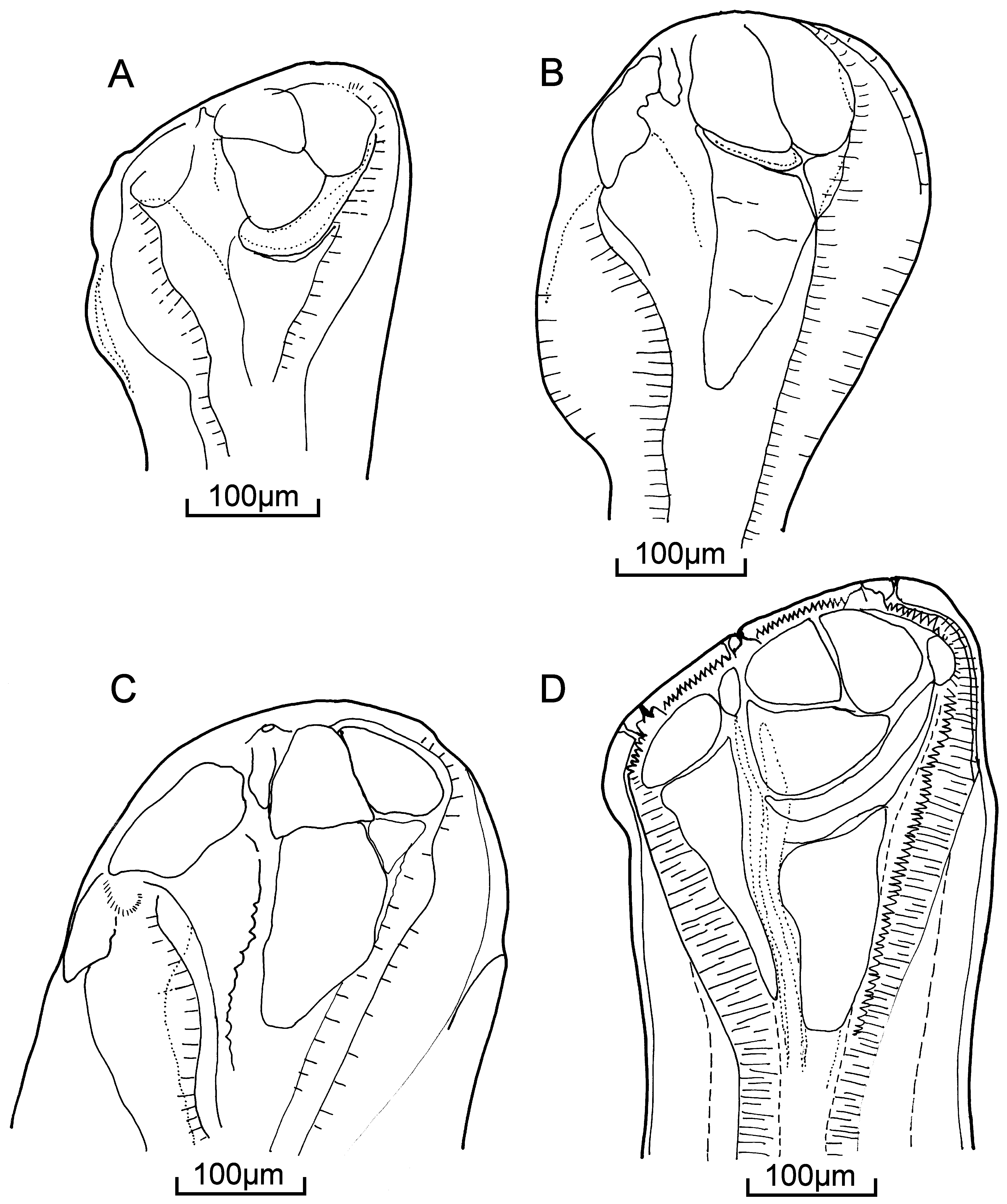
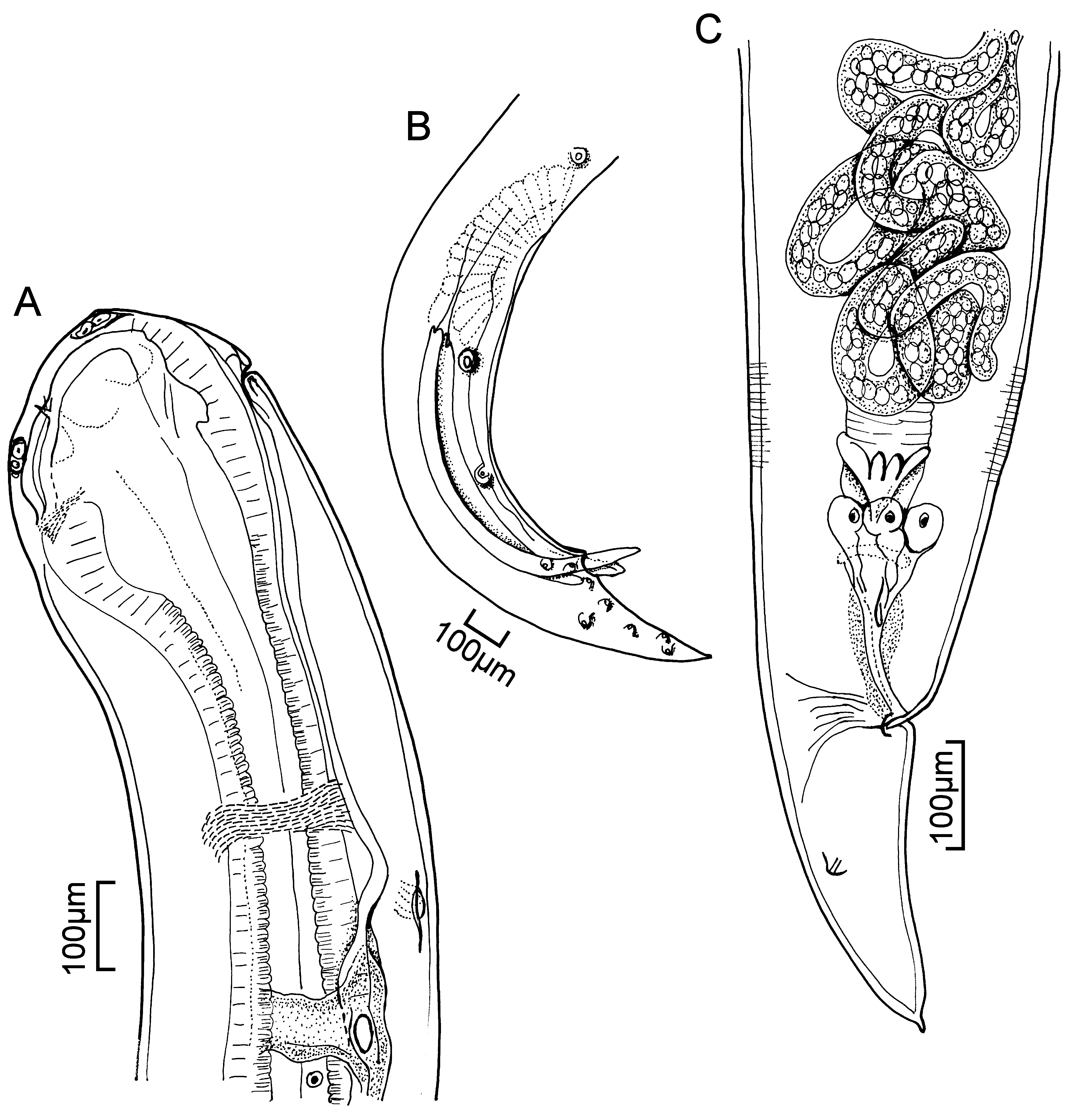
Description (after Choudhury & Dick 1996a). With characteristics of the genus. Body moderately robust, cephalic end rounded, cuticle with transverse striations. Posterior part of body often curved ventrally. Narrow lateral alae beginning in cervical region, terminating at anal region. Mouth opening spindle-shaped, with three conspicuous papillae on either side—the central one likely an amphid. Mouth opening bordered by transparent cuticular collarette bearing weak longitudinal rib-like structures (“teeth”). Inner circle of three small papillae present. Two bud-shaped deirids, one on either side, often asymmetrical, adjacent to lateral alae, and posterior to nerve ring. Tail conical, gently tapering, bearing mucron. Hemizonid posterior to nerve ring. Oesophagus muscular, clavate, expanded anteriorly to form pseudobuccal capsule. Bilaterally symmetrical cephalic plates present, separated by narrow sutures and attached to thickened cuticular plates of pseudobuccal capsule ( Fig. 86 View FIGURE 86 C). Oesophageal lumen lined by cuticle, each side of triradiate lumen thickened to give appearance of “longitudinal rods” embedded into oesophageal musculature. Oesophago-intestinal junction guarded by cuticular folded valve. Excretory pore at about mid-level of pseudobuccal capsule ( Fig. 87 View FIGURE 87 A). Excretory duct long, cuticular, extending posteriorly along ventral body wall past nerve ring to first half of oesophagus, with prominent nucleus present immediately posterior to base of excretory duct ( Fig. 87 View FIGURE 87 A).
Males: 13.5 (9.6–16.9) long; maximum width 0.40 (0.24–0.59). Nerve ring 0.585 (0.526–0.640) from anterior end. Oesophagus 1.35 (1.10–1.55) long. Tail 0.428 (0.301–0.564) long. Single testis originating posteriorly, extending anteriorly and looping back on itself. Posterior pre-cloacal sucker bordered by radially situated muscle bands, 1.70 (1.25–2.10) from posterior end ( Fig. 87 View FIGURE 87 B). Spicules equal or slightly sub-equal, gouge shaped with median serrated margins; left spicule 1.12 (0.90–1.30) long, right one 1.14 (0.87–1.35) long. Gubernaculum with Y-shaped sclerotized body, 0.139 (0.101–0.172) long. Eleven pairs of sessile caudal papillae: four pairs pre-cloacal, one pair ad-cloacal, and six pairs post-cloacal, of which one pair are phasmids. Median papilla immediately anterior to anterior margin of cloacal opening ( Fig. 87 View FIGURE 87 B).
Females: 15.1 (9.9–18.6) long; maximum width 0.43 (0.33–0.53). Nerve ring 0.613 (0.564–0.677) from anterior end. Oesophagus 1.42 (1.15–1.60) long. Tail 0.380 (0.245–0.498) long. Ovaries coiled. Anterior ovary at level of, or posterior to, oesophago-intestinal junction, in anterior third of body. Posterior ovary in posterior fifth of body ( Fig. 87 View FIGURE 87 C). Oviducts extending beyond vulvar region, reflexing and giving rise to thin-walled uteri containing eggs. Vulval opening a transverse slit. Eggs ovoid, thin-shelled, 0.067 x 0.041–0.079 x 0.049.
Site: intestinal lumen
Host: Acipenser fulvescens
Distribution: Canada, Central Canada, Hudson Bay Drainage, Manitoba, Ontario, Saskatchewan Records: Bangham & Hunter 1939 (ON); Bangham 1951 (ON); Bangham 1955 (ON); Anthony 1974 (ON); Dechtiar & Christie 1988 (ON); Choudhury et al. 1990 (MB, SK); Swanson et al. 1991 (MB); Choudhury & Dick 1993 (CC); Choudhury & Dick 1996a (MB, SK); Choudhury & Dick 1996b (CA); Choudhury & Dick 1998 (HBD)
No known copyright restrictions apply. See Agosti, D., Egloff, W., 2009. Taxonomic information exchange and copyright: the Plazi approach. BMC Research Notes 2009, 2:53 for further explanation.
|
Kingdom |
|
|
Phylum |
|
|
Class |
|
|
Order |
|
|
InfraOrder |
Oxyuridomorpha |
|
SuperFamily |
Spiruroidea |
|
Family |
|
|
Genus |