
Truttaedacnitis pybusae Anderson, 1992
|
publication ID |
https://doi.org/ 10.11646/zootaxa.4185.1.1 |
|
publication LSID |
lsid:zoobank.org:pub:0D054EDD-9CDC-4D16-A8B2-F1EBBDAD6E09 |
|
DOI |
https://doi.org/10.5281/zenodo.5626962 |
|
persistent identifier |
https://treatment.plazi.org/id/038FB248-FF29-FF2C-89B9-C18423209DF0 |
|
treatment provided by |
Plazi |
|
scientific name |
Truttaedacnitis pybusae Anderson, 1992 |
| status |
|
** Truttaedacnitis pybusae Anderson, 1992
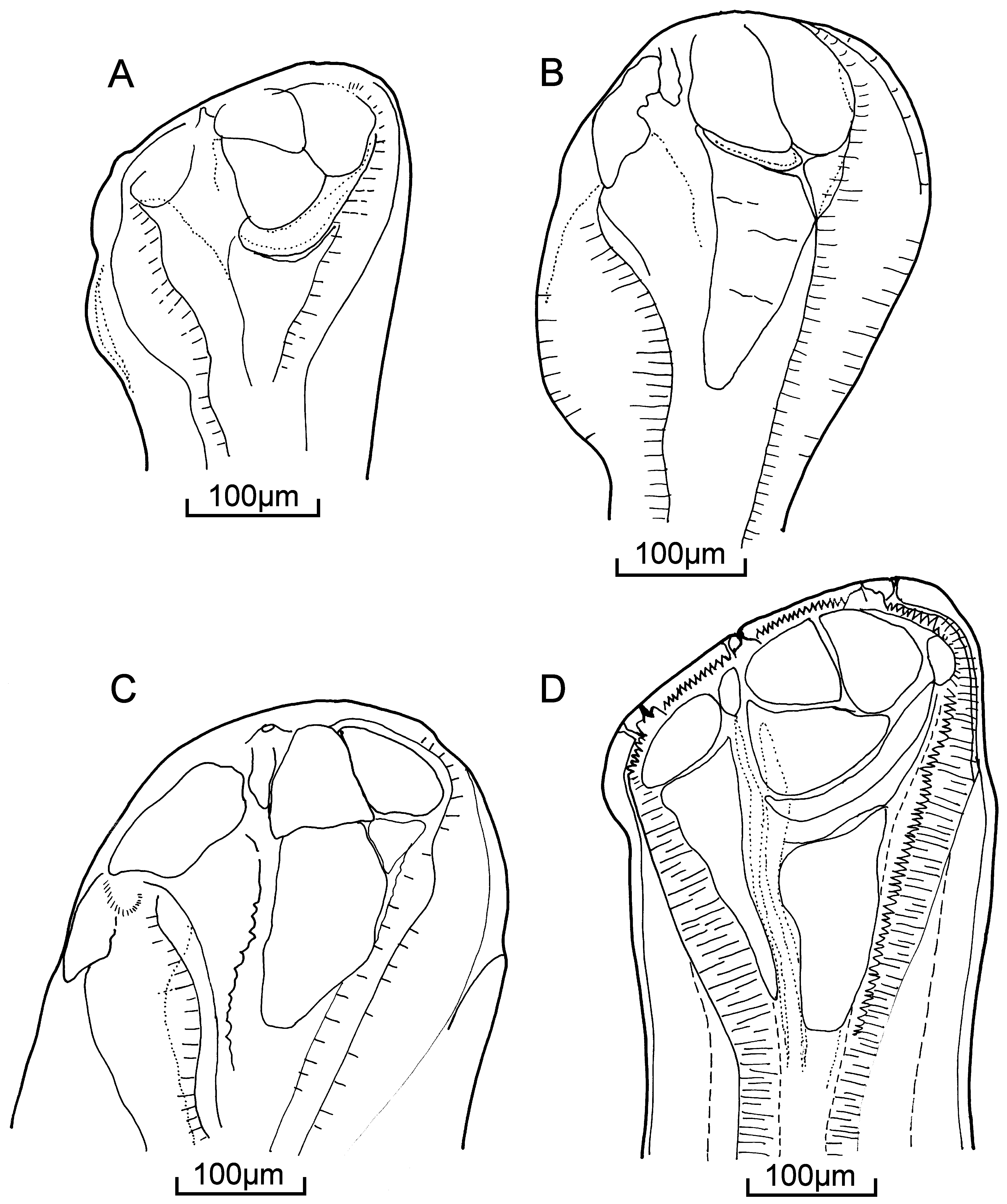
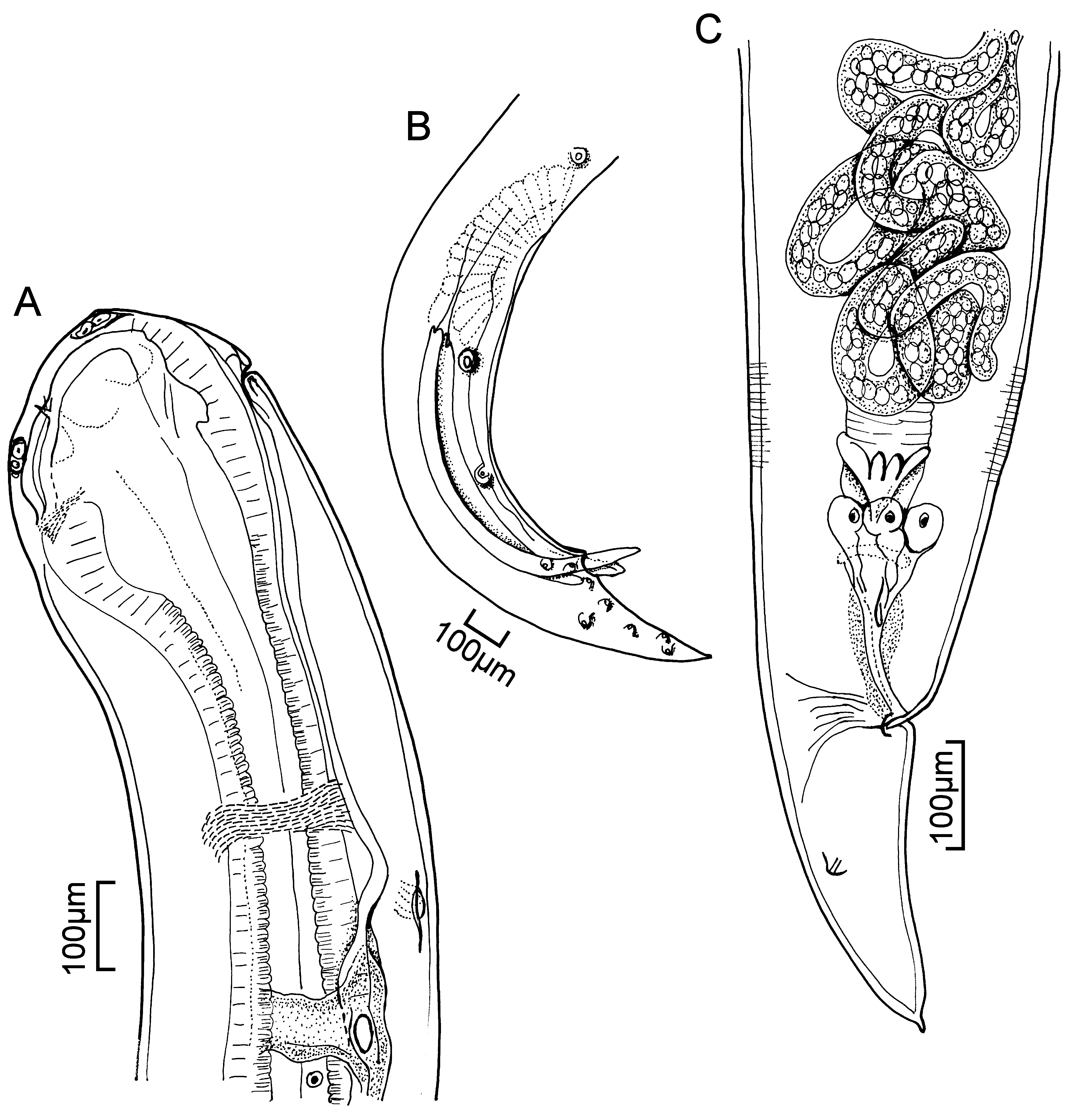
Synonym: Truttaedacnitis stelmioides of Pybus, Uhazy & Anderson, 1978 Description (after Pybus et al. 1978a). With characteristics of the genus. Slender worms with blunt anterior end and pointed, conical tail with small mucron. Cuticle smooth or with fine transverse striations. Lateral alae extend from level of nerve ring to posterior border of pre-cloacal sucker. Deirids behind nerve ring either directly opposite each either or one slightly anterior to the other. Excretory pore behind nerve ring, at same level as, or anterior to, at least one deirid. Mouth opening slit-like or triangular, dorso-ventral in position, surrounded by thin collarette containing 100 to 120 conspicuous cuticular ”teeth”. Cephalic papillae comprise four prominent double submedian papillae. Fused papillae of unequal size; larger papilla containing central cuticular rod. Amphids lateral. Six small papillae located near oral margin. Oesophagus clavate and muscular, expanding anteriorly around conical pseudobuccal capsule. Dorsal oesophageal gland nucleus prominent near posterior end oesophagus; gland opens in dorsal angle of mouth opening. Subventral oesophageal gland nuclei inconspicuous, opening at nerve ring level. Cuticle lining each angle of triradiate oesophagus thickened into paired longitudinal rods extending from near oral margin to anterior oesophago-intestinal valve. Oesophageal rods diverge anteriorly to form pseudobuccal cavity. Cuticle lining capsule thickened to form cephalic plates separated by thin sutures ( Fig. 86 View FIGURE 86 D). Two to four coelomocytes in body cavity.
Males: 10.2 (8.1–12.9) long; maximum width 0.31 (0.26–0.37). Nerve ring 0.370 (0.324–0.465) from anterior end. Oesophagus 0.97 (0.86–1.15) long. Tail 0.287 (0.237–0.367) long. Single testis in posterior third of body, looping anteriorly then reversing. Muscular pre-cloacal sucker lacks cuticularized rim, 8.8 (7.1–11.3) from anterior end. Spicules equal or sub-equal, 0.502 (0.382–0.650) long; cross section of spicules shows three longitudinal hollow areas running entire length. Arrangement of caudal papillae similar to that of T. clitellarius above ( Fig. 87 View FIGURE 87 B). Gubernaculum Y-shaped, 0.087 (0.074–0.104) long.
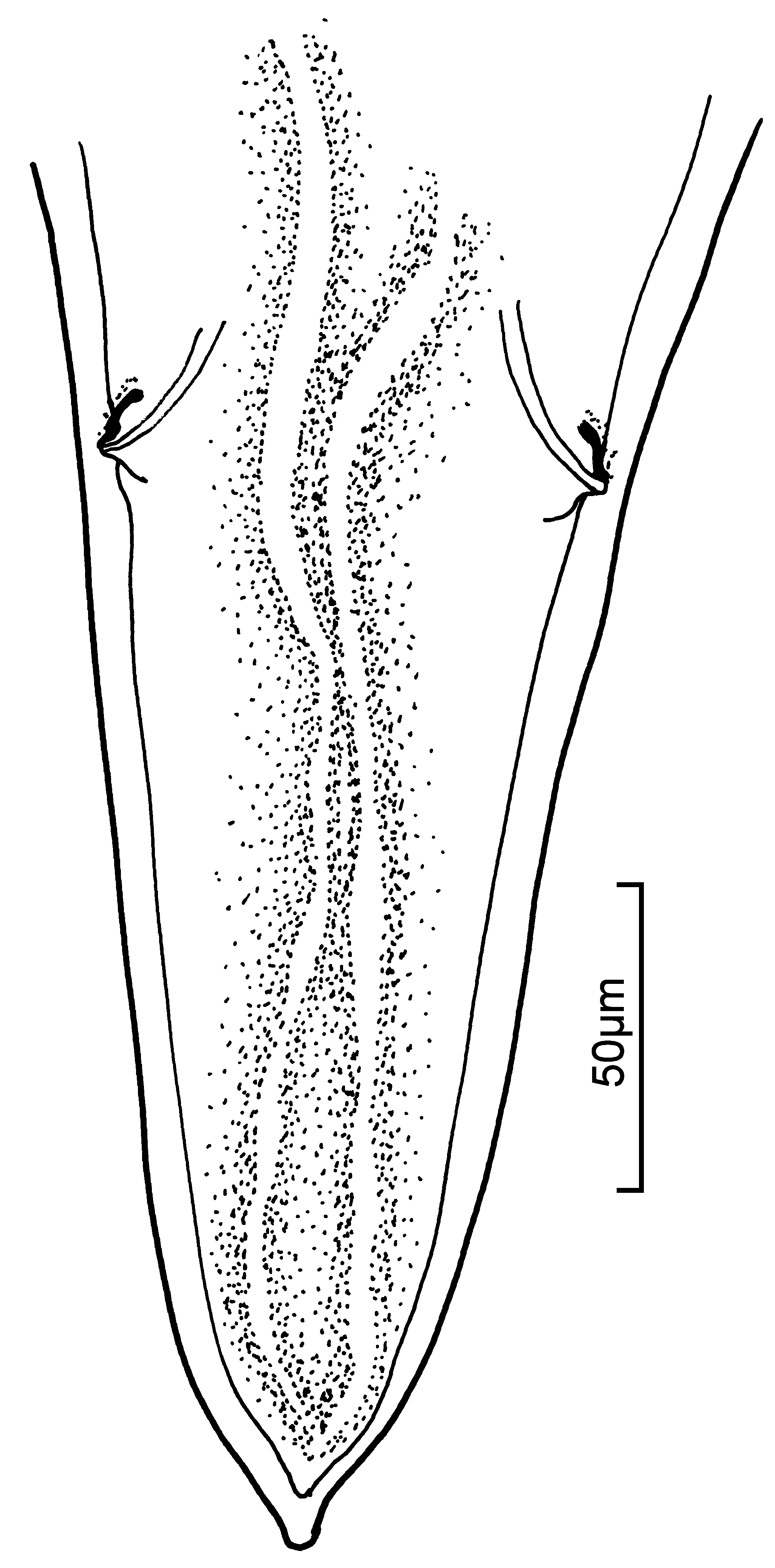
Females: 11.6 (9.5–14.3) long; maximum width 0.34 (0.26–0.44). Nerve ring 0.397 (0.350–0.470) from anterior end. Oesophagus 1.040 (0.937–1.200) long. Anterior ovary in middle third of body, the posterior one in posterior third. Oviducts looping beyond vulva, then reversing and expanding into uteri. Vulva located behind midbody, anterior lip larger than posterior. Eggs near vagina in morula stage, 0.063–0.087 x 0.051–0.068. Tail bears one pair lateral papilliform phasmids ( Fig. 88 View FIGURE 88 ).
Larvae: Pybus et al. (1978b) and Anderson (1996) discussed the transmission of T. pybusae , including the morphometry of larvae in lamprey ammocoetes and “transformers”. The biology of the nematode appears to be closely linked to the metamorphosis and maturation of lampreys.
Sites: gills, gonads, intestinal lumen, kidney, liver, mesenteries
Host: Lethenteron appendix
Distribution: Ontario
Records: Pybus et al. 1978a; Pybus et al. 1978b; Anderson 1996
No known copyright restrictions apply. See Agosti, D., Egloff, W., 2009. Taxonomic information exchange and copyright: the Plazi approach. BMC Research Notes 2009, 2:53 for further explanation.
|
Kingdom |
|
|
Phylum |
|
|
Class |
|
|
Order |
|
|
InfraOrder |
Oxyuridomorpha |
|
SuperFamily |
Spiruroidea |
|
Family |
|
|
Genus |
|
Kingdom |
|
|
Phylum |
|
|
Class |
|
|
Order |
|
|
InfraOrder |
Oxyuridomorpha |
|
SuperFamily |
Spiruroidea |
|
Family |
|
|
Genus |