
Cucullanus annulatus Margolis, 1960
|
publication ID |
https://doi.org/10.11646/zootaxa.4185.1.1 |
|
publication LSID |
lsid:zoobank.org:pub:0D054EDD-9CDC-4D16-A8B2-F1EBBDAD6E09 |
|
DOI |
https://doi.org/10.5281/zenodo.5626974 |
|
persistent identifier |
https://treatment.plazi.org/id/038FB248-FF2C-FF29-89B9-C13524009DB8 |
|
treatment provided by |
Plazi |
|
scientific name |
Cucullanus annulatus Margolis, 1960 |
| status |
|
Cucullanus annulatus Margolis, 1960
Synonym: Cucullanus of Arai (1967 partim)
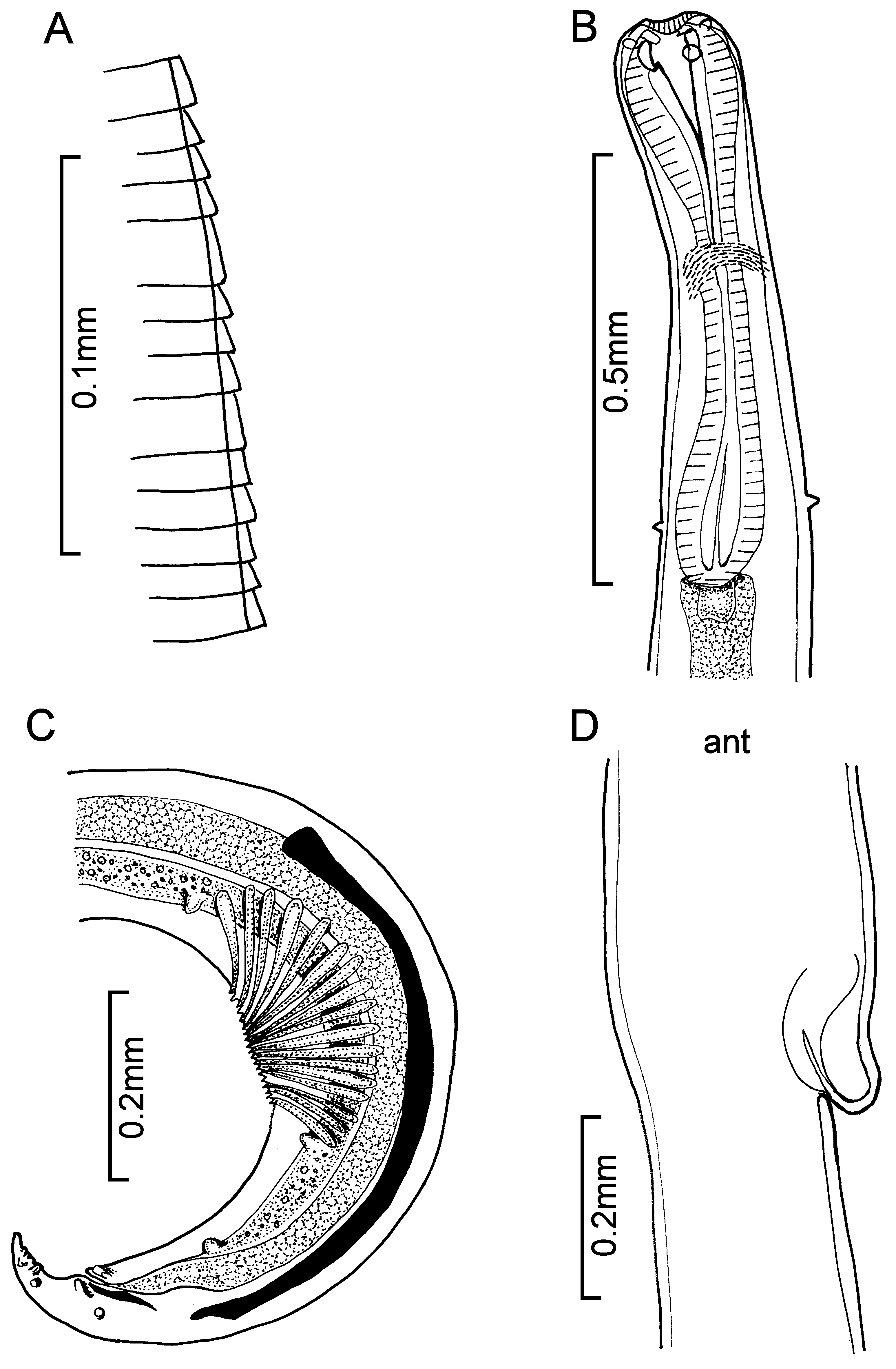
Description (after Margolis 1960). With characteristics of the genus.
Males: slender, 2.21–5.50 long, anterior end bent dorsad to varying degrees, and tail curved ventrad. Head slightly swollen, 0.12–0.18 wide. Body narrows behind head towards mid-oesophageal region then widens again to 0.10–0.25 just behind oesophagus. Body tapers gradually from post-oesophageal region to cloaca and then abruptly to pointed extremity; tail 0.083–0.13 long. Cuticle, 0.002–0.005 thick, appears devoid of transverse striations in posterior half, and very fine striations are barely discernible about mid-length of body, more anteriorly striations become more definite and farther apart; ventrally from a short distance posterior to the oesophago-intestinal junction to the anterior oesophageal swelling they are more properly termed annulations ( Fig. 91 View FIGURE 91 A). Attached to
cuticular collar surrounding mouth is a continuous membranous flange with many small teeth on internal face.
Head with two pairs of cephalic papillae and one pair of lateral amphids. Internally there is a chitinous framework
around mouth opening. Oesophagus 0.43–0.70 long, enlarged anteriorly, narrows at midlength then expands again
to a swelling less pronounced than the anterior one ( Fig. 91 View FIGURE 91 B). Two-lobed valve projects from oesophagus into
intestine. Intestine opens into short rectum which receives ejaculatory duct to form a cloaca. Nerve ring 0.21–0.30
from anterior end. Lateral cervical papillae 0.026–0.097 anterior to oesophago-intestinal junction. Excretory pore
0.057–0.17 behind oesophagus. Testis originates at about level of anterior margin of pre-cloacal sucker, follows
straight course anteriorly then turns back on itself to join seminal vesicle. Pre-cloacal sucker in lateral view appears
as fan-shaped group of muscles that when contracted take the form of cup-shaped sucker; it is 0.14–0.29 long and
its posterior margin is 0.18–0.37 in front of cloaca. Eleven pairs of caudal papillae. Slender spicules, 0.47–0.76
long, pointed distally and slightly enlarged proximally ( Fig. 91 View FIGURE 91 C). Gubernaculum 0.065–0.085 long. Females: egg-bearing specimens 5.16–9.43 long. Dorsal flexure of head not as pronounced as in males. Width
at head and posterior end of oesophagus 0.12–0.15 and 0.15–0.21 respectively. Tail pointed, 0.15–0.24 long.
Cuticle 0.004–0.008 thick. Cuticular annulations, mouth and associated structures like those of males. Nerve ring
0.28–0.33 from anterior end. Lateral cervical papillae 0.07–0.16 anterior to posterior end of oesophagus. Another
pair of lateral papillae occurs about mid-length of the tail. Excretory pore 0.10–0.29 posterior to oesophagus.
Vulva, overhung by prominent anterior lip, 3.1–6.1 from anterior end ( Fig. 91 View FIGURE 91 D). Vagina directed anteriorly joins
divergent uteri. Eggs, thin-shelled, 0.072–0.085 x 0.042–0.050.
Site: intestinal lumen
Hosts: Microstomus pacificus (4); Parophrys vetulus (1, 4); Platichthys stellatus (2, 3);
Distribution: Pacific
Records: 1. Margolis 1960; 2. Arai 1967a; 3. Arai 1969; 4. Kabata & Whitaker 1984
No known copyright restrictions apply. See Agosti, D., Egloff, W., 2009. Taxonomic information exchange and copyright: the Plazi approach. BMC Research Notes 2009, 2:53 for further explanation.
|
Kingdom |
|
|
Phylum |
|
|
Class |
|
|
Order |
|
|
InfraOrder |
Oxyuridomorpha |
|
SuperFamily |
Spiruroidea |
|
Family |
|
|
Genus |