
Ricanula curva, Zhang & Wang & Stroiński & Qin, 2021
|
publication ID |
https://doi.org/ 10.11646/zootaxa.5047.3.7 |
|
publication LSID |
lsid:zoobank.org:pub:2FDA4955-4718-4A34-8053-09ACA9BB0241 |
|
DOI |
https://doi.org/10.5281/zenodo.5540961 |
|
persistent identifier |
https://treatment.plazi.org/id/038FEA2D-9A4C-FFA0-19AA-4B5AFD0C33DD |
|
treatment provided by |
Plazi |
|
scientific name |
Ricanula curva |
| status |
sp. nov. |
Ricanula curva sp. nov.
( Figs 4–6 View FIGURE 4 View FIGURE 5 View FIGURE 6 , 11 View FIGURE 11 )
Etymology. The name is derived from the Latin word ‘ curvus ’, referring to ventral processes of aedeagus being Scurved in lateral view; apical part of ventral processes of periandrium strongly curved laterally in dorsal and ventral view.
Diagnosis. The species is similar to R. peronata sp. nov., but differs from the latter by having apical part of ventral processes of periandrium strongly curved laterally in dorsal and ventral view ( R. peronata —ventral processes boot-shaped in ventral view); ventral processes of aedeagus S-curved in lateral view ( R. peronata —ventral processes of aedeagus oriented ventrally in lateral view).
Description. Measurements. Length (inclu. teg.): male 6.7–8.4 mm, female 7.2–9.6 mm.
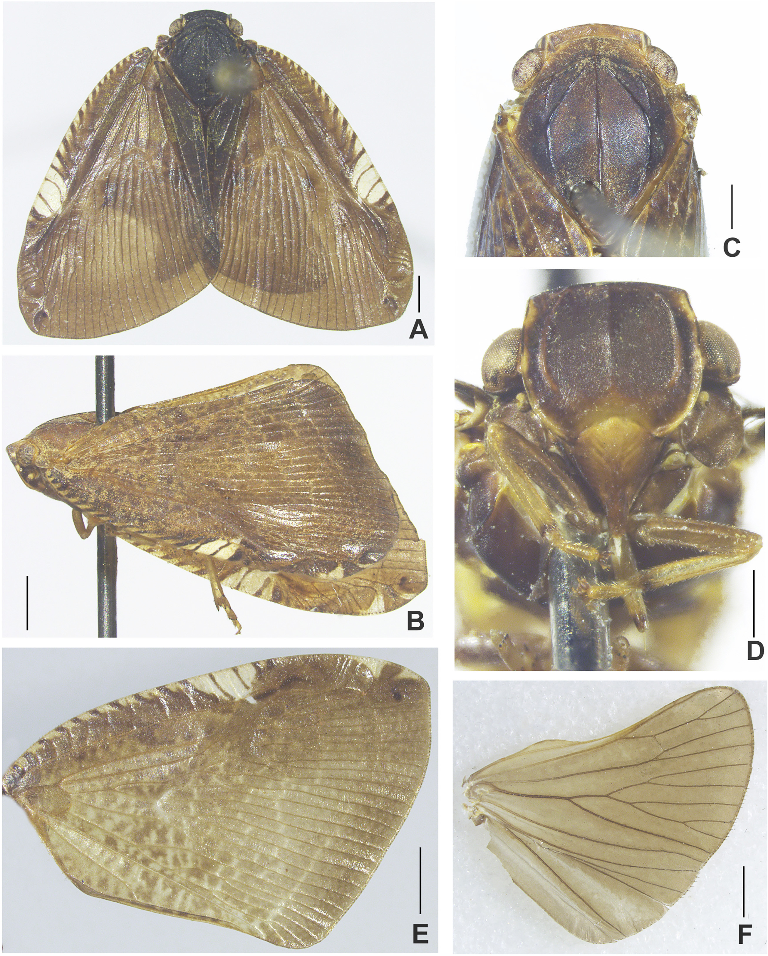
Head. Vertex ( Figs 4A, C View FIGURE 4 ) with or without median carina. Frons: lateral carinae of frontal disc finishing basally at the same level as median carina, ending about the level of antennae.
Thorax. Pronotum ( Figs 4A, C View FIGURE 4 ) with small round depressions submedially on each lateral side (in some specimens weakly visible). Mesonotum: lateral carinae of mesonotum ( Figs 4A, C View FIGURE 4 ) reaching posterior margin; anterolateral carinae connected with anterior margin.
Tegmen: postero-apical part of tegmen with two eye-spot black cells, posterior margin almost straight. Longitudinal veins ScRA and RP arising as short common stem from basal cell, MP veins forked on basal cell. Claval veins Pcu and A 1 fused before midlength of CuP vein. Hind wing with r-m transverse veinlets present in distal part of wing ( Fig. 4F View FIGURE 4 ).
Hing legs: Basitarsomere of metatarsus with 8 apical teeth. Metatibiotarsal formula 2/6/8.
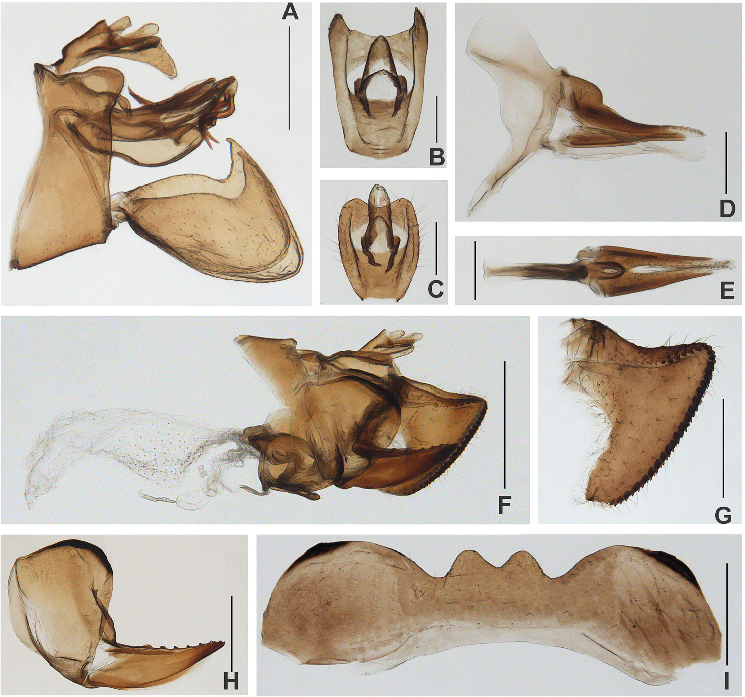
Male terminalia ( Figs 5A–B View FIGURE 5 , 6A–I View FIGURE 6 ). Anal tube (in dorsal view, Fig. 5B View FIGURE 5 ) nearly as rectangle; posterior margin strongly concave, basal margin slightly convex, lateral margins straight; anus placed before midlength, paraproct slightly surpassing the posterior margin. Pygofer (in lateral view, Fig. 5A View FIGURE 5 ): dorso-posterior angle with process. Genital styles ( Fig. 5A View FIGURE 5 ) broadly triangular in lateral view; ventral margin convex; dorsal margin weakly convex, with small concavity before spine-like process.
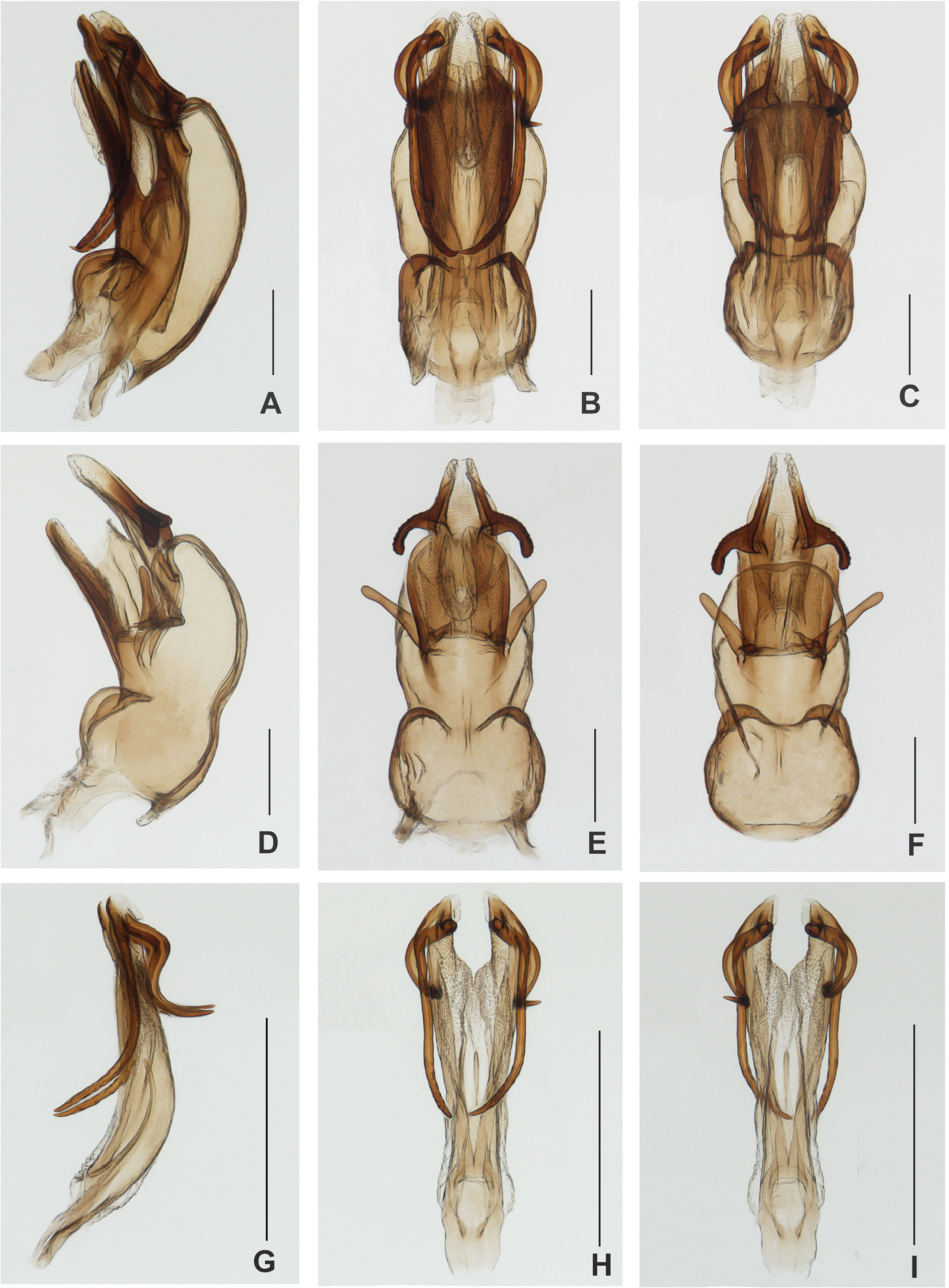
Phallic complex ( Figs 6A–C View FIGURE 6 ): Periandrium ( Figs 6D–F View FIGURE 6 ) with ventral processes, apical part of ventral processes of periandrium curved laterally (in dorsal and ventral view), ventral periandrium distinctly convex in middle; dorsal periandrium with U-shaped structure with membranous apical part sclerotized base in dorsal view; lateral margin of periandrium with rod-shaped processes in lateral view.
Aedeagus ( Figs 6G–I View FIGURE 6 ) with two pairs of processes. Median split asymmetrical: ventral split present only in 1/5; dorsal split very deep, reaching almost basal part. All processes single armed: lateral processes longer than ventral processes, about 2/3 of aedeagus, curved dorsally at 2/3 of its length; ventral processes S-curved ventrally in lateral view.
Female terminalia ( Figs 5C–I View FIGURE 5 ). Pregenital sternite: posterior margin medially with two processes, margin between processes with strong and deep incision ( Fig. 5I View FIGURE 5 ).
Anal tube (in dorsal view, Fig. 5C View FIGURE 5 ) elongate, with posterior part wider than basal one; basal margin almost straight, posterior margin widely concave medially, lateral margins arcuate; anus placed a bit before midlength, paraproct surpassing the posterior margin.
Gonoplac posterior margin with two rows of teeth.
Coloration. General color brown to dark brown ( Figs 4A–B View FIGURE 4 ). Lateral margins of frons yellow, area alongside frontoclypeal suture yellow, clypeus brown with yellow patch medially under frontoclypeal suture, rostrum yellowish with brown apex ( Fig. 4D View FIGURE 4 ). Eyes ( Fig. 4B View FIGURE 4 ) sordid brown, ornamented with irregular black brown patches. Gena ( Fig. 4B View FIGURE 4 ) brown with two yellow spots. Tegmen ( Figs 4A–B, E View FIGURE 4 ) brown to dark brown; costal margin with about 13–16 transverse brown stripes from base to a little beyond middle, between the transverse brown stripes filled with light yellow stripes, tegmen sub-medially with a large flavescent spot marked by 2 central transverse brown lines. Wings ( Fig. 4F View FIGURE 4 ) brown, each side of A 2 with a grayish narrowed band longitudinally. Abdomen and terminalia brown.
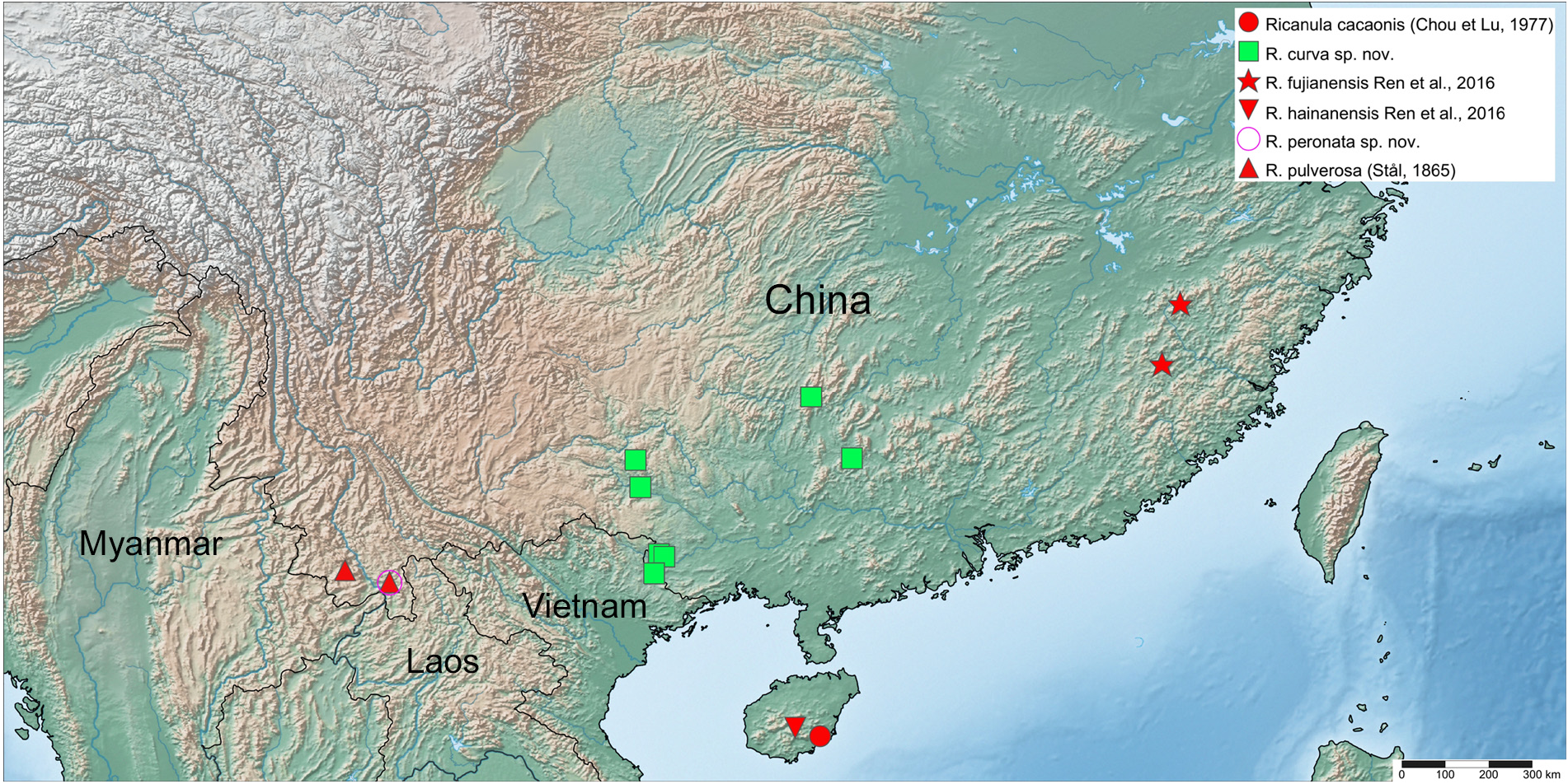
Type material. Holotype, male, China: 28 May 1982, Guangxi, Tianlin, Langping, coll. Jikun Yang.
Paratypes (13 males, 21 females, China) : 4 males, 2 females: 11 Apr. 1978, Guangxi, Baise, Yangwei , coll. Xianyu Qian ; 1 male: 12 May 1980, Guangxi, Longzhou, Longhu , coll. Zhuyin Wang ; 1 male: 17 May 1982, Guangxi, Longzhou, Nonggang , 240m, coll. Fasheng Li ; 2 males: 29 May 1982, 1 female: 30 May 1982, Guangxi, Tianlin, Langping , coll. Jikun Yang ; 1 female: 25 Jun. 1982, Guangxi, Longsheng , coll. Jikun Yang ; 3 males, 14 females: 3 Jun. 1984, 1 female: 9 Jun. 1984, Guangxi, Lingtian commune, coll. Zhengliang Wu et Xiaolin Lu ; 1 male, 1 female: 6 May 1993, 1 male, 1 female: 8 May 1993, Guangxi, Longzhou, Daqing Mountain , coll. Sikong Liu.
Distribution. China (Province Guangxi).
No known copyright restrictions apply. See Agosti, D., Egloff, W., 2009. Taxonomic information exchange and copyright: the Plazi approach. BMC Research Notes 2009, 2:53 for further explanation.
|
Kingdom |
|
|
Phylum |
|
|
Class |
|
|
Order |
|
|
Family |
|
|
Genus |