
Oblitosaurus bunnueli, Sánchez-Fenollosa & Verdú & Cobos, 2023
|
publication ID |
https://doi.org/10.1093/zoolinnean/zlad076 |
|
publication LSID |
lsid:zoobank.org:pub:9A4BC231-A3D0-4335-AFCB-8A99DEF3C953C |
|
DOI |
https://doi.org/10.5281/zenodo.10513266 |
|
persistent identifier |
https://treatment.plazi.org/id/039087FB-3918-5138-BF84-4C12FF28C47D |
|
treatment provided by |
Plazi |
|
scientific name |
Oblitosaurus bunnueli |
| status |
gen. et sp. nov. |
Oblitosaurus bunnueli gen. et sp. nov.
( Figs 2–8 View Figure 2 View Figure 3 View Figure 4 View Figure 5 View Figure 6 View Figure 7 View Figure 8 )
Etymology: The specific name bunnueli honours Luis Buñuel, a prestigious Spanish film director born in the province of Teruel.
Holotype: A dentary tooth (CPT-1440), a right ungual pollex from manus (CPT-1444), and an almost complete less hindlimb: femur (MAP-8290), tibia (MAP-8291), fibula (MAP-8292), astragalus (MAP-8293), calcaneum (MAP-8294), metatarsal IV (MAP-8295), phalanx 1 of digit II (MAP-8296), phalanx 3 of digit IV (MAP-8297), ungual phalanx of digit II (MAP-8298), and ungual phalanx of digit IV (MAP-8299).
Locality and horizon: Barrihonda-El Humero (RD-10) site in the municipality of Riodeva, province of Teruel, Aragón, Spain. South-Iberian Basin, Villar del Arzobispo Formation, Riodeva Facies Association, Upper Jurassic (upper Kimmeridgian–Tithonian).
Diagnosis: Ankylopollexian ornithopod diagnosed by the following autapomorphies (* when dubious due to taphonomic distortions): femur with (i*) a wide and robust lesser trochanter that almost covers the lateral surface of the greater trochanter; and (ii) a sub-circular distal medial condyle; with (iii) a pronounced and narrow lateromedial ridge on its ventral surface; tibia with (iv) an internal condyle projected dorsally above the dorsal margin of the cnemial crest; and metatarsal IV with (v) sub-triangular (or ellipsoid) morphology of the proximal articulation facet.
Description
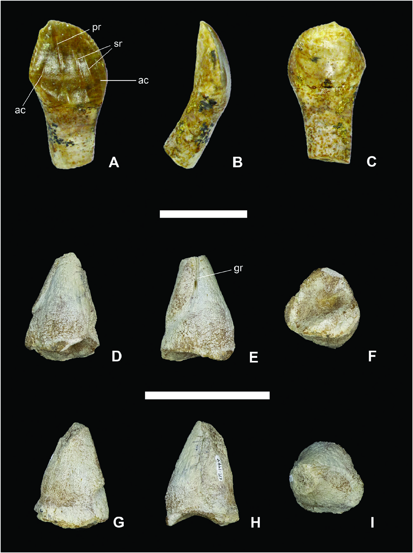
Dentition: CPT-1440 ( Fig. 2A–C View Figure 2 ) is a less dentary tooth. This well-preserved tooth is strongly labially curved. The crown is shield-shaped and the enamel is restricted to the lingual surface. The primary ridge is posteriorly offset and bifurcates toward the root. Two incomplete and less prominent secondary ridges are positioned mesially to the primary ridge. The one nearest to the primary ridge also bifurcates at the end of the crown. In addition, there are two faint and shorter accessory ridges located distally and mesially, respectively. There are no ridges on the nonenamelled labial face of the crown. The carinae present tongueshaped denticles with smooth margins from mid-crown height to apex. There is a worn facet, almost restricted to the labial surface. There is no cingulum separating the crown and root on the lingual surface. The root is incomplete and circular in shape in the section near the tip. There is a shallow depression on each side of the teeth (more distally evident) that extends from the root to the crown to accommodate contiguous growing teeth.
Manual phalanges: The right ungual phalanx of the digit I CPT-1444 ( Fig. 2D–I View Figure 2 ) is robust, short, sub-conical, and pointed (despite the tip is not being preserved). Furthermore, it has a sub-circular and concave proximal articular facet, which suggests a certain degree of mobility at the articulation with the proximal phalanx. There are two nail grooves: a curved and deep medial groove, and a much fainter (almost inappreciable) lateral groove.
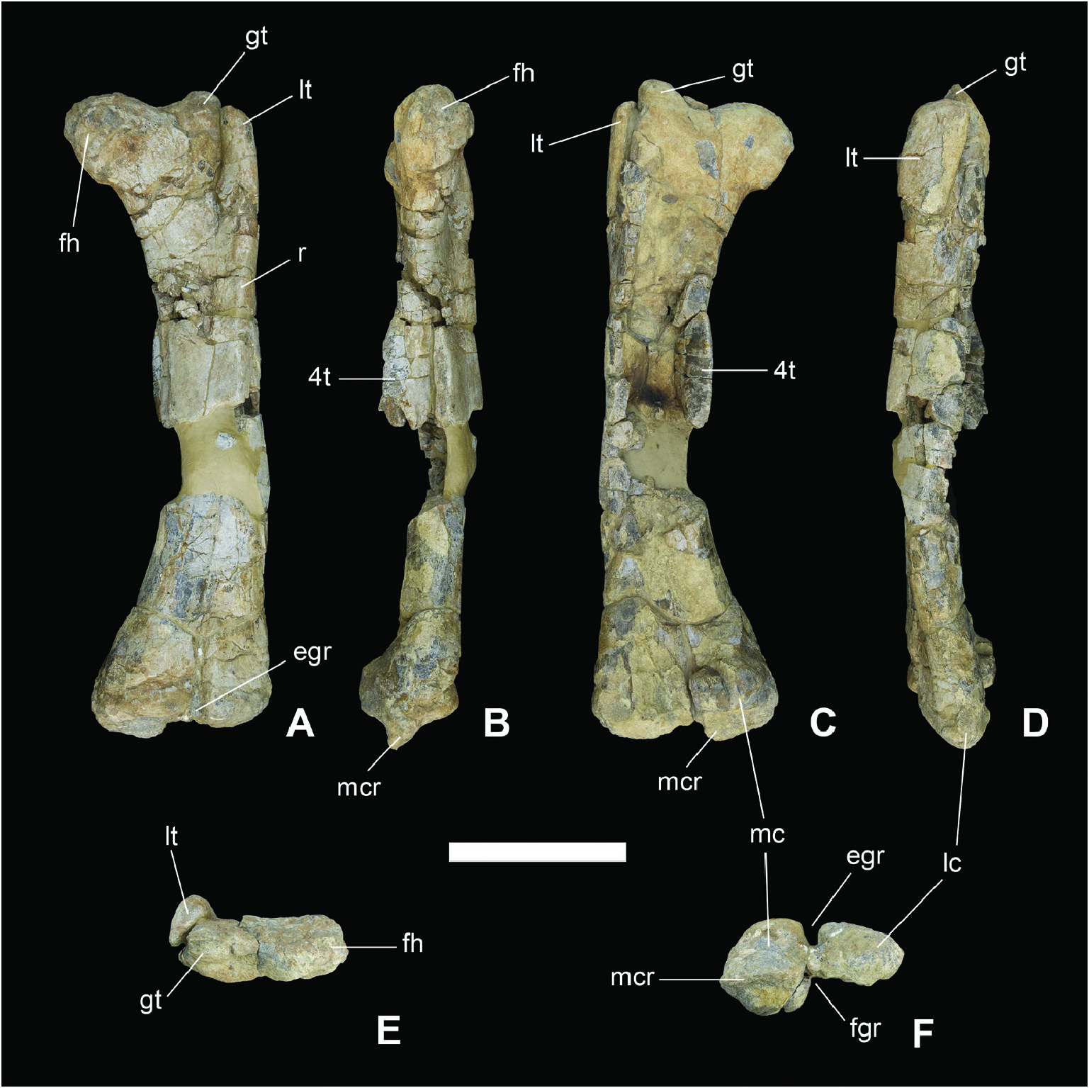
Femur: MAP-8290 ( Fig. 3 View Figure 3 ) is an almost entire less femur but lacking part of the diaphysis below the fourth trochanter. It is robust and slightly curved posteriorly in medial/lateral view. Both epiphyses are lateromedially expanded relative to the diaphysis. The femoral head is bulbous and broad with a shallow concavity on the posterior face, separated from the greater trochanter by a wide depression. The greater trochanter is dorsally convex and projects dorsolaterally above the lesser trochanter. The spike-like lesser trochanter is wide and robust, oblique in dorsal view, and covers almost the entire lateral surface of the greater trochanter ( Fig. 3D View Figure 3 ). Moreover, it is separated from the greater trochanter by a wide cless. The fourth trochanter is fragmented and incomplete at the distal end; however, the preserved part is large, sub-triangular, and located in the proximal-midpart of the diaphysis. There is a sharp but low ridge running diagonally from the lesser trochanter to the distal end, but it does not reach the medial condyle. At the distal end, the medial and lateral condyles are separated by a deep U-shaped intercondylar extensor groove. The medial condyle is sub-circular in ventral view ( Fig. 3F View Figure 3 ), with no protrusion extending to the intercondylar flexor groove, and has a marked but narrow transversal ridge, which projects ventrally ( Fig. 3B View Figure 3 ). The posterior half of the lateral condyle is not preserved.
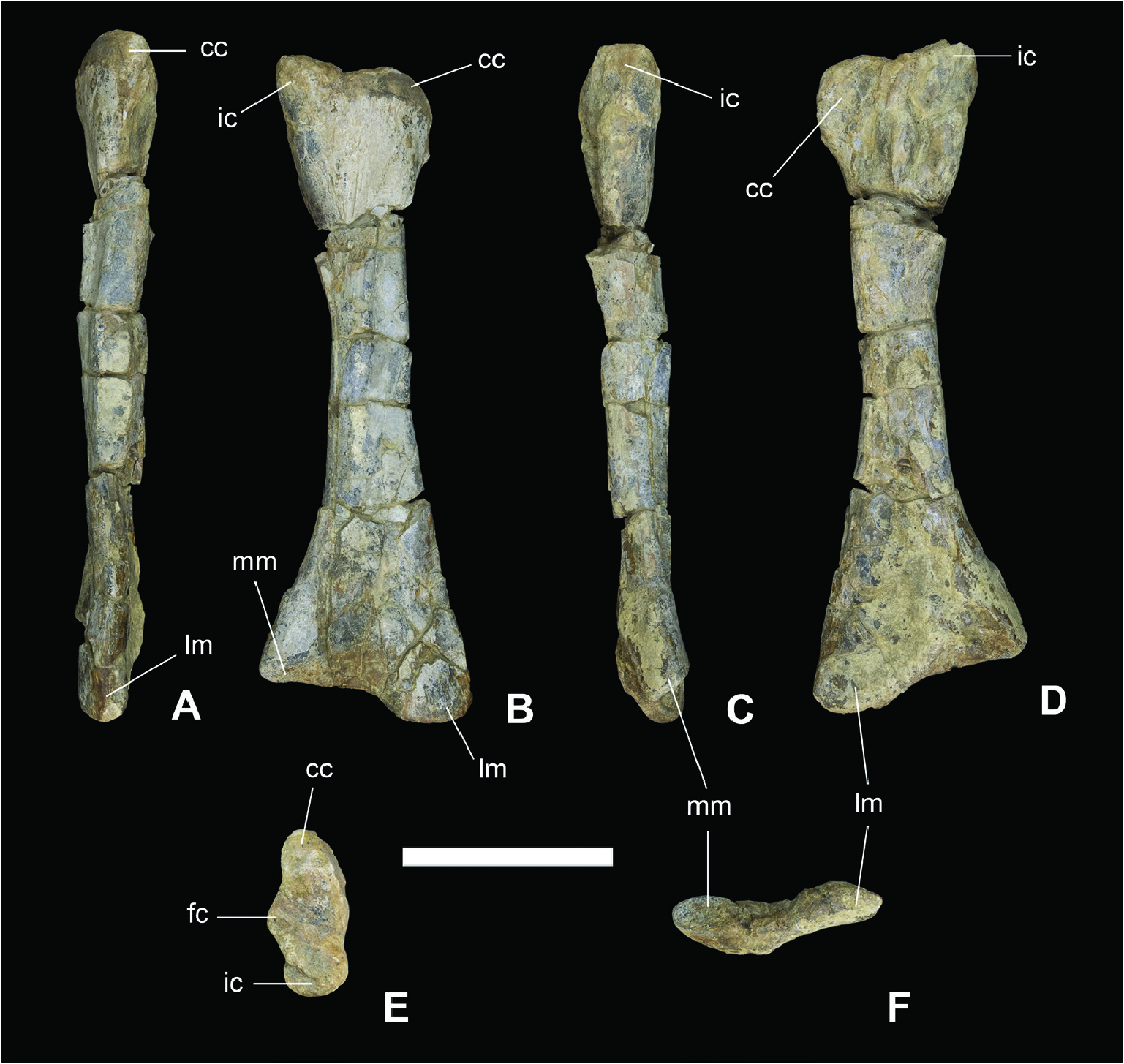
Tibia: MAP-8291 ( Fig. 4 View Figure 4 ) is an almost complete less tibia, shorter than the femur. The typical rotation present in the proximal end of the tibia in ornithopods cannot be observed here probably due to taphonomic reasons. The anterior, posterior, and dorsal surfaces of the proximal epiphysis are slightly eroded, but the cortical bone can be observed. However, the medial and lateral surfaces are eroded; therefore, its morphology should be considered with caution. The proximal epiphysis is slightly expanded both anteroposteriorly and lateromedially ( Fig. 4E View Figure 4 ). The cnemial crest is short, curved, robust, and laterally separated from the fibular (or lateral) condyle by a gentle concave margin. The eroded fibular condyle is located in the median position. The medial edge of the proximal end is gently sinuous. Moreover, the internal condyle projects dorsally above the cnemial crest ( Fig. 4B, D View Figure 4 ). The diaphysis is large, presumably cylindrical, and expands drastically toward the distal end. The distal end is much wider lateromedially than the proximal end anteroposteriorly. In particular, the lateral malleolus is wider and projects more distally than the medial one. Fibula: MAP-8292 ( Fig. 5 View Figure 5 ) is a slender less fibula that is shorter than the tibia. Both the proximal and distal regions are expanded, especially the proximal region, which has an angular cranial process. The fibula is twisted along its length. The lateral surface of the proximal end is slightly convex, whereas the medial surface is concave to receive the fibular condyle of the tibia. The dorsal margin of the fibula slopes slightly from posteroventral to anterodorsal in lateral view.
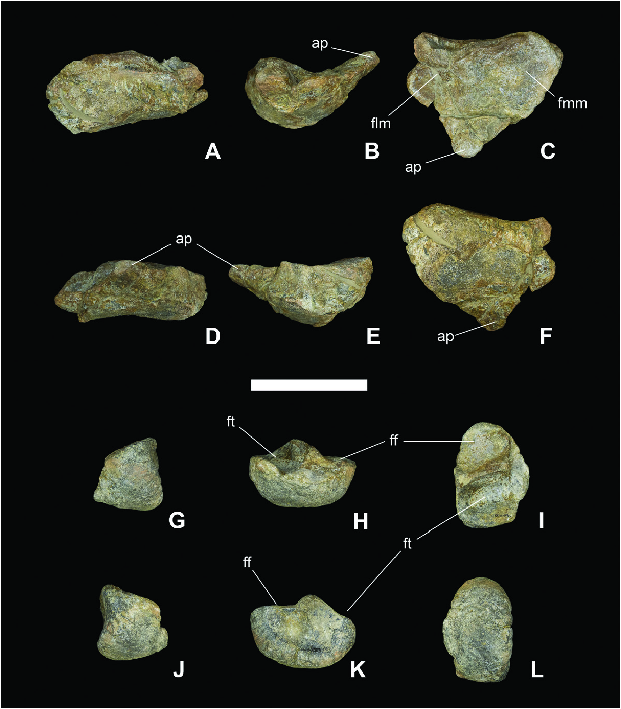
Astragalus: MAP-8293 ( Fig. 6A–F View Figure 6 ) is a complete less astragalus with a general sub-triangular shape. The dorsal surface has two slightly concave facets for the medial and lateral malleoli of the tibia. These facets are separated by a ridge, and the medial facet is wider both anteroposteriorly and lateromedially than the lateral facet. A prominent triangular ascending process is observed on the posterior margin ( Fig. 6C View Figure 6 ). Both the anterior and posterior margins do not face each other enclosing articular facets, so the astragalus is very open in dorsal view. In contrast to the dorsal surface, the ventral surface is convex.
Calcaneum : MAP-8294 ( Fig. 6G–L View Figure 6 ) is a complete less calcaneus with an overall hemispherical shape. The dorsal surface is divided into fibular and tibial facets, separated from each other by a pronounced oblique lateromedial ridge. The fibular facet is larger than the tibial facet. The tibial facet has a strongly concave surface and the fibular facet has an almost straight one. Both facets present straight margins in lateral view. There is a depression on the lateral side, with a gentle ridge separating it into two halves. The anteroventral surface is markedly convex and thick.
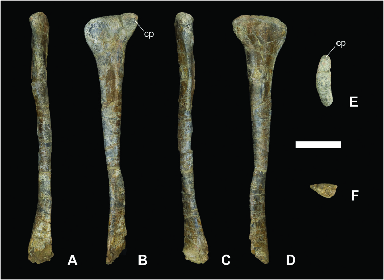
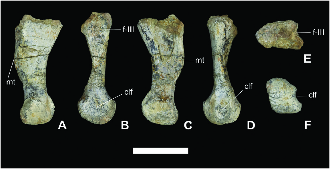
Metatarsal IV: MAP-8295 ( Fig. 7 View Figure 7 ) comprises a long and slender less metatarsal IV. In anterior and posterior views, the medial margin is convex, forming the medial tuberosity, while the lateral margin is concave. Both the proximal and distal ends are expanded. The former has a concave, anteroposteriorly compressed, and sub-triangular articular facet ( Fig. 7E View Figure 7 ) with a lateromedial width much greater than the anteroposterior, so the lateral edge diverges from the medial one in anterior and posterior views ( Fig. 7A, C View Figure 7 ). Moreover, there is a concave facet in the medial margin for articulating with the metatarsal III. In lateral and medial views, the anterior margin of the diaphysis is straight, but the posterior margin is concave. The distal end is sub-circular in lateral and medial views, but it is sub-rectangular in distal view, being longer than wide. Both the medial and lateral collateral ligament foveae at the distal end are deeply depressed, especially on the lateral side ( Fig. 7F View Figure 7 ). The condyles are not well-defined.
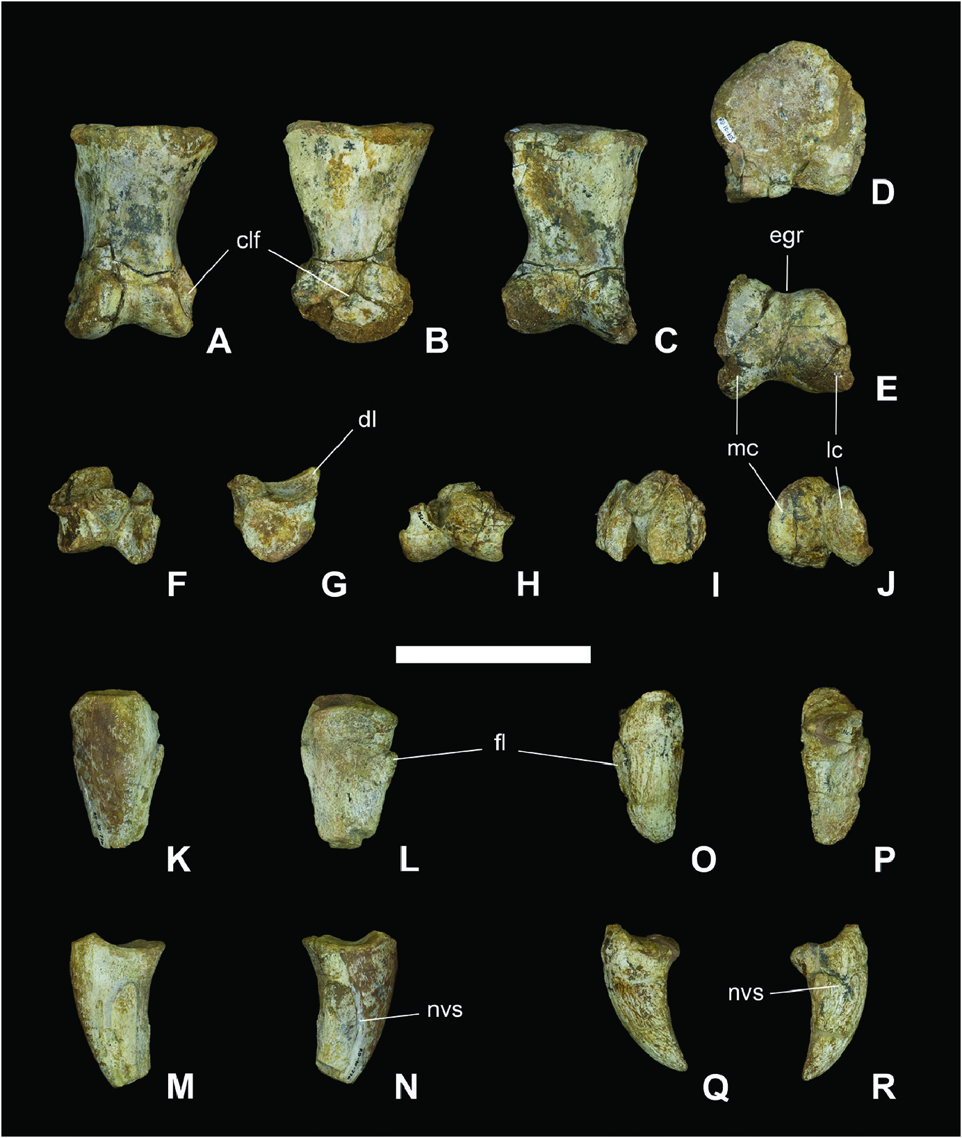
Pedal phalanges: Four phalanges belonging to the less pes have been recovered. These include the phalanx 1 of digit II (MAP-8296), ungual phalanx of digit II (MAP-8298), phalanx 3 of digit IV (MAP-8297), and ungual phalanx of digit IV (MAP-8299). The phalanx 1 of digit II (MAP-8296, Fig. 8A–E View Figure 8 ) is longer than tall, with an elongated diaphysis. This phalanx is keyholdershaped in both lateral and medial views. Its proximal articular facet is concave, with an almost straight plantar margin and convex dorsal margin. On the other hand, the distal end has two prominent condyles separated by a deep extensor and flexor intercondylar grooves in the median position. The medial condyle is more prominent than the lateral condyle. There are two well-developed collateral ligament foveae on both sides of the distal end.
The ungual phalanx of digit II (MAP-8298, Fig. 8K–N View Figure 8 ) is elongated, slightly curved, and presumably pointed (the tip is not preserved). This phalanx has a sub-circular (resembling an irregular and rounded pentagon) and concave proximal articular facet. The lateral and medial surfaces have neurovascular sulci along their entire length, with a flap.
In contrast to the phalanx 1 of digit II, the phalanx 3 of digit IV (MAP-8297, Fig. 8F–J View Figure 8 ) has no diaphysis, and it is proximodistally compressed. The proximal articular facet is concave and a dorsal lip arises from the dorsal margin. The condyles are well-developed at the distal end.
Finally, the ungual phalanx of digit IV (MAP-8299, Fig. 8O–R View Figure 8 ) has a concave and sub-circular proximal articular facet. Unlike the ungual phalanx of digit II (MAP-8298), it is shorter, slender, more curved and has shallower medial and lateral neurovascular sulci. Moreover, it has flaps and is pointed at the distal end.
No known copyright restrictions apply. See Agosti, D., Egloff, W., 2009. Taxonomic information exchange and copyright: the Plazi approach. BMC Research Notes 2009, 2:53 for further explanation.