
Cycloporus australis Prudhoe 1982
|
publication ID |
https://doi.org/ 10.11646/zootaxa.4652.2.5 |
|
publication LSID |
lsid:zoobank.org:pub:951C3124-F209-4E02-B489-695CBF71543D |
|
persistent identifier |
https://treatment.plazi.org/id/039187B7-D26D-3B4E-FF35-FEC26664FF7E |
|
treatment provided by |
Plazi |
|
scientific name |
Cycloporus australis Prudhoe 1982 |
| status |
|
Cycloporus australis Prudhoe 1982
( Figure 4 View FIGURE 4 , 5 View FIGURE 5 )
Material examined: One specimen (BNHS-Pclad 141, 5.75 x 4.10 mm) dissected as sagittal sections for reproductive structure (25 slides), collected on 03-12-2013 at Mandvi in Ratnagiri district, Maharashtra. One specimen (BNHS-Pclad 176, 5.95 x 3.37 mm) dissected as sagittal sections for reproductive structure (18 slides), collected 21-11-2014 at Alawa in Ratnagiri district, Maharashtra .
Distribution: South Australia ( Prudhoe 1982)
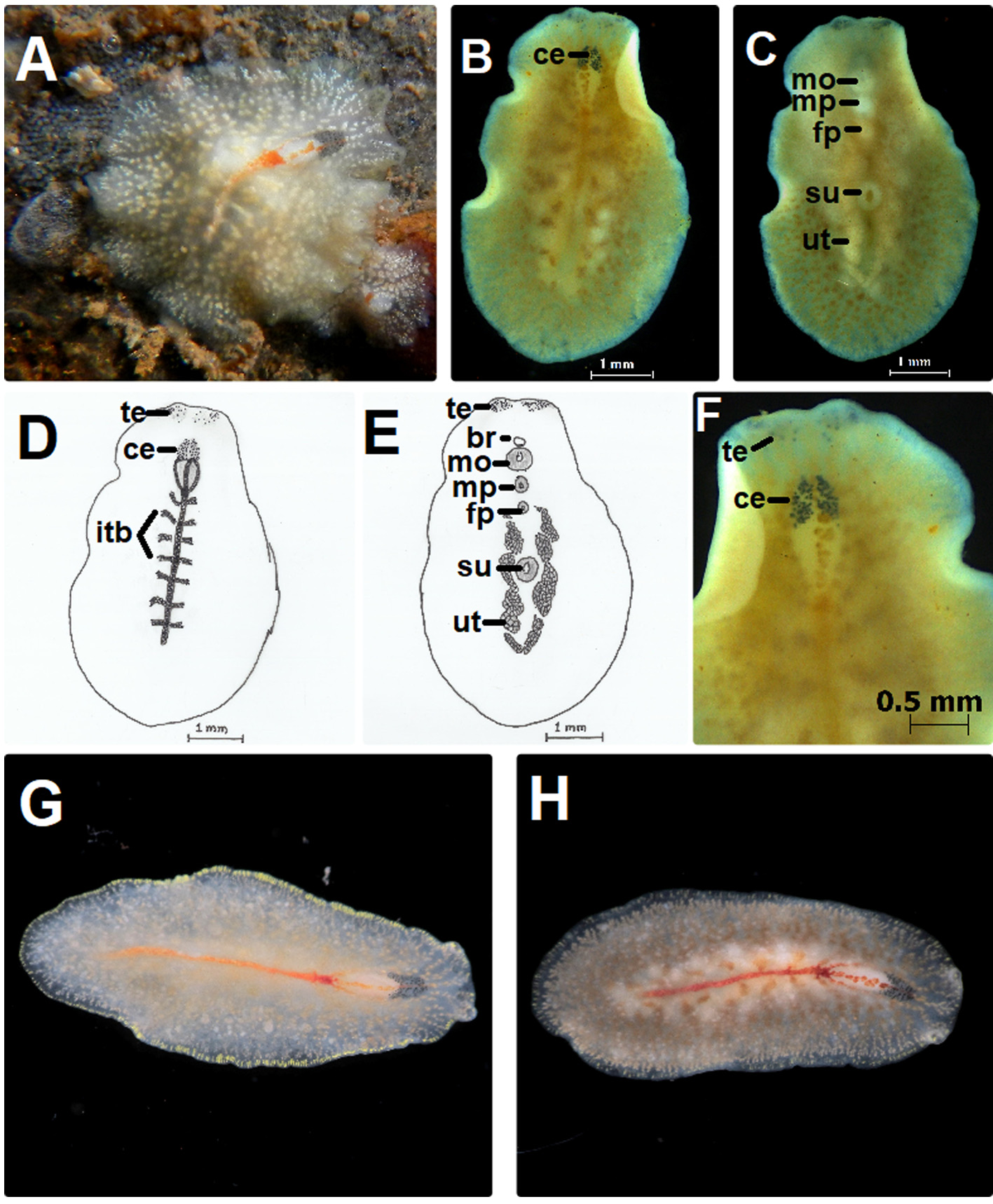
Diagnosis: Translucent body sometime appears buff brownish, median intestinal branch is prominent red and trifurcates at the pharyngeal region, one branch passes over the pharynx and cerebral region and two lateral branches pass sideways to these organs. About 8–9 intestinal branches.
Description: Colour: Translucent body appears buff brownish, or tinge of brown in some specimens ( Fig. 4A, G, H View FIGURE 4 ). White spots are prominent and seen over the dorsal surface which perhaps represent scattered, dorsally arranged ovaries. Margin is clear with yellow lateral stripes or dots representing peripheral glands. Median main intestinal branch is prominently red. This branch trifurcates at the rear end of the tubular pharynx, forming dark red spot and runs anteriorly with median branch exactly over the pharynx and cerebral region; two lateral branches pass sideways to these organs; posteriorly it ends prior to the margin ( Fig. 4A, D, G View FIGURE 4 , and H). Ventrally translucent to slightly creamish.
Form: Oval-elongated.
Pseudotentacles: Small bumps, sometimes slightly raised ( Fig. 4A View FIGURE 4 ), measuring 0.19–0.30 mm.
Eyespots: Cerebral eyespot (53–58) separated into two groups elongated and broad (0.49 mm x 0.33 mm) at the base, arranged semi-circularly over the anterior margin of the tubular pharynx ( Fig. 4F View FIGURE 4 ). Located 0.69 mm away from the anterior margin. Tentacular eyespots (38–42) in dorsal groups scattered or clustered together, ventral groups contain 65–74 eyespots.
Digestive system: Pharynx short and tubular, 0.67–0.72 mm long. Mouth opens 0.99–1.44mm from the anterior margin. Intestine with around 8–9 lateral branches ( Fig. 4B, D View FIGURE 4 ). Main intestine from base of the pharynx to posterior end measures 2.43 mm long. The lateral intestinal branches are organised rather loosely, and lateral peripheral glands as conspicuous yellow stripes seen over the translucent margin.
Gonopores: Male gonopore present immediately behind the pharynx and 1.48–1.79 mm from anterior margin, measuring 0.17 mm. Female gonopore 0.35 mm behind the male gonopore and 1.94–2.25 mm from anterior margin, measuring 0.12–0.15mm ( Fig. 4C, E View FIGURE 4 ).
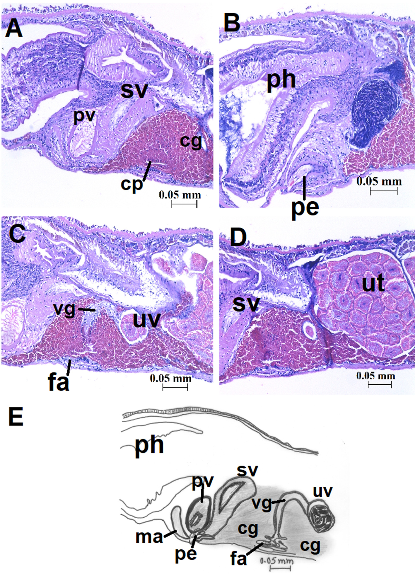
Male reproductive system: In male apparatus, oval thick walled seminal vesicle (0.20 mm x 0.13 mm) found dorsal to the prostatic vesicle and parallel to the pharynx ( Fig. 5A View FIGURE 5 ). Oval prostatic vesicle (0.11 mm x 0.13 mm) is placed between the pharyngeal chamber and ejaculatory duct. Stylet, 0.069 mm pointed and bent at the tip, enters wide semi-circular male atrium via penis pocket ( Fig. 5B View FIGURE 5 ).
Female reproductive system: female atrium wide, oval (0.069 mm wide) and opens into vagina with shallow lateral invagination (0.022 mm) for cement pouches. Cement glands densely disposed ( Fig. 5C View FIGURE 5 ). Vagina slightly extends (0.082 mm) dorsally, turns posterior to uterine spaces ( Fig. 5C View FIGURE 5 ). Uterine vesicle (0.19 mm x 0.15 mm) is visible ( Fig. 5C View FIGURE 5 ). Uteri filled with oocytes appear on either side of the median line and is visible through ventral surface ( Fig. 4C, E View FIGURE 4 , 5D View FIGURE 5 )
Taxonomic remarks: round/oval body, tubular pharynx, small bumps of tentacles, lateral branches of intestine range within 4-10 and multiple uterine vesicles place these specimens into the genus Cycloporus as defined in Newman and Canon (2002).
Prudhoe (1982) mentioned the close resemblance of this species with C. gabriellae and C. maculatus . However, he differentiated C. australis in terms of colouration and greater number of eyes. Cycloporus maculatus also shows an elongate cluster, formed by cerebral and tentacular eyespots, distinguishing it from C. australis . It also differs from other Cycloporus species due to the anterior triad of intestinal branch.
Hallez (1893), while describing C. maculatus from the Mediterranean, mentioned the juvenile forms with red intestinal branches but this colouration was not observed in the adult forms. The colour of the main intestinal branches is not recorded in the original description of C. australis . However, all the specimens in the present study showed a distinct red colour.
The specimens were always observed over or around the compound ascidian.
No known copyright restrictions apply. See Agosti, D., Egloff, W., 2009. Taxonomic information exchange and copyright: the Plazi approach. BMC Research Notes 2009, 2:53 for further explanation.