
Pyura vannamei Monniot C. 1994
|
publication ID |
https://doi.org/10.11646/zootaxa.4459.3.1 |
|
publication LSID |
lsid:zoobank.org:pub:5C5A86AC-6FA4-46AD-9A89-068E9119DD28 |
|
DOI |
https://doi.org/10.5281/zenodo.5966673 |
|
persistent identifier |
https://treatment.plazi.org/id/039187CA-B948-3C12-FF6B-5767BB943F1E |
|
treatment provided by |
Plazi |
|
scientific name |
Pyura vannamei Monniot C. 1994 |
| status |
|
Pyura vannamei Monniot C. 1994 View in CoL
Figs 19 View FIGURE 19 , 20 View FIGURE 20
Monniot C. 1994, and synonymy; Collin et al 2005, Brazil
Stations: AB 108, 155, 179, 189, 193, 301. AM 05, 07. AR 100, 101, 103, 188, 352; AR 353. (MNHN S2 PYU502).
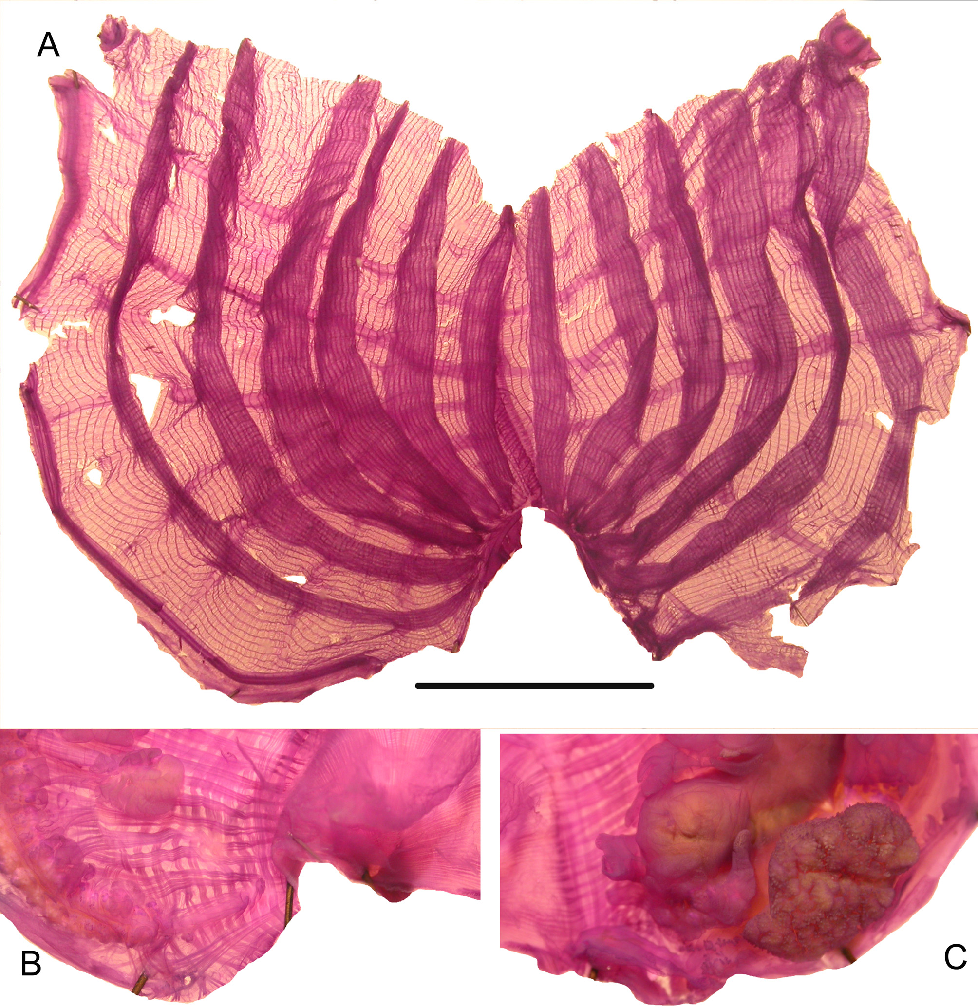
The body is red partly or entirely covered with epibionts ( Fig. 19B View FIGURE 19 ), the colour remaining some time in formalin to become brown. The well separated siphons have tunic tubercles, but no spinules have been found. The tunic surface is corrugated on the body and has a leathery consistency. Extracted from the tunic the siphons have a circular red line at the rim and a membranous velum. The musculature is complete with strong siphonal sphincters and regularly crossed and large fibres on the body ( Fig. 19A View FIGURE 19 ). The oral tentacles are stout with a wide base ( Fig. 19A,C View FIGURE 19 ) and tiny secondary branching. The prepharyngeal band forms a deep V ( Fig. 19C View FIGURE 19 ). The dorsal tubercle is variable, either in a U anteriorly opened or a sideways S curve with one horn inrolld ( Fig. 19A,C View FIGURE 19 ) The branchial sac has 6 high folds ( Fig. 20A View FIGURE 20 ). One formula on the right side, in a specimen 4cm large, is: E- 8 (20) 7 (20) 7 (22) 8 (21) 7 (22) 7 (18) 2-DL.
The dorsal lamina has numerous pointed languets. The endostyle forms a short loop at its anterior end. The longitudinal vessels end in papillae against the oesophagus entrance. The digestive tract is long with an enlarged rectum ( Figs 19A View FIGURE 19 , 20C View FIGURE 20 ). The anus has 8 round lobes which are often rolled up ( Fig. 20C View FIGURE 20 ). The digestive gland is dark with a large papillated mass on the stomach and some other papillae on the oesophagus ( Fig. 19A View FIGURE 19 ). The external side of the gut loop is lined by numerous foliated endocarps ( Fig. 19A View FIGURE 19 ). There is a very long gonad on each side, the left one contained inside the gut loop. The gonad lobes are applied to each other in a dense double series and wear endocarps( Fig. 19A View FIGURE 19 ). The genital papillae are joined and short ( Fig. 20B View FIGURE 20 ). Dorsally to the right gonad, and well separated from it, are 3 to 5 cushion like endocarps on the body wall ( Fig. 19A View FIGURE 19 ). There are no other endocarps elsewhere on the body wall.
This species has many characters in common with P. vittata : the red colour, general body shape and size, same branchial sac, an enlarged rectum. It differs by the presence of endocarps on the body wall, absence of spinules on the siphons, the right gonad extending less anteriorly and applied against the endostyle.
The geographic distribution of P. vannamei is limited to the western tropical Atlantic Ocean, but it may have been confused with P. vittata .

No known copyright restrictions apply. See Agosti, D., Egloff, W., 2009. Taxonomic information exchange and copyright: the Plazi approach. BMC Research Notes 2009, 2:53 for further explanation.
|
Kingdom |
|
|
Phylum |
|
|
Class |
|
|
Order |
|
|
Family |
|
|
Genus |