
Microcosmus exasperatus Heller, 1878
|
publication ID |
https://doi.org/10.11646/zootaxa.4459.3.1 |
|
publication LSID |
lsid:zoobank.org:pub:5C5A86AC-6FA4-46AD-9A89-068E9119DD28 |
|
DOI |
https://doi.org/10.5281/zenodo.5966663 |
|
persistent identifier |
https://treatment.plazi.org/id/039187CA-B95F-3C00-FF6B-5604BF593E55 |
|
treatment provided by |
Plazi |
|
scientific name |
Microcosmus exasperatus Heller, 1878 |
| status |
|
Microcosmus exasperatus Heller, 1878 View in CoL
Figs 6 View FIGURE 6 , 7 View FIGURE 7
Heller, 1878, Jamaica; Van Name: 1921 and synonymy; Monniot C.:1983, Martinique; Rocha et al: 2012a, Brazil.
Stations: AB 149, 452. AM 34. AR 101, 314, 558. (MNHN S2 MIC 204)
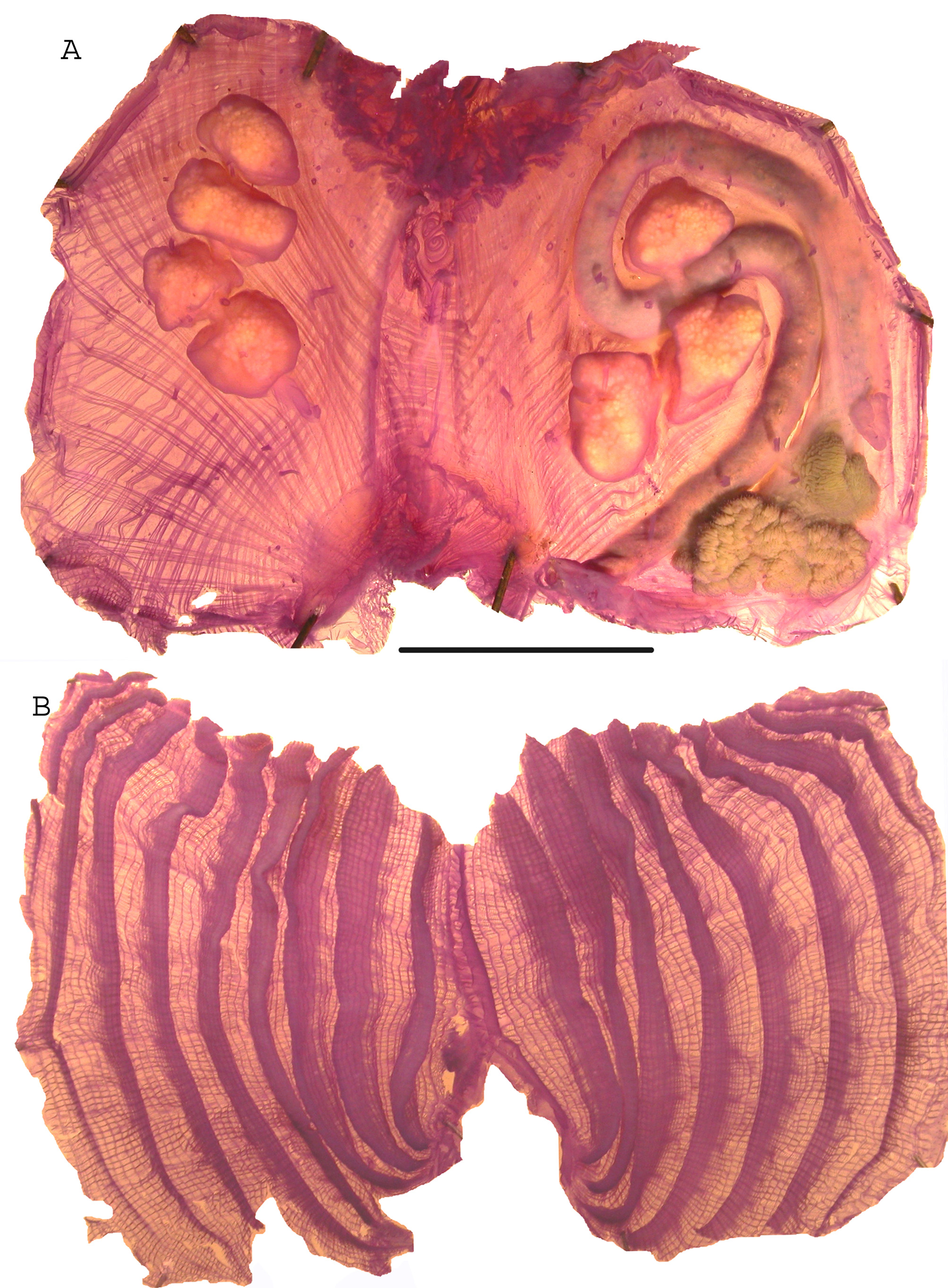
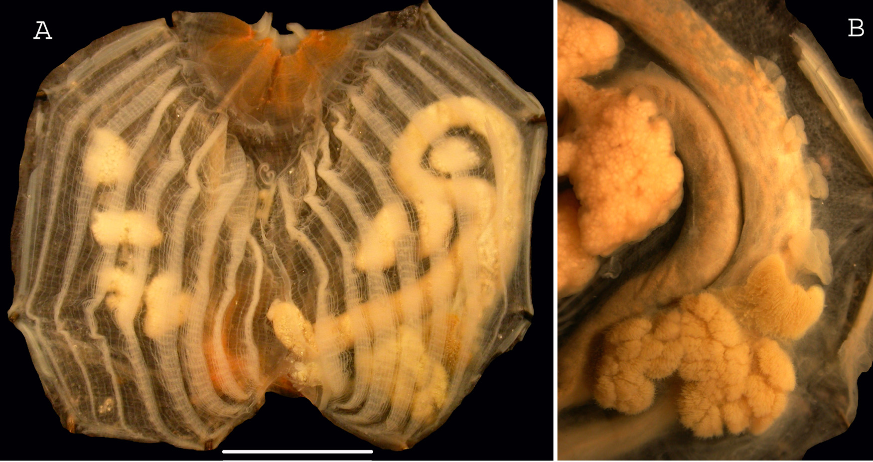
The body is fixed by its ventral part, partially covered with epibionts with protruding siphons well apart. The tunic is red and wrinkled externally and red also internally on the siphons. There are dense spinules on the siphons. The body wall is thick with crossed bundles of muscle fibres ( Fig. 6A View FIGURE 6 ). There is a thin oral velum adhering to the tunic lining which has brown longitudinal lines. The 12-14 largest oral tentacles are bushy, three times ramified. The prepharyngeal band forms a wide and deep V including the dorsal tubercle with rolled horns ( Fig.7A View FIGURE 7 ). The dorsal lamina has a plain edge. There is an average of 9 branchial folds on each side ( Fig. 6B View FIGURE 6 , 7A View FIGURE 7 ), the most ventral thinner and incomplete disappearing ventrally; a formula of a 3.2cm large specimen is on the right side: E- 1 (12) 3 (15) 3 (17) 4 (17) 4 (23) 3 (23) 4 (24) 4 (22) 3 (20) 2-DL.
There are parastigmatic vessels and irregularly some papillae on the transverse vessels. The second curve of the long digestive loop is wide open ( Fig. 6A View FIGURE 6 , 7A View FIGURE 7 ), the rectum being close to the stomach. The hepatic gland has two parts ( Fig. 7B View FIGURE 7 ) each with convoluted papillae wearing button like papillae. After the hepatic gland on the ascending limb of the intestine are 1 to 4 endocarps ( Fig. 6A View FIGURE 6 , 7B View FIGURE 7 ). These endocarps were not mentioned in previous descriptions. There are no endocarps on the body wall.
There is one gonad on each side the left one crossing the intestine ( Fig. 6A View FIGURE 6 , 7A View FIGURE 7 ). They contain 3 or 4 well separated lobes linked by a longitudinal gonoduct. The male and female papillae are short and linked. Numerous thread-like papillae cover the entrance of the atrial siphon and the large velum.
From its first record from Jamaica M. exasperatus was described in detail from several Caribbean locations ( Van Name 1921; 1945) and from Martinique ( Monniot C. 1983). This species is common in the whole western Atlantic ( Rocha et al 2012a) and its distribution is also worldwide, from the Mediterranean Sea ( Ramos et al 2013), the Indian Ocean ( Monniot C. 2002), and from the western Pacific Ocean ( Kott 1985; Monniot C. 1989).
No known copyright restrictions apply. See Agosti, D., Egloff, W., 2009. Taxonomic information exchange and copyright: the Plazi approach. BMC Research Notes 2009, 2:53 for further explanation.
|
Kingdom |
|
|
Phylum |
|
|
Class |
|
|
Order |
|
|
Family |
|
|
Genus |