
Fusarium desaboruense N. Maryani, Sand.
|
publication ID |
https://doi.org/ 10.3767/persoonia.2019.43.02 |
|
DOI |
https://doi.org/10.5281/zenodo.5613549 |
|
persistent identifier |
https://treatment.plazi.org/id/0391CB21-0B53-866B-FCDC-FE070F0C905C |
|
treatment provided by |
Plazi |
|
scientific name |
Fusarium desaboruense N. Maryani, Sand. |
| status |
|
Fusarium desaboruense N. Maryani, Sand. View in CoL -Den., L. Lombard, Kema & Crous, sp. nov.
— MycoBank MB828961;
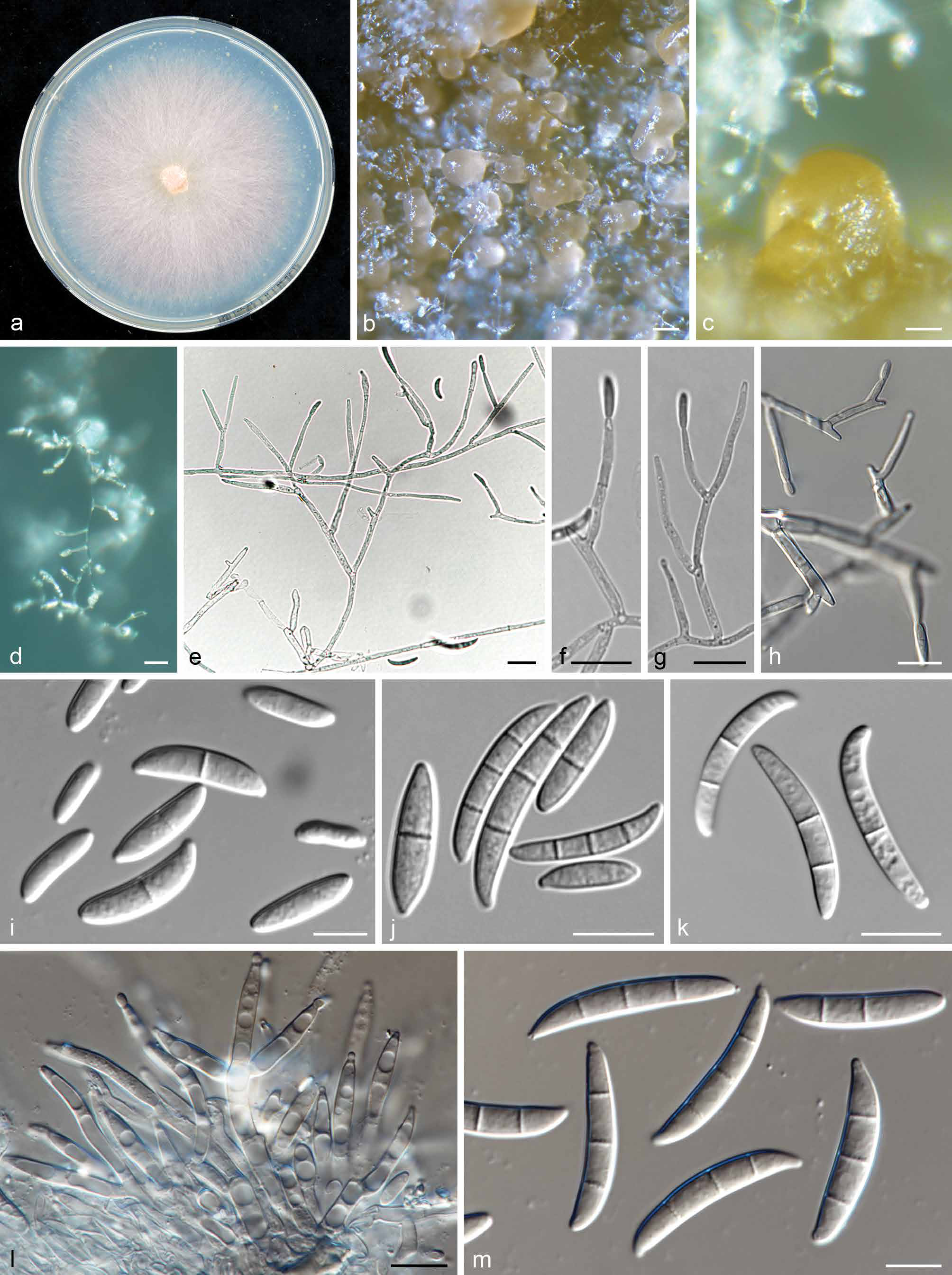
Fig. 7 View Fig
Etymology. Name refers to Desa Boru,the village from where this species was collected in Indonesia.
Typus. INDONESIA, Desa Boru, Kecamatan Waigate, Sikka Flores , East Nusa Tenggara (E122°22'7" S8°36'49"), on infected pseudostem of Musa sp. var. Pisang Kepok ( ABB), 17 Aug. 2015, N. Maryani (holotype specimen and culture, InaCC F 951 , preserved in metabolically inactive state) GoogleMaps .
Sporulation abundant from conidiophores carried on aerial mycelium and from sporodochia. Conidiophores on aerial mycelium abundant on PDA and SNA, less frequent on CLA, septate, sparingly or profusely branching irregularly or sympodially, rarely reduced to solitary conidiogenous cells, formed laterally on aerial hyphae; conidiogenous cells mono- or polyphialidic, acute, subulate or subcylindrical, smooth- and thin-walled (6–)15–33(–44) × (2–)2.5–4(–7) µm (av. 21.5 × 3 µm), formed terminally, singly or in whorls on conidiophores or intercalary, proliferating percurrently, periclinal thickening inconspicuous or absent; conidia of two types: a) (microconidia) ovoid to el- lipsoid, smooth- and thin-walled, (10–)11–16(–18) × (4–)6(–7) μm (av. 13 × 5 µm), 0–1-septate, arranged in false heads on monophialides; and b) (macroconidia) falcate and multiseptate, apical cells papillate, basal cells indistinct or foot-shaped, 1–3-septate, formed on polyphialides: 1-septate conidia 22.5– 26(–27) × 3.4–4 µm; 2-septate conidia (21.5–)22–26 × 3–4.5 µm; 3-septate conidia (23–)24.5–34(–37) × 3–4.5 µm; av. (21.5–)22–30.5(–37) × 3–4.5 µm. Sporodochia formed abundantly on CLA after 7 d, pale orange to orange. Conidiophores in sporodochia unbranched, rarely laterally branched up to two times; conidiogenous cells monophialidic, smooth- and thin-walled (15.5–)16.5–24(–29) × (2.5–) 3–4 µm (av. 20 × 3.5 µm), solitary, terminal or lateral, or in terminal groups of up to three conidiogenous cells, with minute collarettes and periclinal thickening; sporodochial conidia falcate, apical cells gently curved, papillate, basal cells gently curved, foot-shaped, 1–3(–4)-septate: 1-septate conidia (14.5–)15–20.5(–22) × 3.5–4.5 µm; 2-septate conidia (20.5–)21.5–24 × 3.5–4.5(–5) µm; 3-septate conidia (21–)24–29(–31.5) × (3.5–)4–5(–5.5) µm; 4-septate conidia 34 × 5.5 µm; av. (14.5–)20–28(–34.5) × (3.5–)4–5(–5.5) µm. Chlamydospores not observed.
Culture characteristics — Colony on PDA showing optimal growth at 25 °C with an average growth rate of 4.9–5.2 mm /d. Colony reverse, pale violet becoming white towards the margins, turning violet with age and pigmented. Colony surface cottony, pale violet, becoming white with age, immersed mycelium becoming purple and lacking exudates.Aerial mycelium abundant, cottony, with abundant sporulation.
Geography & Host — Sikka Flores, East Nusa Tenggara, Musa sp. var. Pisang Kepok (ABB).
Pathogenicity — Not pathogenic on Cavendish (AAA).
Additional materials examined. INDONESIA, Desa Boru, Kecamatan Wai- gate, Sikka Flores , East Nusa Tenggara (E122°22'7" S8°36'49"), on infected pseudostem of Musa sp.var. Pisang Kepok ( ABB), 17 Aug. 2015, N. Maryani (InaCC F 950, InaCC F 952) GoogleMaps .
Notes — Morphologically very similar to F. sacchari ( Leslie
& Summerell 2006) and F. subglutinans ( Nelson et al. 1983) , except that this species produces sporodochia abundantly un- der regular culturing conditions. Fusarium desaboruense can be distinguished by the septation of its macroconidia (1–4-septate) and microconidia (1–3-septate), not observed in F. saccari ( Leslie & Summerell 2006) . Phylogenetic analyses of partial rpb2 gene sequences recognised this species as distinct from F. sacchari with strong support of BP 99 %.
| ABB |
Asian Bacterial Bank |
| N |
Nanjing University |
| F |
Field Museum of Natural History, Botany Department |
No known copyright restrictions apply. See Agosti, D., Egloff, W., 2009. Taxonomic information exchange and copyright: the Plazi approach. BMC Research Notes 2009, 2:53 for further explanation.
|
Kingdom |
|
|
Phylum |
|
|
Class |
|
|
Order |
|
|
Family |
|
|
Genus |