
Histricostoma tuberculatum ( Koch & Berendt, 1854 )
|
publication ID |
https://doi.org/ 10.3897/zookeys.16.224 |
|
publication LSID |
lsid:zoobank.org:pub:DB5973A9-8CF6-400B-87C4-7A4521BD3117 |
|
DOI |
https://doi.org/10.5281/zenodo.3791606 |
|
persistent identifier |
https://treatment.plazi.org/id/0392774B-7851-106B-B6DD-56DEFB89FD7F |
|
treatment provided by |
Plazi |
|
scientific name |
Histricostoma tuberculatum ( Koch & Berendt, 1854 ) |
| status |
|
¡ Histricostoma tuberculatum ( Koch & Berendt, 1854) View in CoL
Figs 1-2 View Figures 1-5 , 6-8 View Figures 6-9
Synonymy. See Dunlop (2006, p. 179).
Holotype. MfN, Berendt collection, repository nr. 7248, redescribed by Dunlop (2006), from Baltic amber (Palaeogene, Eocene); precise locality unclear.
Additional material. MfN, MB.A. 1652 (also bears a label “Ser. 8/11”) and MB.A. 1653. Bitterfeld amber, probably from the site of the Goitsche open-cast Mine near Bitterfeld, Sachsen-Anhalt, Germany; Palaeogene (Oligocene: Chattian).
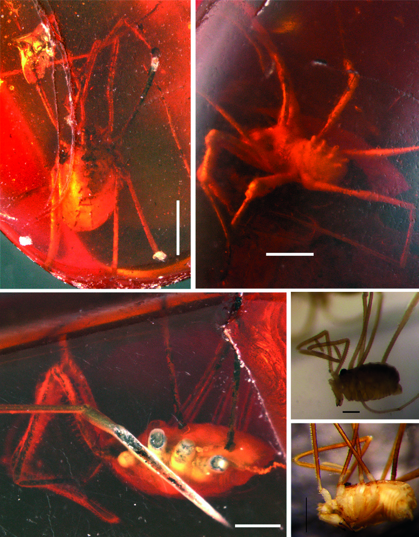
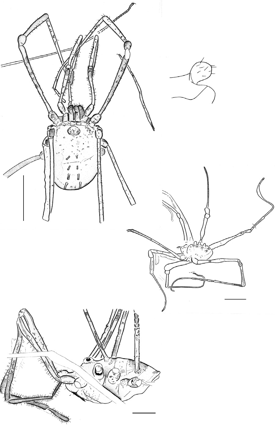
Description. MB.A. 1652 ( Figs 1 View Figures 1-5 , 6 View Figures 6-9 ) is a fairly complete specimen in dorsal view. Body compact, smoothly oval, length 1.48 mm; maximum width ca. 1.0 mm. Prosoma and opisthosoma completely fused together with little or no evidence of tagmosis between these body regions, of divisions of the prosomal dorsal shield and/ or of opisthosomal segmentation. Ocular tubercle flattened, diameter of each lens c. 0.1 mm. Slight bilobation to the anterior margin of the prosomal dorsal shield. Basal article of chelicera projects forwards, maximum length 0.26 mm, more distal articles largely tucked under body obscuring details, but quite setose on their anterior margin. Basal article distally with a globular and setose apophysis ( Fig. 7 View Figures 6-9 ). Pedipalps elongate and slender. Palpal coxae project forwards up to 0.19 mm. Subsequent articles with lengths (in mm) of trochanter, 0.28; femur, 0.85; patella, 0.70; tibia, 0.78; tarsus, 0.33; giving a total post-coxal length of 2.94. Palpal trochanter oval, bearing numerous very short setae. Femur longer, curving mesally slightly and widening distally, and with longer inward-pointing setae; especially on the mesal surface. Patella and tibia also with some longer setae. Tarsus slightly swollen and quite densely setose. Leg 1 almost complete, other legs attached to body as femora only; with leg 4 quite poorly preserved. Leg 1 article lengths (in mm): trochanter, 0.22; femur, 1.59; patella, 0.26; tibia, 0.69; metatarsus, 1.50; tarsus at least 0.56. Tarsus with at least one long and one short distal article but distal end and any claw equivocal. Femora 2 and 3 with lengths of 1.87 and 1.44 mm respectively. Most leg articles elongate, slender and widening slightly distally, but patella short and globular. Femora typically with three pseudo-articulations, revealed as concavities in the limb outline and/or as paler bands about a third to a half of the way along the article. Trochanters often with short hairs (as in the pedipalps) and femora with a mixture of short hairs and robust, but very short spines along the length of the article. Setation seemingly weak or absent on more distal leg articles, but reappearing as a few longer setae towards the distal end. At least two disarticulated distal limb regions also cross the body, presumably deriving
from the same animal. Both end in a single curved claw and one is clearly an annulate tarsus with at least 15 individual elements. Opisthosoma smooth, without ornament save for four pairs of prominent spines standing perpendicular to the body surface. Spines c. 0.2 mm high, ending in slightly swollen and rounded tips. Anterior three pairs more or less parallel, c. 0.2 mm apart, posterior (fourth) pair a little larger and more widely separated, c. 0.3 mm apart. Ventral surface of body unknown.
MB.A. 1653 ( Figs 2 View Figures 1-5 , 8 View Figures 6-9 ) is an almost complete specimen, but one which is largely covered with a white emulsion, which makes features of the body and especially the proximal regions of the legs appear much thicker than they would have been in life. Body oval, length 1.52 mm. Eye tubercle distinct, but lacking details, while differentiation into a prosoma and opisthosoma unusually clearly expressed for a nemastomatid harvestman. Opisthosoma bears four pairs of erect spines arranged in two sub-parallel rows. Chelicerae and pedipalps largely equivocal. Legs relatively complete. Leg 1 with article lengths (in mm) of: femur, ca. 1.2; patella, 0.37; tibia, 0.77; metatarsus and tarsus, ca. 1.8. Total length ca. 4.1 mm. Metatarsus–tarsus boundary indistinct, but tarsus with some degree of distal division into tarsomeres. Leg 2 incomplete, lengths of trochanter 0.28 and femur 2.62 mm. Femur noticeably slender. Leg with article lengths (in mm) of: trochanter, 0.23; femur, 1.23; patella preserved at an angle which prevents measurement; tibia, 0.63; metatarsus and tarsus, ca. 2.77. Metatarsus–tarsus boundary indistinct. Leg 4 especially well preserved with article lengths (in mm) of: trochanter, 0.33; femur, 1.90; patella, 0.35; tibia, 0.80; metatarsus, 1.4; tarsus, ca. 2.3. Total length ca. 7 mm. Tarsus subdivided into four long and ten short tarsomeres; tarsus ends in a single claw.
Remarks.? Histricostoma tuberculatum ( Koch & Berendt, 1854) has now been recorded from both Baltic and Bitterfeld amber. All these fossils are tentatively assignable to Histricostoma Kratochvíl, 1958 based on the pillar-like opisthosomal spines. Howev- er, other nematostomid genera can show similar armature, thus the slight uncertainty about this referral. Unfortunately without penis morphology, which has so far not been recorded in amber fossils, an unequivocal referral to Histricostoma , or any other genus remains difficult. An alternative could be Mediostoma Kratochvíl, 1958 , although for us? H. tuberculatum is more ‘ Histricostoma ‘ like than ‘ Mediostoma ‘ like in term of the form and position of the thorns; the absence of specific microsculpture elements on the leg femora and scutum, and the position of the ocular tubercle. We concede that the shape of the apohysis matches the Mediostoma type, but this in isolation is insufficient – some other nemastomatid gerera express a similar shape (e.g. Vestiferum Martens, 2006 , Nemastomella Mello-Leitão, 1936 ) – and variation in apophysis shape within Mediostoma species can be high ( Martens 2006: 186).
Interestingly the opisthosomal spines are less pronounced in the holotype from Baltic amber ( Dunlop 2006: fig. 6C) – which also has a slightly broader, somewhat pear-shaped, opisthosoma – when compared to the new Bitterfeld material. However, as Martens (1978) noted, in Recent species spination is less pronounced in females compared to males. Minor differences in the degree of spination would thus be a poor species character to adopt and it is conceivable that the differences observed here are principally sexually dimorphic. The unusually clear dividing line between the pro- and opisthosoma in MB.A. 1653 may result from the emulsion layer highlighting a furrow between the prosoma and opisthosoma, which is normally present in many adult nemastomatids. In juveniles such tagmosis is also clearly present since the scutum magnum remains divided into two parts (e.g. Rambla 1968: fig. 4), although here they are clearly separate sclerites. This cannot be resolved in the emulsion-covered MB.A. 1653.
Also of note is the fact that in this fossil species the legs and pedipalps are elongate and slender. This is untypical for males of Recent species, where the female pedipalp is usually the more slender (cf. Martens 2006: fig. 26). However, the presence of an apophysis on the chelicera of MB.A. 1652 ( Fig. 7 View Figures 6-9 ) is characteristic for males. It is conceivable that we could be dealing with an andromorphic female; see e.g. Chemini (1984) for an example of this phenomenon in Recent nemastomatids. Further support for this hypothesis comes from the absence of any thorns on the distal end of the pedipalp patella ( Fig. 6 View Figures 6-9 ) which are normally present in Histricostoma males ( Staręga 1976b; Martens 1978, 2006; P. Mitov pers. obs.). In any case, the distal end of the pedipalp femur bears concavities – something typical for Recent Histricostoma species and provides further grounds for referring these fossils to this genus. Other potentially useful characters, such as the form and concavity of the secretion area of the cheliceral gland, are equivocal in these fossils.
| MfN |
Museum für Naturkunde |
No known copyright restrictions apply. See Agosti, D., Egloff, W., 2009. Taxonomic information exchange and copyright: the Plazi approach. BMC Research Notes 2009, 2:53 for further explanation.