
Conualevia marcusi, Collier & Farmer, 1964
|
publication ID |
https://doi.org/10.1046/j.1096-3642.2002.00039.x |
|
DOI |
https://doi.org/10.5281/zenodo.4634252 |
|
persistent identifier |
https://treatment.plazi.org/id/03927F0E-FFA3-6007-FCB8-FBBE6A64D585 |
|
treatment provided by |
Carolina |
|
scientific name |
Conualevia marcusi |
| status |
|
CONUALEVIA MARCUSI COLLIER & FARMER, 1964 View in CoL View at ENA
( FIGS 34D View Figure 34 , 44-46 View Figure 44 View Figure 45 View Figure 46 )
Conualevia marcusi Collier & Farmer, 1964: 381–383 View in CoL , fig. 1C- H, pl. 2.
Type material
HOLOTYPE (by original designation): 6 km south of Puertecitos , Baja California, Mexico 1963, 15 mm preserved length, leg. C. L. Collier ( CASIZ 018370 ) . PARATYPES: 6 km south of Puertecitos , Baja California, Mexico 1963, one specimen, 10 mm preserved length, leg. C. L. Collier ( CASIZ 018371 ) .
Additional material
Puerto Refugio, Isla Ángel de la Guarda, Baja California, Mexico 1963, one specimen, 18 mm preserved length, leg. C. L. Collier ( CASIZ 018372 ). Centro de Aquicultura, Bahía Tortugas, Baja California Sur, 1 July 1984, one specimen, 10 mm preserved length, leg. T. M. Gosliner ( CASIZ 071531 ). 80 km south of Puertecitos , Baja California, Mexico, 10 April 1973, two specimens, 8–9 mm preserved length, leg. G. McDonald ( CASIZ 069116 ) .
External morphology
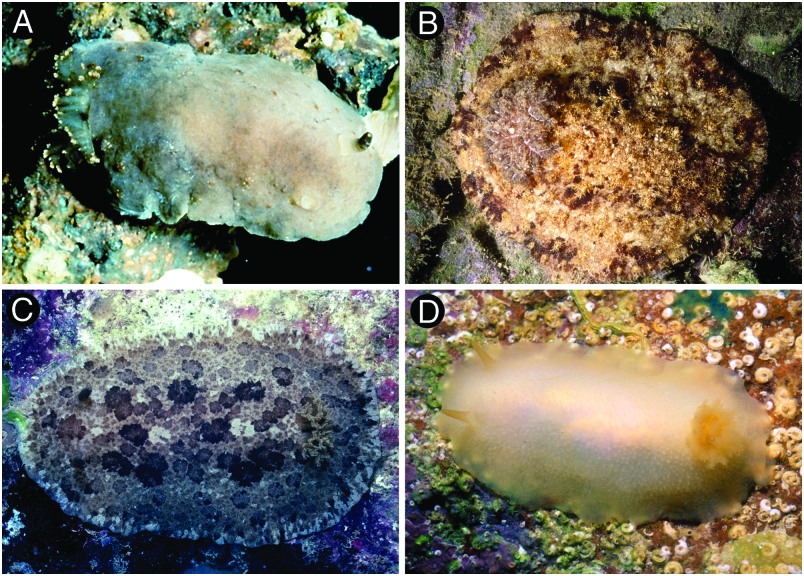
The general colour of the living animals is uniformly cream or pale yellow ( Fig. 34D View Figure 34 ). The rhinophores and gill are yellow or cream, somewhat darker than the dorsum. The viscera are visible through the dorsal skin. The whole dorsum is covered with small, rounded tubercles ( Fig. 44D View Figure 44 ). The largest tubercles are situated in the central region of the body. The rhinophoral and branchial sheaths have tubercles similar to those on the rest of the dorsum. There are seven unipinnate branchial leaves, forming a circle. The anal papilla is situated in the centre of the branchial circle of leaves. The rhinophores are elongate and almost smooth, with several irregular and inconspicuous lamellae ( Fig. 45A View Figure 45 ).
Ventrally the anterior border of the foot is grooved but not notched ( Fig. 45E View Figure 45 ). There are no oral tentacles, but two blunt prolongations on both sides of the mouth area.
Anatomy
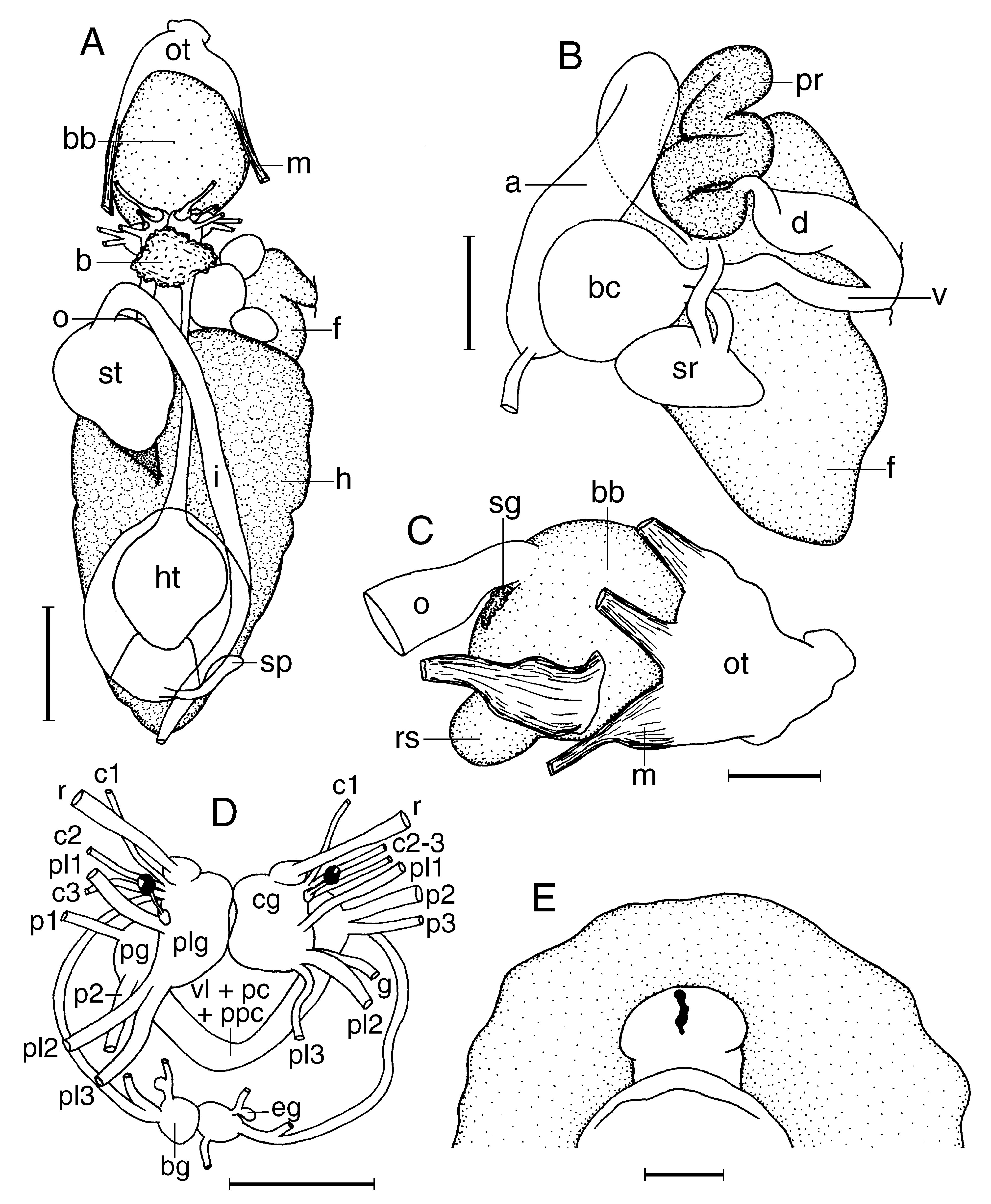
The posterior end of the glandular portion of the oral tube has six strong retractor muscles ( Fig. 46C View Figure 46 ) which attach to the body wall. The oval, muscular buccal bulb has two large additional muscles attached; two short salivary glands connect with it at each side of the oesophageal junction. The buccal bulb is longer than the glandular portion of the oral tube. The labial cuticle is smooth. The radular formula is 33 ¥ 51.0. 51 in a 10-mm long specimen. Rachidian teeth are absent. The lateral teeth are hamate and lack denticles ( Fig. 44A View Figure 44 ). The teeth from the middle portion of the half-row are larger than those closer to the medial portion of the radula ( Fig. 44B View Figure 44 ). The outermost teeth are smaller and also lack denticles ( Fig. 44C View Figure 44 ). The oesophagus is short and connects directly to the stomach ( Fig. 46A View Figure 46 ).
The ampulla is very long and folded ( Fig. 46B View Figure 46 ). It branches into a short oviduct and the prostate. The oviduct enters the female gland mass near to its centre. The prostate is tubular and connects with a short duct that narrows and expands again into the large ejaculatory portion of the deferent duct. The penis is unarmed ( Fig. 45B View Figure 45 ). The muscular deferent duct opens into a common atrium with the vagina. The vagina is long. At its proximal end it joins the bursa copulatrix. From the bursa copulatrix leads another duct connecting to the uterine duct and the seminal receptacle. The bursa copulatrix is oval in shape, about twice as large as the seminal receptacle.
In the central nervous system ( Fig. 46D View Figure 46 ) the cerebral and pleural ganglia are fused and distinct from the pedal ganglia. There are three cerebral nerves leading from each cerebral ganglion and three pleural nerves leading from each pleural ganglion. There is no separate abdominal ganglion on the right side of the visceral loop. The buccal ganglia are near to the rest of the central nervous system, joined to the cerebral ganglia by two relatively short nerves. Gastrooesophageal, rhinophoral and optical ganglia are present. The pedal ganglia are clearly separated, having two nerves leading from the left ganglion and three from the right one. The pedal and parapedal commissures are enveloped together with the visceral loop.
The circulatory system ( Fig. 46A View Figure 46 ) consists of a large heart and a blood gland situated in front of the central nervous system.
Remarks
Conualevia marcusi appears to be different from Conualevia alba , the other member of the genus, by its external morphology and anatomy. According to Collier & Farmer (1964), C. alba is a much thinner animal than C. marcusi , and more delicate in appearance and the mantle glands of C. alba are more evident. In addition, the rhinophores of C. alba are longer relative to their width than those of C. marcusi , and C. alba has half as many branchial leaves as C. marcusi . Anatomically, the main difference between these two species is the arrangement of the bursa copulatrix and the seminal receptacle, which are on opposing sides in C. marcusi and on the same side in C. alba .


| T |
Tavera, Department of Geology and Geophysics |
No known copyright restrictions apply. See Agosti, D., Egloff, W., 2009. Taxonomic information exchange and copyright: the Plazi approach. BMC Research Notes 2009, 2:53 for further explanation.
|
Kingdom |
|
|
Phylum |
|
|
Class |
|
|
Order |
|
|
Family |
|
|
Genus |
Conualevia marcusi
| Valdés, Ángel 2002 |
Conualevia marcusi
| Collier CL & Farmer WM 1964: 383 |