
Aphelodoris antillensis, BERGH, 1879
|
publication ID |
https://doi.org/10.1046/j.1096-3642.2002.00039.x |
|
DOI |
https://doi.org/10.5281/zenodo.5106569 |
|
persistent identifier |
https://treatment.plazi.org/id/03927F0E-FFC0-607B-FF65-FC766E57D102 |
|
treatment provided by |
Carolina |
|
scientific name |
Aphelodoris antillensis |
| status |
|
APHELODORIS ANTILLENSIS BERGH, 1879 View in CoL
( FIGS 4G View Figure 4 , 26 View Figure 26 , 27 View Figure 27 )
Aphelodoris antillensis Bergh, 1879: 108–113 View in CoL .
Doris bistellata Verrill, 1900: 548 View in CoL , pl. 66, fig. 2.
Type material
The type material of Aphelodoris antillensis could not be located at ZMUC (K. Jensen, pers. comm.) and is presumed lost .
Additional material
Off ferry dock , Puerto Morelos, South of Cancún, Quintana Roo, Mexico, 28 March 1985, one specimen, 10 mm preserved length, leg. T. M. Gosliner ( CASIZ 071876 ). Burger King Reef , near Soto’s Reef , South of West Bay, Grand Cayman Island, Cayman Islands, 8 May 1991, one specimen, 18 mm long, leg. J. Hamann ( CASIZ 077289 ) .
External morphology
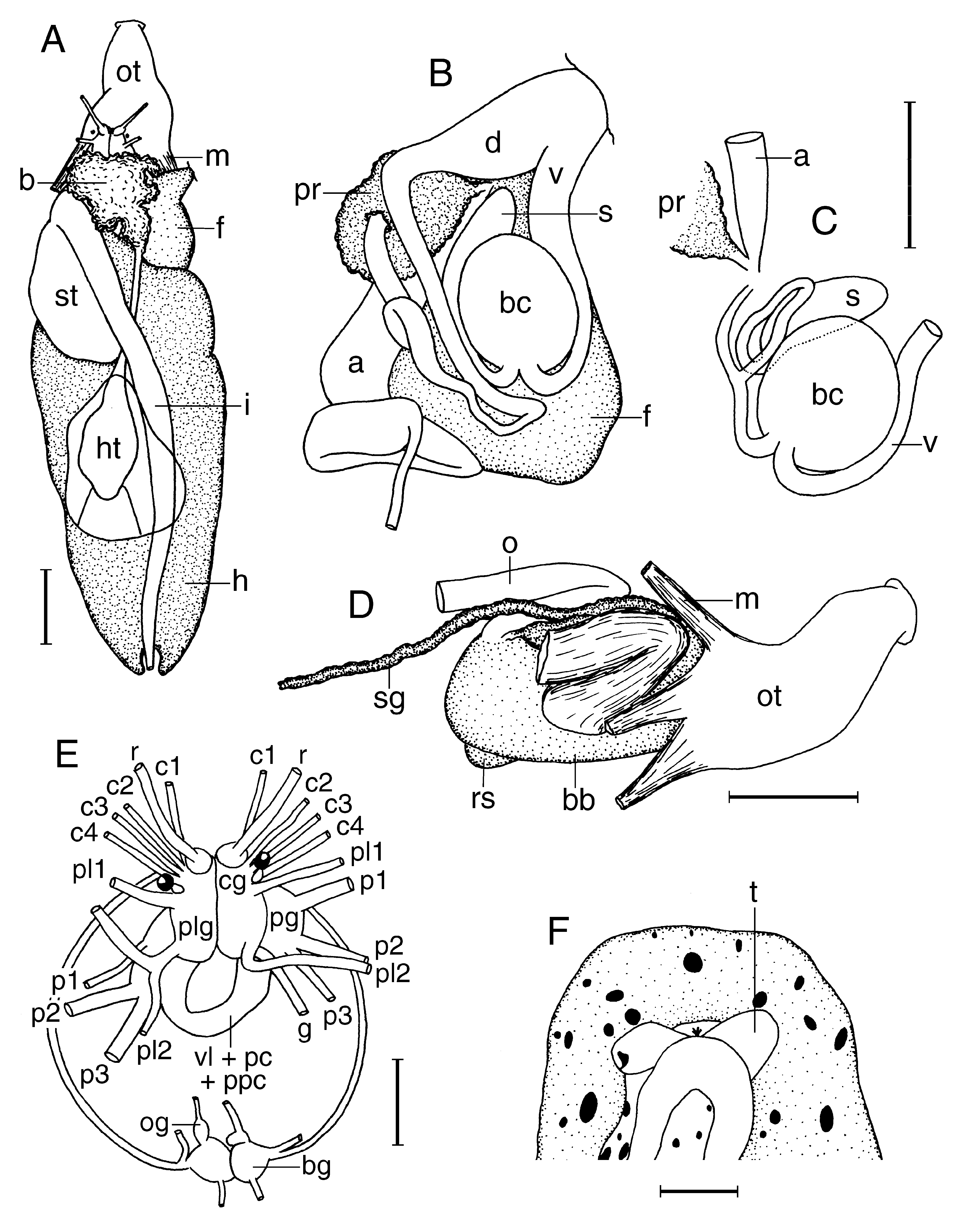
The background colour is translucent pale cream, with numerous opaque white, yellow and brown spots ( Fig. 4G View Figure 4 ). The arrangement, size and abundance of these spots is extremely variable. This variability has been described and illustrated in detail by Hamann (1992). The rhinophores and gill are also translucent pale cream, having brown, yellow or opaque white spots, which vary in size and arrangement. The dorsum is smooth, bearing a few low and soft tubercles. The rhinophoral and branchial sheaths have no tubercles. There are six bipinnate branchial leaves, forming a circle. The anal papilla is situated in the centre of the branchial circle of leaves. The rhinophores are elongate, having nine lamellae in a 10-mm preserved length specimen.
Ventrally there are two large, blunt and grooved oral tentacles ( Fig. 26F View Figure 26 ). The anterior border of the foot is grooved but not notched.
Anatomy
The posterior end of the glandular portion of the oral tube has six strong retractor muscles ( Fig. 26D View Figure 26 ), which attach to the body wall. The oval, muscular buccal bulb has two large additional muscles attached; two long salivary glands connect with it at each side of the oesophageal junction. The buccal bulb is as long as the glandular portion of the oral tube. The labial cuticle is smooth. The radular formula is 31 ¥ 43.0. 43 in a 10-mm preserved length specimen. Rachidian teeth are absent. The innermost lateral teeth are triangular, having a long cusp with 5–6 denticles ( Fig. 27A View Figure 27 ). The following teeth are smooth. The teeth from the middle portion of the half-row are larger than those closer to the medial portion of the radula ( Fig. 27B View Figure 27 ). The outermost teeth are smaller and also lack denticles ( Fig. 27C View Figure 27 ). The oesophagus is short and connects directly to the stomach.
The ampulla is very long and convoluted ( Fig. 26B View Figure 26 ). It branches into a short oviduct and the prostate. The oviduct enters the female gland mass near to its centre. The prostate is short and flattened. It connects with a long duct that narrows and expands again into the large ejaculatory portion of the deferent duct. The muscular deferent duct opens into a common atrium with the vagina. The vagina is long. At its proximal end it joins the bursa copulatrix. From the bursa copulatrix leads another duct connecting to the uterine duct and the seminal receptacle ( Fig. 26C View Figure 26 ). The bursa copulatrix is oval in shape, about four times as large as the seminal receptacle.
In the central nervous system ( Fig. 26E View Figure 26 ) the cerebral and pleural ganglia are fused and distinct from the pedal ganglia. There are four cerebral nerves leading from each cerebral ganglion and two pleural nerves leading from each pleural ganglion. There is no separate abdominal ganglion on the right side of the visceral loop. The buccal ganglia are near to the rest of the central nervous system, joined to the cerebral ganglia by two relatively short nerves. Gastrooesophageal, rhinophoral and optical ganglia are present. The pedal ganglia are clearly separated, having three nerves each one. The pedal and parapedal commissures are enveloped together with the visceral loop.
The circulatory system ( Fig. 26A View Figure 26 ) consists of a large heart and a blood gland situated behind the central nervous system.
Remarks
This common Caribbean species was described by Bergh (1879) based on several preserved specimens from St. Thomas, Virgin Islands. Ev. Marcus & Er. Marcus (1963) illustrated and described living animals for the first time. Hamann (1992) redescribed A. antillensis and synonymized it with Doris bistellata Verrill, 1900 .


| ZMUC |
Zoological Museum, University of Copenhagen |
No known copyright restrictions apply. See Agosti, D., Egloff, W., 2009. Taxonomic information exchange and copyright: the Plazi approach. BMC Research Notes 2009, 2:53 for further explanation.
|
Kingdom |
|
|
Phylum |
|
|
Class |
|
|
Order |
|
|
Family |
|
|
Genus |
Aphelodoris antillensis
| Valdés, Ángel 2002 |
Doris bistellata
| Verrill AE 1900: 548 |
Aphelodoris antillensis
| Bergh R 1879: 113 |