
Thordisa rubescens, Behrens & Henderson, 1981
|
publication ID |
https://doi.org/ 10.1046/j.1096-3642.2002.00039.x |
|
DOI |
https://doi.org/10.5281/zenodo.5110235 |
|
persistent identifier |
https://treatment.plazi.org/id/03927F0E-FFC7-6062-FF21-FA536F92D5F4 |
|
treatment provided by |
Carolina |
|
scientific name |
Thordisa rubescens |
| status |
|
THORDISA RUBESCENS BEHRENS & HENDERSON, 1981 View in CoL ( FIGS 4F View Figure 4 , 21-23 View Figure 21 View Figure 22 View Figure 23 )
Thordisa rubescens Behrens & Henderson, 1981: 120– 124 View in CoL , figs 1-7, 13, 14.
Type material
Big Kelp Reef, Paradise Cove, Los Angeles County, California, USA, 17 October 1979, 67 mm preserved length, leg. R. Henderson ( CASIZ 015860).
Additional material
Off Los Angeles, Los Angeles County, California, USA., June 1989, one specimen, 47 mm preserved length, leg. R. Fay ( CASIZ 068976).
External morphology
The background colour of the living animals is bright red-orange ( Fig. 4F View Figure 4 ). The dorsum is covered with gold flecks forming a halo around the branchial pit, a middorsal stripe and half crescents posterior to the rhinophores. The intensity of this pattern varies between individuals. In some specimens there are small black and opaque white spots. There is a black spot on top of the largest dorsal papillae. The rhinophores are orange to brown, with several irregular white spots and a white apex. The branchial leaves are the same colour as the dorsum. The whole dorsum is covered with soft and inflated papillae of various shapes and sizes ( Fig. 21D View Figure 21 ). The papillae are contracted when the animal is under stress ( Behrens & Henderson, 1981), and are surrounded by irregularly protruding spicules. Some larger papillae are randomly distributed among the others. The rhinophoral and branchial sheaths have papillae similar to those on the rest of the dorsum. There are six tripinnate branchial leaves. The anal papilla is situated in the centre of the branchial circle of leaves. The rhinophores are elongate, having 20 lamellae in a 47-mm preserved length specimen.
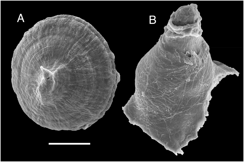
Ventrally the anterior border of the foot is grooved and notched ( Fig. 22F View Figure 22 ). The oral tentacles are conical.
Anatomy
The posterior end of the glandular portion of the oral tube has six strong retractor muscles ( Fig. 22D View Figure 22 ) which attach to the body wall. The oval, muscular buccal bulb has two large additional muscles attached. Two short salivary glands connect with the buccal bulb at each side of the oesophageal junction. The buccal bulb is as long as the glandular portion of the oral tube. The labial cuticle is smooth. The radular formula is 39 ¥ 40.0. 40 in a 47-mm long specimen. Rachidian teeth are absent. The lateral teeth are hamate and lack denticles ( Fig. 21A View Figure 21 ). The teeth from the middle portion of the half-row are larger than those closer to the medial portion of the radula ( Fig. 21B View Figure 21 ). The outermost teeth are smaller and have a number of small denticles ( Fig. 21C View Figure 21 ). The oesophagus is long and connects directly to the stomach.
The ampulla is long and folded ( Fig. 22C View Figure 22 ). It branches into a short oviduct and the prostate. The oviduct enters the female gland mass near to its centre. The prostate is large and flattened. It has two different portions that are clearly distinguishable in colour and texture. The prostate connects with a long duct that expands into the ejaculatory portion of the deferent duct ( Fig. 22B View Figure 22 ). The penis is armed with a series of large hooks, which have a wide and flat base and a curved cusp ( Fig. 23A View Figure 23 ). The muscular deferent duct opens into a common atrium with the vagina. At the vaginal connection with the atrium there are two small accessory glands attached, and two small sacs each containing a short and irregular hard structure ( Fig. 23B View Figure 23 ). At its proximal end the vagina joins the bursa copulatrix. From the bursa copulatrix leads another duct connecting to the uterine duct and the seminal receptacle. The bursa copulatrix is oval in shape, about 15 times as large as the elongate seminal receptacle.
In the central nervous system ( Fig. 22E View Figure 22 ) the cerebral and pleural ganglia are fused and distinct from the pedal ganglia. There are three cerebral nerves leading from each cerebral ganglion and three pleural nerves leading from each pleural ganglion. There is no separate abdominal ganglion on the right side of the visceral loop. The buccal ganglia are near to the rest of the central nervous system, joined to the cerebral ganglia by two relatively long nerves. Gastro-oesophageal, rhinophoral and optical ganglia are present. The pedal ganglia are clearly separated, having two nerves leading from each one. The pedal and parapedal commissures are enveloped together with the visceral loop.
The circulatory system ( Fig. 22A View Figure 22 ) consists of a large heart and two blood glands situated in front of and behind the central nervous system.
Remarks
This is a well-known species of Thordisa described from California by Behrens & Henderson (1981). It was included in the analysis because it is the only species described so far that has penial hooks. Other features of this species agree with the original description of the genus (see Behrens & Henderson, 1981).


| R |
Departamento de Geologia, Universidad de Chile |
No known copyright restrictions apply. See Agosti, D., Egloff, W., 2009. Taxonomic information exchange and copyright: the Plazi approach. BMC Research Notes 2009, 2:53 for further explanation.
|
Kingdom |
|
|
Phylum |
|
|
Class |
|
|
Order |
|
|
Family |
|
|
Genus |
Thordisa rubescens
| Valdés, Ángel 2002 |
Thordisa rubescens
| Behrens & Henderson 1981: 120 - 124 |