
Peltodoris atromaculata, BERGH, 1880
|
publication ID |
https://doi.org/10.1046/j.1096-3642.2002.00039.x |
|
DOI |
https://doi.org/10.5281/zenodo.5110237 |
|
persistent identifier |
https://treatment.plazi.org/id/03927F0E-FFDD-607A-FCE2-FA4B6BBAD002 |
|
treatment provided by |
Carolina |
|
scientific name |
Peltodoris atromaculata |
| status |
|
PELTODORIS ATROMACULATA BERGH, 1880 View in CoL View at ENA
( FIGS 4H View Figure 4 , 28 View Figure 28 , 29 View Figure 29 )
Peltodoris atromaculata Bergh, 1880: 45–46 View in CoL .
Type material
SYNTYPE: Naples , Italy, spring of 1880, one specimen, 34 mm preserved length ( ZMUC GAS-2054 )
Additional material
Islas Medas, La Escala, west coast of Gerona, Spain, three specimens, 25–34 mm preserved length, leg. T. M. Gosliner ( CASIZ 099147). Cala Salada, Ibiza, Spain, one specimen, 49 mm preserved length, leg. A. Valdés ( CASIZ 119474). 1 km east of Caloura, Ilha São Miguel, Azores, Portugal, eight specimens, 44–67 mm preserved length, leg. T. M. Gosliner ( CASIZ 072584).
External morphology
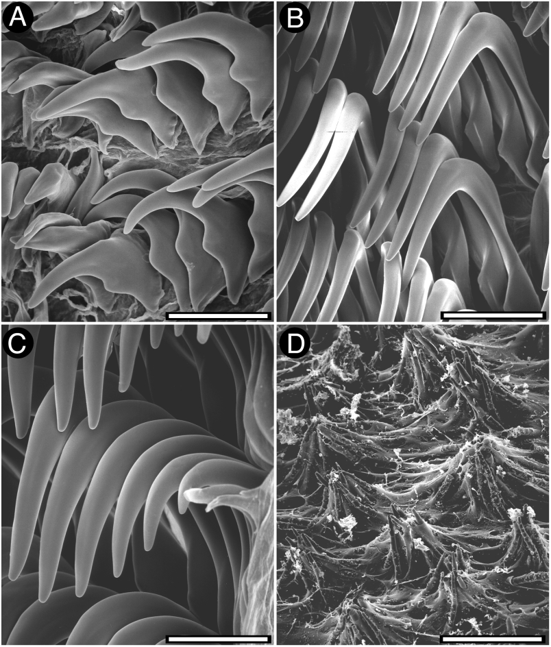
The general colour of the living animals is whitish to pale cream ( Fig. 4H View Figure 4 ). There is a number of dark brown or black large patches distributed on the dorsum, varying in shape and size. The rhinophores and gill are white or pale cream. The branchial leaves have some small dark brown or black spots. The whole dorsum is covered with small, conical tubercles, which have spicules protruding on their dorsal surface ( Fig. 28D View Figure 28 ). The largest tubercles are situated in the central region of the body. The rhinophoral and branchial sheaths have tubercles similar to those of the rest of the dorsum. There are six tripinnate branchial leaves, forming a circle. The anal papilla is situated in the centre of the branchial circle of leaves. The rhinophores are elongate, having 22 lamellae in a 52-mm preserved length specimen.
Ventrally there are two short and conical oral tentacles ( Fig. 29F View Figure 29 ). The anterior border of the foot is grooved and notched.
Anatomy
The posterior end of the glandular portion of the oral tube has six strong retractor muscles ( Fig. 29D View Figure 29 ) which attach to the body wall. The oval, muscular buccal bulb has two additional muscles attached; two long salivary glands connect with it at each side of the oesophageal junction. The buccal bulb is longer than the glandular portion of the oral tube. The labial cuticle is smooth. The radular formula is 22 ¥ 48.0. 48 in a 54-mm preserved length specimen. Rachidian teeth are absent. The inner lateral teeth are short, having a long, curved cusp and lacking denticles ( Fig. 28A View Figure 28 ). They also have a secondary, short and blunt cusp situated behind the main cusp. The teeth from the middle portion of the half-row are hamate, long and larger than those closer to the medial portion of the radula ( Fig. 28B View Figure 28 ). The outermost teeth are smaller and also smooth ( Fig. 28C View Figure 28 ). The oesophagus is short and connects directly to the stomach ( Fig. 29A View Figure 29 ).
The ampulla is long and thin, and branches into a short oviduct and the prostate ( Fig. 29C View Figure 29 ). The oviduct enters the female gland mass near to its centre. The prostate is flattened, long, folded and granular ( Fig. 29B View Figure 29 ), with two differentiated portions distinguishable by their colour and texture. It connects with a long duct that narrows and expands again into the small ejaculatory portion of the deferent duct. The muscular deferent duct opens into a common atrium with the vagina. The vagina is long. Near to its proximal end it joins the bursa copulatrix. From the bursa copulatrix leads another duct that connects to the seminal receptacle and the uterine duct. The bursa copulatrix is oval in shape, about 10 times as large as the seminal receptacle.
In the central nervous system ( Fig. 29E View Figure 29 ) the cerebral and pleural ganglia are fused and distinct from the pedal ganglia. There are four cerebral nerves leading from the left cerebral ganglion and three from the right one, and three pleural nerves leading from each pleural ganglion. There is a separate abdominal ganglion on the right side of the visceral loop. The buccal ganglia are near to the rest of the central nervous system, joined to the cerebral ganglia by two relatively long nerves. Gastro-oesophageal, rhinophoral and optical ganglia are present. The pedal ganglia are clearly separated, having four nerves leading from each one. The pedal and parapedal commissures are enveloped together with the visceral loop.
The circulatory system ( Fig. 29A View Figure 29 ) consists of a large heart and a two blood glands situated in front of and behind the central nervous system.


| ZMUC |
Zoological Museum, University of Copenhagen |
| T |
Tavera, Department of Geology and Geophysics |
No known copyright restrictions apply. See Agosti, D., Egloff, W., 2009. Taxonomic information exchange and copyright: the Plazi approach. BMC Research Notes 2009, 2:53 for further explanation.
|
Kingdom |
|
|
Phylum |
|
|
Class |
|
|
Order |
|
|
Family |
|
|
Genus |
Peltodoris atromaculata
| Valdés, Ángel 2002 |
Peltodoris atromaculata
| Bergh R 1880: 46 |