
Myzostoma kymae, Summers, Mindi M., Al-Hakim, Iin Inayat & Rouse, Greg W., 2014
|
publication ID |
https://doi.org/ 10.11646/zootaxa.3873.4.1 |
|
publication LSID |
lsid:zoobank.org:pub:84F8465A-595F-4C16-841E-1A345DF67AC8 |
|
DOI |
https://doi.org/10.5281/zenodo.6138519 |
|
persistent identifier |
https://treatment.plazi.org/id/039287ED-AD53-FFC5-CF9C-F968FB48FCE8 |
|
treatment provided by |
Plazi |
|
scientific name |
Myzostoma kymae |
| status |
sp. nov. |
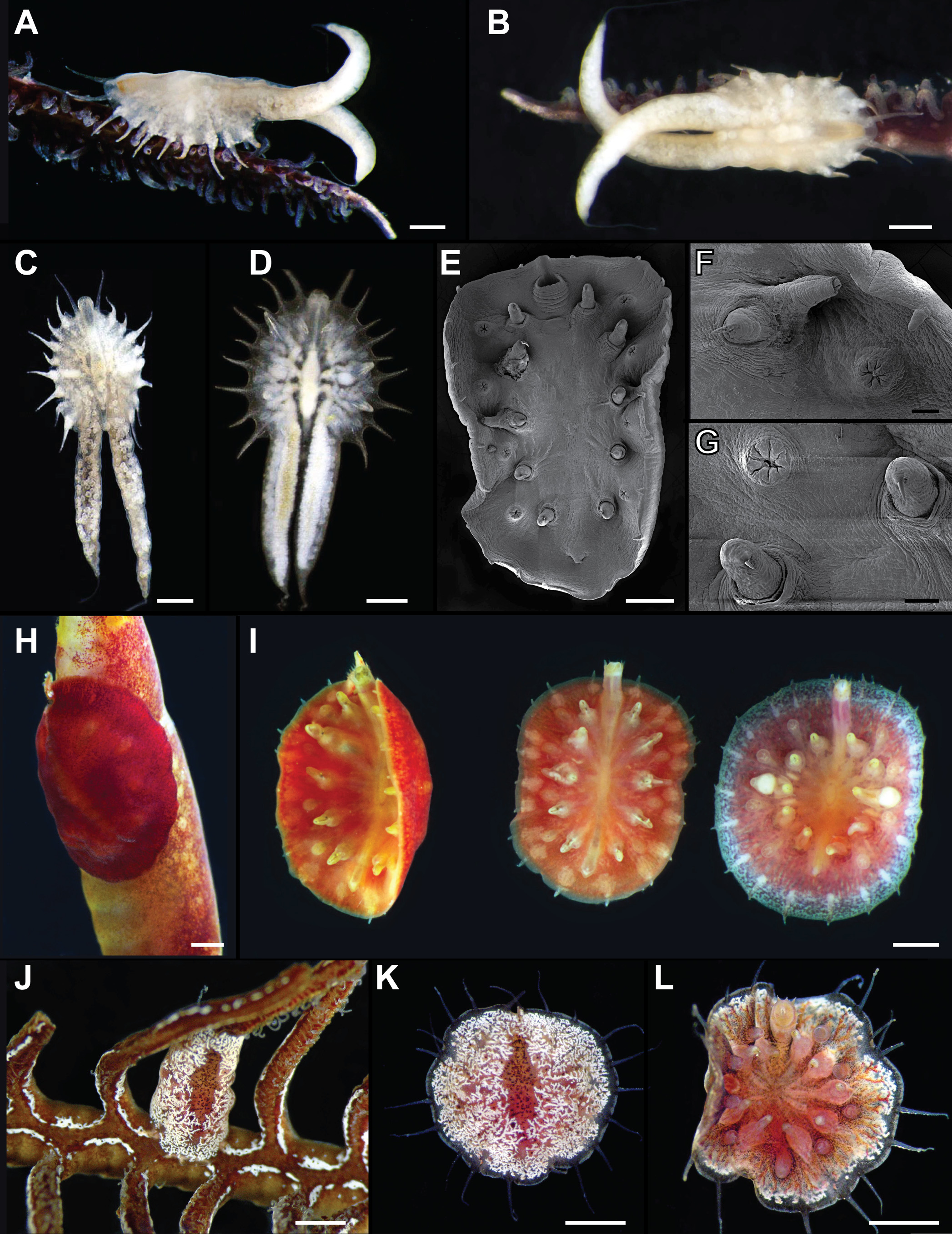
Myzostoma kymae n. sp. Summers & Rouse
Fig. 5 View FIGURE 5 E–I
Holotype: SIO-BIC A3681 hologenophore (1 spm: 95% ethanol). Madang Harbor, Papua New Guinea (5°12'27.63"S, 145°48'32.45"E), 3– 17 m. Collected at night using scuba on 4 December 2012 by MMS and GWR. Genbank (COI—KM014197).
Host. Alloeocomatella n. sp. (AH Clark) ( Comatulidae , Comatulida , Crinoidea). MNHN-IE 2013-8027 (dried voucher) & SIO-BIC E5862 (tissue subsample in 95% ethanol). Genbank (COI—KJ874980).
Paratypes: SIO-BIC A3682 paragenophore (2 spms: 1—95% ethanol; 1—mounted for SEM). Same host and locality as holotype.
Etymology. Named for Kym Vercoe, sister-in-law of GWR, in honor of her birthday.
Diagnosis and description. Holotype body oval-shaped, ~ 4 mm long, 2 mm wide following fixation. Dorsal longitudinal ridge; otherwise smooth surface ( Fig. 5 View FIGURE 5 H). Body margin with 20 short, triangular cirri ( Fig. 5 View FIGURE 5 I). Mouth and cloaca on ventral surface. Mouth and cloaca sub-terminal, in line with lateral organs. Proboscis with ~20 papillae. Paired penes in line with third pair of parapodia. Five pairs of parapodia, midway between midline and body margin. Lateral organs closer to parapodia than body margin. Color bright red in life, faded in preservative.
Remarks. This species most resembles Myzostoma viride Atkins, 1927 , which was described from the Great Barrier Reef ( Australia) associated with Comanthus annulatus (= Comanthus parvicirrus ). This description was of three green-colored specimens with a white anterior-posterior ridge on the dorsal surface and red extended proboscis with papillae. In two of the specimens studied here, the anterior and posterior margins were broadly rounded, while in the third the body tapered to a point posteriorly. We collected yellow specimens, with a dorsal ridge from Comatella nigra (Carpenter) at Lizard Island, Australia. We suggest that these species are most closely allied to the green specimens described, and that the host of Atkin’s specimens may have been Comatella (most of Atkin’s species were described from Comanthus annulatus ). Myzostoma kymae n. sp. differs from these specimens assigned to Myzostoma viride in color, placement of mouth (nearer to the body margin in M. viride ), lack of clear dorsal ridge, host use, and molecular data ( Myzostoma cf. viride published in Summers & Rouse (2014)). The paratype of M. kymae n. sp. is slightly larger, brown, and the proboscis has fewer (~7) papillae.
No known copyright restrictions apply. See Agosti, D., Egloff, W., 2009. Taxonomic information exchange and copyright: the Plazi approach. BMC Research Notes 2009, 2:53 for further explanation.