
Heligmonina kotoensis, Diouf & Daouda & Durette-Desset, 2005
|
publication ID |
https://doi.org/ 10.5281/zenodo.5402359 |
|
persistent identifier |
https://treatment.plazi.org/id/039287F8-FFB2-7D13-0D02-FB8AC1D2FA2B |
|
treatment provided by |
Marcus |
|
scientific name |
Heligmonina kotoensis |
| status |
sp. nov. |
Heligmonina kotoensis n. sp.
( Fig. 1 View FIG )
TYPE MATERIAL. — Holotype, allotype, 15. VI.2001, I. A. H. Daouda coll. ( MNHN 668 View Materials KQa) . Paratypes: 15. VI.2001, I. A. H. Daouda coll., 10, 10 ( MNHN 668 View Materials KQb) ; 21, 17 ( ZIT / IFAN 01 View Materials I), coparasites of Neoheligmonella lamaensis n. sp .
ETYMOLOGY. — After Koto: the name of the forest vil-
New Trichostrongylina (Nematoda) from Benin
TYPE LOCALITY. — Koto, Republic of Benin.
HOST. — Mastomys natalensis (Smith, 1834) ( Muridae , Murinae).
SITE. — Small intestine (SI 1-SI 2).
DESCRIPTION
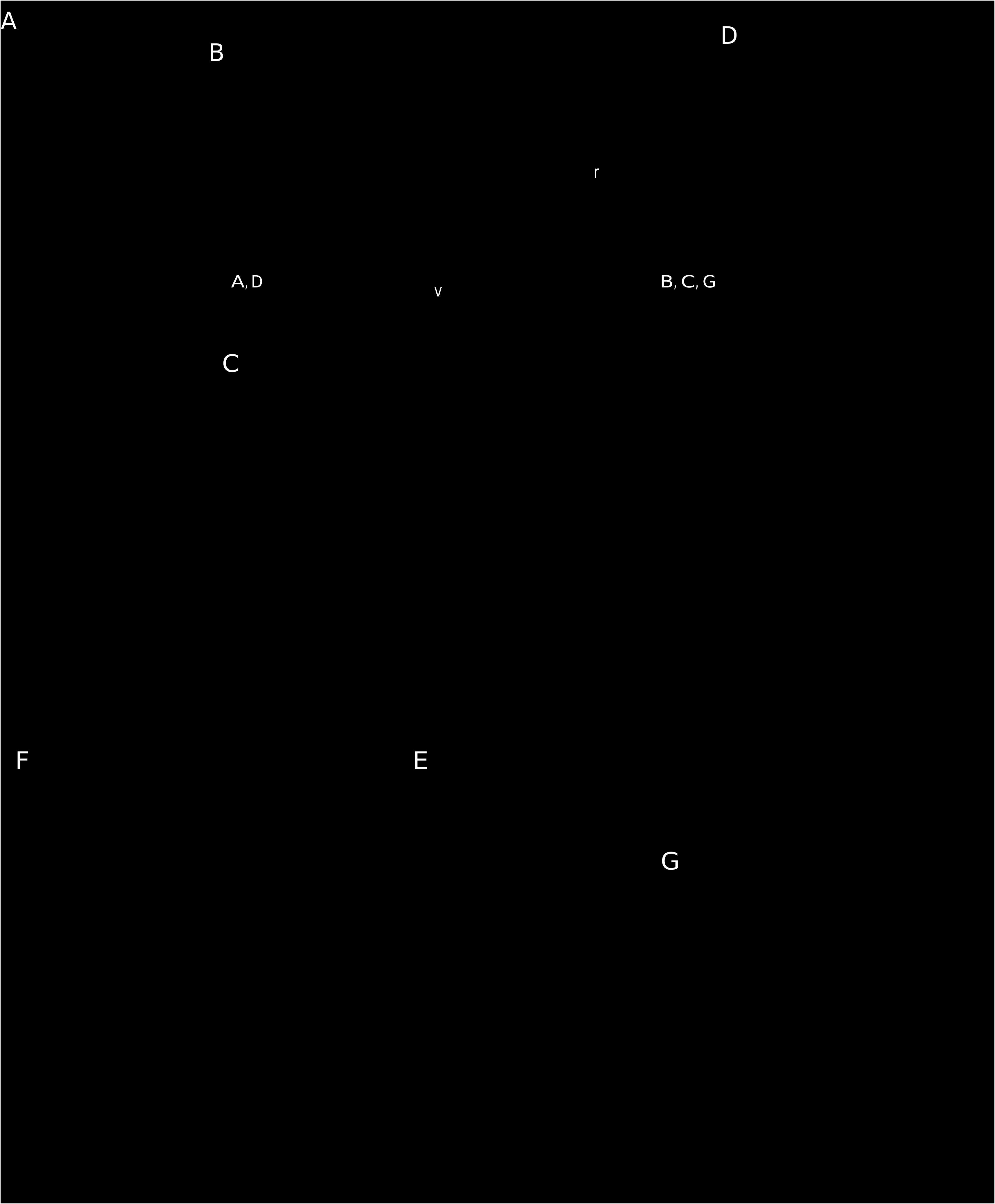
Small nematodes with body uncoiled or slightly coiled along ventral side. Excretory pore situated about at mid-region of oesophagus. Deirids posterior to excretory pore and very distant from it ( Fig. 1A View FIG ). Vestibule twice as long as sphincter. Uterus very short less than 18% of body length.
Synlophe (studied in one male and one female paratypes)
In both sexes, cuticule bearing longitudinal, uninterrupted ridges, all appearing at different levels between cephalic vesicle and end of muscular oesophagus and disappearing anterior to caudal bursal in male. In female, ventral ridges disappearing anterior to vulvar opening, dorsal ridges posterior to vulva. Number of ridges: 11 (six dorsal, four ventral and left hypertrophied) at mid-body ( Fig. 1B, C View FIG ). Double gradient of size decreasing from left to right on ventral side, and from right to left on dorsal side ( Fig. 1B, C View FIG ). In both sexes, tips of ridges orientated from right to left with axis of orientation inclined at about 70° to sagittal axis ( Fig. 1B, C View FIG ). In vulvar region, ridges orientated perpendicularly to body ( Fig. 1G View FIG ).
Holotype male
2.0 mm long, 150 μm wide. Cephalic vesicle 43 μm long, 26 μm wide. Nerve ring, excretory pore and deirids situated at 75 μm, 80 μm and 130 μm from apex, respectively. Oesophagus 225 μm long, with anterior muscular part 85 μm and posterior glandular part 140 μm ( Fig. 1A View FIG ).
| VI |
Mykotektet, National Veterinary Institute |
| ZIT |
Grusinian Academy of Sciences |
No known copyright restrictions apply. See Agosti, D., Egloff, W., 2009. Taxonomic information exchange and copyright: the Plazi approach. BMC Research Notes 2009, 2:53 for further explanation.
|
Kingdom |
|
|
Phylum |
|
|
Class |
|
|
Order |
|
|
Family |
|
|
Genus |