
Allophrys, Forster, 1869
|
publication ID |
https://doi.org/10.11646/zootaxa.4378.3.9 |
|
publication LSID |
lsid:zoobank.org:pub:6D6A7F17-1466-482A-9569-E365F3A7EA07 |
|
DOI |
https://doi.org/10.5281/zenodo.5979350 |
|
persistent identifier |
https://treatment.plazi.org/id/039287FA-CC58-A901-FF30-6686DBB2F81F |
|
treatment provided by |
Plazi |
|
scientific name |
Allophrys |
| status |
|
Key to species of Allophrys View in CoL View at ENA occurring in Vietnam
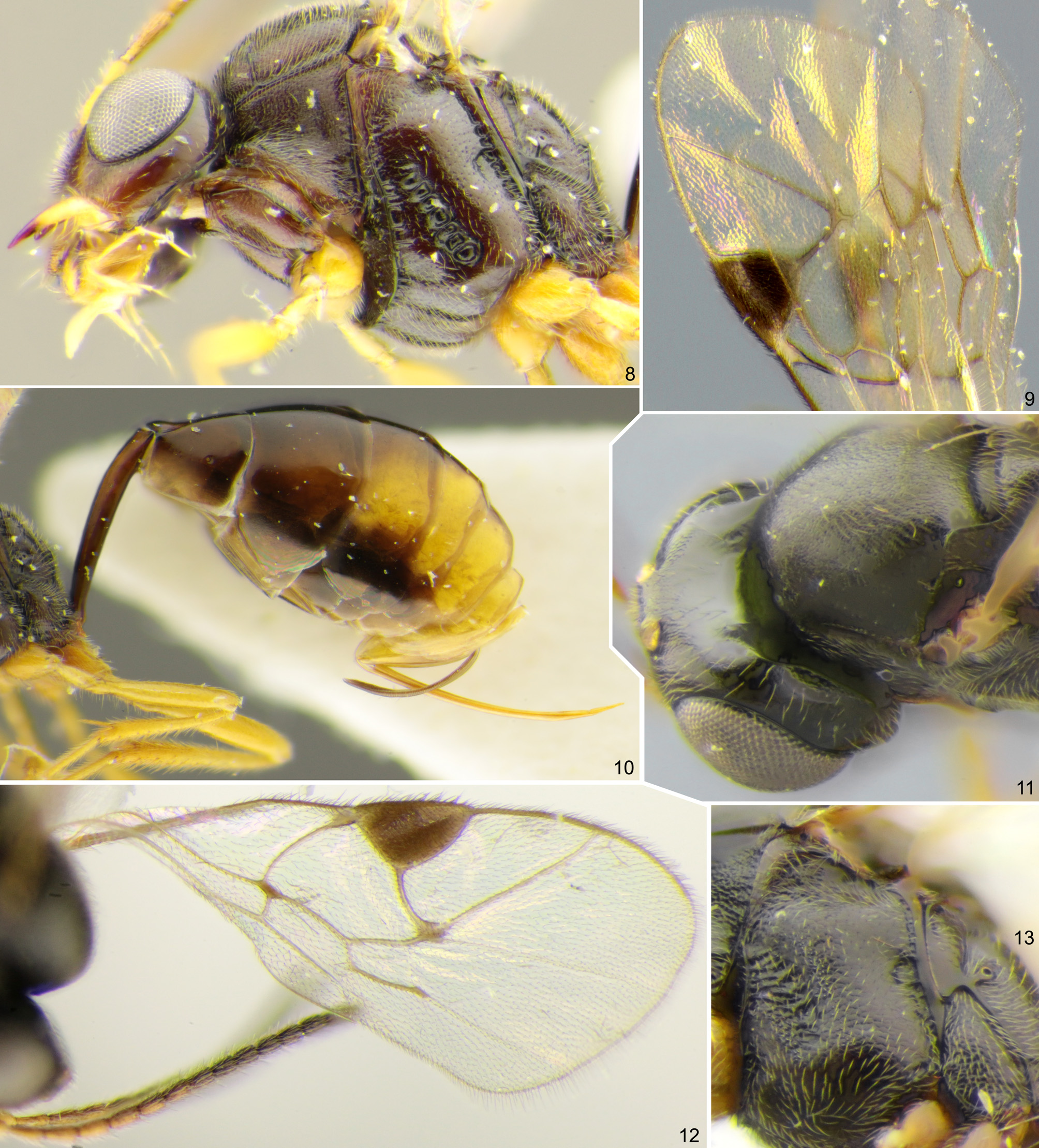
1. Vertex polished ( Fig. 11 View FIGURES 8–13 ). Mesopleuron centrally finely granulate, impunctate, dull ( Fig. 13 View FIGURES 8–13 ). Fore wing with second recurrent vein (2 m-cu) completely absent ( Fig. 12 View FIGURES 8–13 )................................................. A. occipitata Khalaim
- Vertex granulate, dull or weakly shining ( Fig. 2 View FIGURES 1–7 ). Mesopleuron smooth or granulate, but always with fine to rather strong punctures. Fore wing with second recurrent vein (2 m-cu) distinct at least in its posterior half ( Fig. 9 View FIGURES 8–13 )....................... 2
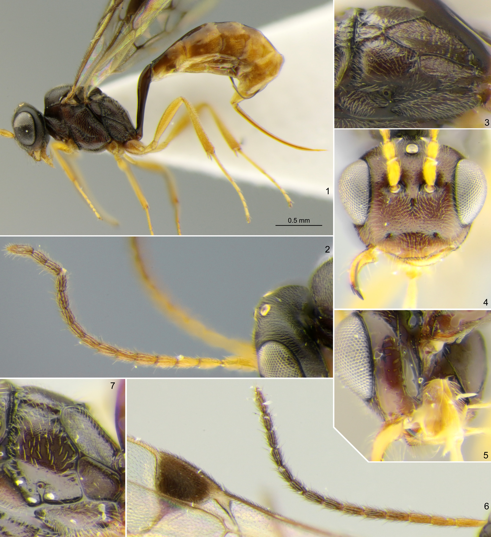
2. Subapical flagellomeres 1.6–1.8× as long as broad ( Fig. 6 View FIGURES 1–7 ). Fore wing with second recurrent vein (2 m-cu) antefurcal ( Fig. 9 View FIGURES 8–13 ). Hind wing with nervellus ( cu 1& cu-a) weakly reclivous, slanted about 70° from horizontal............ A. davichia sp. nov.
- Subapical flagellomeres 1.1–1.4× as long as broad ( Figs 2 View FIGURES 1–7 , 14 View FIGURES 14–18 ). Fore wing with second recurrent vein (2 m-cu) postfurcal or interstitial. Hind wing with nervellus ( cu 1& cu-a) strongly reclivous, slanted about 45° from horizontal ( Fig. 15 View FIGURES 14–18 ).......... 3
3. Basal area of propodeum short, 0.2–0.3× as long as apical area ( Fig. 3 View FIGURES 1–7 ). Malar space 0.7–0.8× as long as basal mandibular width. Flagellum with 14–15 flagellomeres ( Fig. 2 View FIGURES 1–7 ). Legs yellow ( Fig. 1 View FIGURES 1–7 ).......................... A. daklaka sp. nov.
- Basal area of propodeum long, about 0.8× as long as apical area ( Fig. 16 View FIGURES 14–18 ). Malar space 1.0–1.2× as long as basal mandibular width ( Fig. 14 View FIGURES 14–18 ). Flagellum with 13 flagellomeres ( Fig. 14 View FIGURES 14–18 ). At least hind coxa extensively brown basally ( Fig. 15 View FIGURES 14–18 )................................................................................................ A. tonkina sp. nov.

No known copyright restrictions apply. See Agosti, D., Egloff, W., 2009. Taxonomic information exchange and copyright: the Plazi approach. BMC Research Notes 2009, 2:53 for further explanation.
|
Kingdom |
|
|
Phylum |
|
|
Class |
|
|
Order |
|
|
Family |