
Calicnemia uenoi Asahina, 1996
|
publication ID |
https://doi.org/ 10.11646/zootaxa.4232.3.9 |
|
publication LSID |
lsid:zoobank.org:pub:2BC1013F-6106-4C66-964F-AA6C35DED784 |
|
DOI |
https://doi.org/10.5281/zenodo.6049387 |
|
persistent identifier |
https://treatment.plazi.org/id/03938786-FFEE-FFDC-D0ED-1FE3FC66F8FA |
|
treatment provided by |
Plazi |
|
scientific name |
Calicnemia uenoi Asahina, 1996 |
| status |
|
Calicnemia uenoi Asahina, 1996 View in CoL
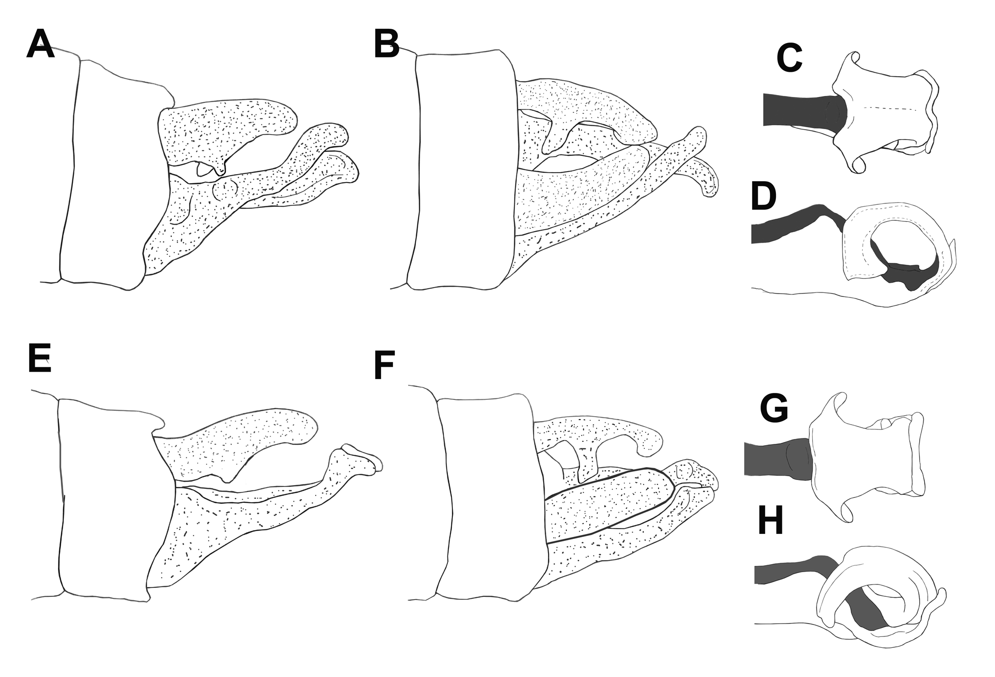
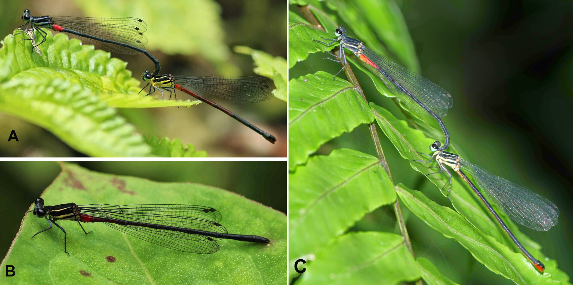
( Figures 5 View FIGURE 5 E–H; 6A–B)
Calicnemia uenoi: Asahina, 1997: 21 View in CoL –22, Figs. 14–17 (Sa Pa, Lao Cai Prov.); Do & Dang, 2007: 57, Distribution map (Sa Pa, Lao Cai Prov.).
Specimens examined. 2♂, 18.V.2015, Sa Pa, Lao Cai Prov., Tom Kompier leg.; 1♂ 1♀, 10.V.2014, Nam Bung, Yen Bai Prov., Tom Kompier leg.
Notes. Asahina (1997) provided a very poor description of C. uenoi with rough figures and without illustrations of genital ligula structure. It was uncertain which group C. uenoi belonged to. Moreover, we could not check the type specimens because the type series of C. uenoi at present cannot be found at the National Museum of Nature and Science, Tokyo, Japan ( Dow et al. 2014). Therefore, we examined several specimens of Calicnemia from its type locality (Sa Pa, Lao Cai Prov.) and compared these with the original description. Coloration and structure of the appendages of our specimens ( Figs. 5 View FIGURE 5 E–H, 6A–B) fit C. uenoi well. In our males the abdomen is black with S2 reddish. Asahina (1997) described the male as having S1–5 reddish, but in fact he only illustrated this for the female. C. uenoi appears to be closely related to C. haksik but differs in several characters of body pattern and structure of anal appendages (see Diagnosis). We provide here an improved description of both male and female and provide new detailed illustrations of appendages and ligula. Based on the structure of the ligula, C. uenoi belongs to group 2.
Redescription of male & female. Male. Head. Labium, labrum, postclypeus, anterior half of mandibles all shiny black. Anteclypeus dark brown, posterior half of mandibles and genae ivory white. Top of head matte black, the anterior half with grey pruinosity, distinctly so on antefrons, and with two indistinct oval pale spots between base of antennae and posterior ocelli. Antennae dark. Genae, labrum and clypeus with sparse long hairs, densely so on frons and sparse on posterior part of top of head. Eyes blackish on top and greenish below. Thorax ( Fig. 6 View FIGURE 6 A–B). Prothorax matte black, propleuron grey pruinose, posterior lobe with sparse long hairs. Synthorax matte black with two distinct antehumeral stripes. These stripes yellowish at immature stage ( Fig. 6 View FIGURE 6 B) as the orginal description ( Asahina 1997: 21, Fig. 14), but becoming grey pruinose with maturity ( Fig. 6 View FIGURE 6 A). Mesepimeron and mesinfraepisternum matte black. Metepisternum grey pruinose, but anterior of spiracle pale yellow and black along metapleural suture. Metepimeron grey pruinose, but anterior end with some yellow. All thoracic sclerites with sparse long hairs. Legs black with long black spines, greyish pruinosity most evident on femora; coxae brownish yellow with grey pruinose areas. Wings hyaline with black veins, 20 postnodal crossveins in both fore– and hindwings. Pterostigma brown, rhombic, covering 1.5 underlying cells. Abdomen with S1 brownish black, with some pruinosity laterally, S2 bright red, S3 with small red area along anterior suture, otherwise matte black, S4–10 all matte black, appendages black. S1–2 with sparse long hairs, S3–10 increasingly densely covered in short hairs, which are longer on the ventral side. Appendages ( Fig. 5 View FIGURE 5 E–F) entirely black, covered in dense short hairs. Cerci in dorsal view converging, almost equally thick from base to rounded tip. In lateral view with strong ventral tooth directed diagonally inward and with shallowly forked apex (difficult to see in lateral view, but obvious in posterior view) at approximately 2/5th of length, and apex drooping somewhat, along ventral side thinning somewhat until becoming somewhat bulbous and actually curved inward somewhat at apex. Cerci reaching about 3/4th of paraprocts. Paraprocts in lateral view gently narrowing until at 3/4th of their length curving upwards under 45 degree angle and apically suddenly turning down again in a small boot shape. Genital ligula structurally simple, typical of group 2 ( Fig. 5 View FIGURE 5 G–H).
Female. Head as male, but pruinosity less distinct on postfrons. Thorax color pattern as the male, but without pruinosity. Antehumeral stripe, metepisternum and metepimeron yellow, but with thick black line over metapleural suture. Long hairs much more sparse. Legs like male, but without pruinosity and with some yellow on femora. Coxae yellowish. Wings hyaline with black veins, 20 postnodal crossveins in both fore– and hindwings; pterostigma pale brown, rhomboid, shape as in male. Abdomen with S1 yellow, S2–5 orange–red, with indistinct blackish rings along sutures, S6 laterally orange–red, but with blackish dorsum and broader blackish rings at anterior and posterior end, S7–10 blackish, but ventral sclerite of S8, ovipositor and ventral side of S9–10 orange. Cerci very short, conical, black.
Measurements (mm): male, abdomen including anal appendages 35; hindwing 24.5.
female, abdomen including anal appendages 29; hindwing 23.
Diagnosis. C. uenoi can be distinguished from C. haksik as follows:
(1) In the male of C. uenoi , only S2 (and a minute part of S3) reddish ( Fig. 6 View FIGURE 6 A–B), but S 1–3 in C. haksik ( Fig. 6 View FIGURE 6 C).
(2) Cerci of C. haksik with double teeth in lateral view ( Fig. 5 View FIGURE 5 A), but only one blunt tooth in C. uenoi , although its tip shallowly incised in posterior view, ( Fig. 5 View FIGURE 5 E).
(3) Paraprocts of C. uenoi are boot–shaped at tip in lateral view, but in C. haksik the tip of paraprocts is not boot–shaped.
(4) Genital ligula of C. haksik is more strongly incised at mid–dorsum ( Fig. 5 View FIGURE 5 C) than that of C. uenoi ( Fig. 5 View FIGURE 5 G); in C. haksik the lobe of the genital ligula is broader at tip ( Fig. 5 View FIGURE 5 D) than that of C. uenoi ( Fig. 5 View FIGURE 5 H).
(5) In the female, the five anterior abdominal segments of C. uenoi are reddish ( Fig. 6 View FIGURE 6 A), but in C. haksik the abdomen is very dark or blackish dorsally, laterally shading to yellow on anterior segments and to yellow or reddish on venter of S8–10 ( Fig. 6 View FIGURE 6 C).
Distribution. Sa Pa, Lao Cai Prov.; Nam Bung, Yen Bai Prov.
No known copyright restrictions apply. See Agosti, D., Egloff, W., 2009. Taxonomic information exchange and copyright: the Plazi approach. BMC Research Notes 2009, 2:53 for further explanation.
|
Kingdom |
|
|
Phylum |
|
|
Class |
|
|
Order |
|
|
Family |
|
|
Genus |
Calicnemia uenoi Asahina, 1996
| Phan, Quoc Toan, Kompier, Tom & Karube, Haruki 2017 |
Calicnemia uenoi:
| Do 2007: 57 |
| Asahina 1997: 21 |