
Asterocheres sarsi Bandera & Conradi, 2009
|
publication ID |
https://doi.org/ 10.11646/zootaxa.3827.4.6 |
|
publication LSID |
lsid:zoobank.org:pub:2C480B30-8005-4F66-8FB3-5C54A44F68F3 |
|
DOI |
https://doi.org/10.5281/zenodo.5613651 |
|
persistent identifier |
https://treatment.plazi.org/id/03938795-FFDF-1C5C-37C2-9111C2E2974B |
|
treatment provided by |
Plazi |
|
scientific name |
Asterocheres sarsi Bandera & Conradi, 2009 |
| status |
|
Asterocheres sarsi Bandera & Conradi, 2009
( Figs 6–8 View FIGURE 6 View FIGURE 7 View FIGURE 8 )
Material examined. (a) holotype (preserved in ethanol, deposited in ZMO under registration number ZMO F21600a), and 10 females (preserved in ethanol, deposited in ZMO under registration number ZMO F21600b), collected from the bottom-residue of a large collecting-bottle containing a number of different invertebrate animals in RauØ by G. O. Sars. (b) 1 female (labelled as Ascomyzom latum and preserved in ethanol, deposited in ZMUC under registration number ZMUC-CRU-4937), collected from Kapt. Ørssad (58º11’NB 4ºØ, L. 658 ~).
Description of adult female. Body ( Fig. 6 View FIGURE 6 A) cyclopiform, slender with cephalothorax oval and cylindrical urosome. Mean body length from anterior margin of rostrum to posterior margin of caudal rami 740 µm (710-780 µm); maximum width 450 µm (400-480 µm), based on 4 specimens. Prosome comprising cephalothorax (fully incorporating first pedigerous somite) and three free pedigerous somites. Somites bearing legs 2–3 broad; epimeral areas with posterolateral angles rounded (leg 2) or pointed (leg 3) ( Fig. 6 View FIGURE 6 A). Somite bearing leg 4 much smaller and narrower than preceding ones and largely concealed under somite bearing leg 3.
Urosome 4-segmented, comprising leg 5-bearing somite, genital double-somite and two free abdominal somites. Urosome ornamented with large epicuticular spinules arranged in irregular pattern ( Fig. 6 View FIGURE 6 A, C) in all urosomites except for leg-5 bearing somite which shows the spinules in overlapping rows pattern ( Fig. 6 View FIGURE 6 C). Genital double-somite ( Fig. 6 View FIGURE 6 C) slightly wider than long; paired genital apertures bipartite, each comprising lateroventral copulatory pore and dorsolateral gonopore (oviduct opening); lateral margins with setular tufts in distal third (posterior to genital apertures).
Caudal rami ( Fig. 6 View FIGURE 6 C) about twice longer than wide (measured along outer margin); armed with seven setae; seta I present ( Fig. 6 View FIGURE 6 C), minute and displaced onto lateral surface, setae II–VII all arranged around posterior margin with setae II and VII slightly displaced onto dorsal surface. All of them plumose except for seta I which is naked ( Fig. 6 View FIGURE 6 A).
Antennule ( Fig. 6 View FIGURE 6 B) 21-segmented, about 395 µm long. Segmental fusion pattern as follows (Roman numerals indicating ancestral segments): 1(I)-2, 2(II)-2, 3(III)-2, 4(IV)-2, 5(V)-2, 6(VI)-2, 7(VII)-2, 8(VIII)-2, 9(IX–XII)-7, 10(XIII)-1+spine, 11(XIV)-1+spine, 12(XV)-2, 13(XVI)-2, 14(XVII)-2, 15(XVIII)-1, 16(XIX)-1, 17(XX)-2, 18(XXI)-2+ae, 19(XXII–XXIII)-3, 20(XXIV), 21(XXV–XXVIII)-6. Segment 10(XIII) reduced, forming incomplete sclerite partly overlapped by distal expansion of compound segment 9(IX–XII).
Antenna biramous ( Fig. 6 View FIGURE 6 D), about 260 µm long. Coxa unarmed, with tufts of spinules. Basis unarmed, with fine spinule rows in lateral inner margin and longer spinule rows medially as shown in Figure 6 View FIGURE 6 D. Exopod onesegmented, slender, about 2.5 times longer than wide; with two small lateral setae and one long terminal seta. Endopod three-segmented; proximal segment elongated, ornamented with lateral and medial rows of spinules as figured; middle segment produced distally on medial side but articulating with distal segment proximally on lateral side, bearing one naked subterminal seta; distal segment with two pinnate setae, one of them subterminal, and one terminal claw with rows of fine spinules; surface of distal segment with long setules.
Siphon long and slender, about 230 µm long, reaching nearly to posterior margin of intercoxal sclerite of leg 1. Mandible ( Fig. 7 View FIGURE 7 B) comprising stylet-like gnathobase and slender two-segmented palp. Proximal segment of palp longest, ornamented with rows of spinules on lateral and distal margins; distal segment shortest, with two plumose, unequal apical setae. Stylet located in oral cone, formed by anterior labrum and posterior labium. Stylet with denticulate margin subapically ( Fig. 7 View FIGURE 7 B).
Maxillule ( Fig. 7 View FIGURE 7 A) bilobed; praecoxal gnathobase (inner lobe) distinctly larger than palp (outer lobe). Praecoxal endite conical, ornamented with setules proximally and spinules distally on the lateral margin and a row of long setules medially; armed with one short and naked and four long but unequal plumose setae, the three longer with minute spines distally. Palp reduced, about three times shorter than praecoxal endite, with one short naked seta and three longer pinnate setae.
Maxilla ( Fig. 7 View FIGURE 7 D) two-segmented but with partial transverse surface suture on syncoxa (proximal segment) possibly marking plane of praecoxa-coxa fusion; praecoxal portion bearing flaccid aesthetasc-like element medially, representing tubular extension of external opening of maxillary gland; coxal portion unarmed but ornamented with a row of spinules medially as figured. Basis claw-like, more or less straight but recurved towards the apex; armed with one seta at middle length.
Maxilliped ( Fig. 7 View FIGURE 7 C) five-segmented, comprising short syncoxa, long basis and three-segmented endopod. Syncoxa with one seta and a row of spinules distally. Basis with rows of spinules on distal outer and inner margin and one seta at middle length. First endopodal segment ornamented with spinules on lateral margin and armed with two medial setae and one short distal seta; second endopodal segment bearing one long barbed seta; third endopodal segment bearing recurved terminal claw plus additional apical pinnate seta. Distal margin of claw provided with a row of minute spinules.
Swimming legs 1–4 ( Figs. 8 View FIGURE 8 A-D) biramous, with three-segmented rami. Intercoxal sclerite present in legs 1–4, ornamented with patches of spinules in legs 1–3.
Spine and seta formula as Table 3 View TABLE 3 .
Coxae ornamented with spinule rows around outer margin; inner coxal seta absent in leg 1 (ornamented with a crown of spinules as figured), long and plumose in legs 2-3 and short and bare in leg 4. Bases of legs 1-3 with spinules around inner margin; outer seta long and naked in leg 1, long and plumose in legs 2-3 and short and smooth in leg 4. Outer spines of exopodal segments in legs 1-4 bilaterally serrate. Lateral margins of exopodal segments with minute serrations or spinular rows; those of endopodal segments with rows of setules.
Fifth leg ( Fig. 6 View FIGURE 6 C) with protopod incorporated into somite; outer basal seta displaced to laterodorsal surface. Free segment (exopod) elongate-oval, with one short naked seta subterminal and two long plumose setae distally; outer and inner margins with spinules.
Sixth leg ( Fig. 6 View FIGURE 6 C) represented by paired opercular plates closing off gonopores on genital double-somite; armed each with one plumose seta and one spiniform element.
Distribution. Norway ( Sars 1915).
Remarks. This species was poorly described by G. O. Sars (1915) as Ascomyzon latum . However, as Bandera and Conradi (2009b) pointed out, the specimens that Sars stated to be identical to Cyclopicera lata (Brady) and described as Ascomyzon latum were actually different from A. echinicola (= A. violaceus ) and Cyclopicera lata . These authors redescribed C. lata as Asterocheres latus and named the species described by Sars as Ascomyzon latum as Asterocheres sarsi but they did not redescribed this species.
Asterocheres sarsi is characterized by the possession of 21 segments in the female antennule, 2-segmented mandibular palp, oral cone reaching to the posterior margin of intercoxal plate of leg 1, inner seta on coxa of leg 1 absent and body cyclopiform, with cephalotorax oval and cyclindrical urosome and epimeral areas of somite bearing leg 3 with posterolateral angles pointed. These features are only shared by another species, A. eugenioi , described above; however, the length of the caudal rami differs in both species. While A. sarsi presents caudal rami that are twice longer than wide, A. eugenioi has a shorter caudal rami, about 1.5 times longer than wide. In A. sarsi , caudal seta I is present but it is absent A. eugenioi . Asterocheres sarsi shows the antenna, including the claw, much more ornamented with spinules and setules than A. eugenioi ; and the urosomal somites with large epicuticular spinules arranged in irregular pattern in all urosomites except for leg-5 bearing somite which shows the spinules in overlapping rows pattern. This kind of ornamentation has not been observed in the urosome of A. eugenioi .
As for the fifth leg, in A. eugenioi the seta of the protopodal segment and those of the free segment are naked. However, in A. sarsi the seta of the protopodal segment is plumose, the two longer setae belonging to the free segment are barbed and the shorter one is naked.
The stylet of the mandible also serves to separate these species. A. sarsi has a stylet with the tip sharply pointed; in contrast, A. eugenioi possesses a stylet with the margin multi-denticulated subapically.
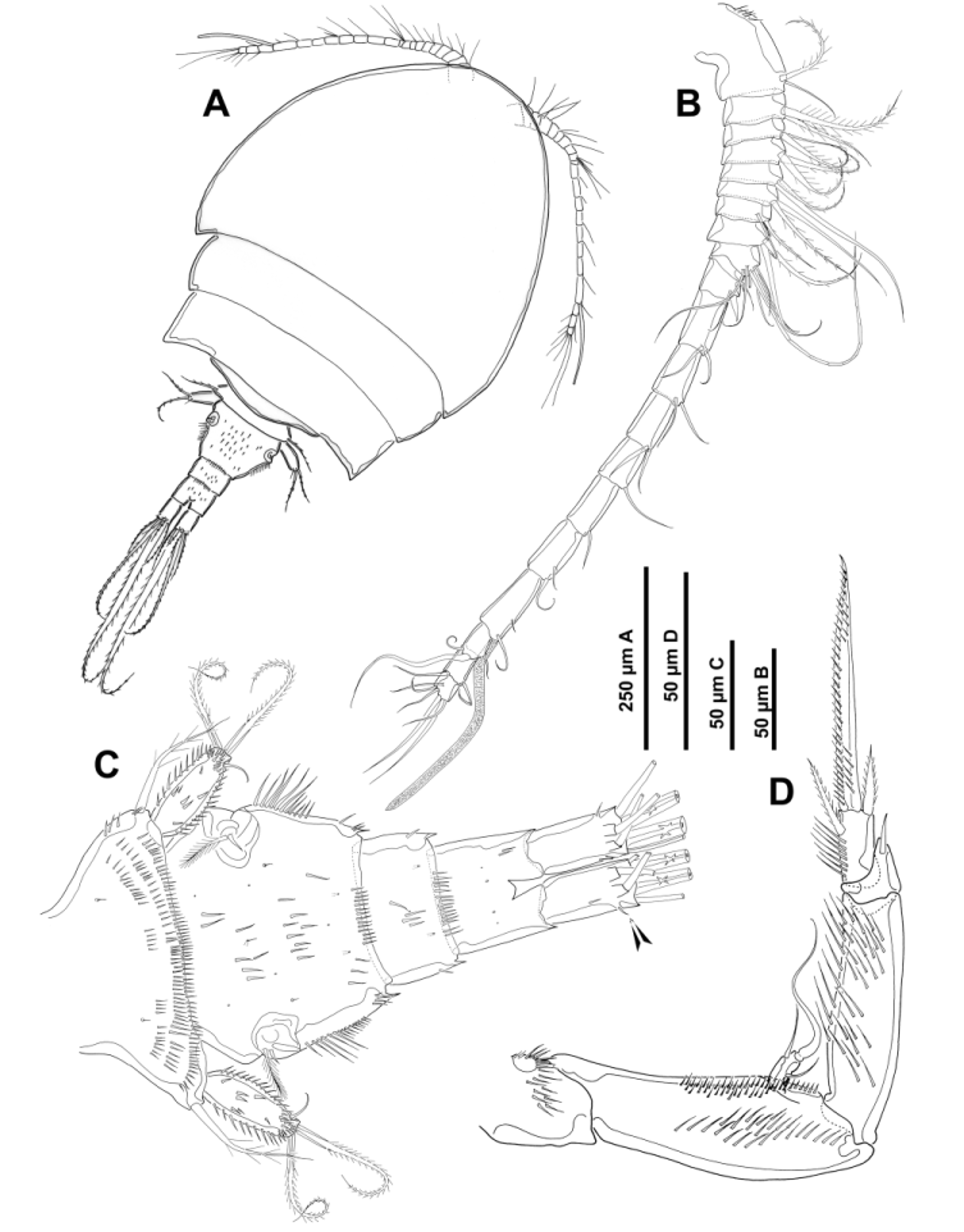

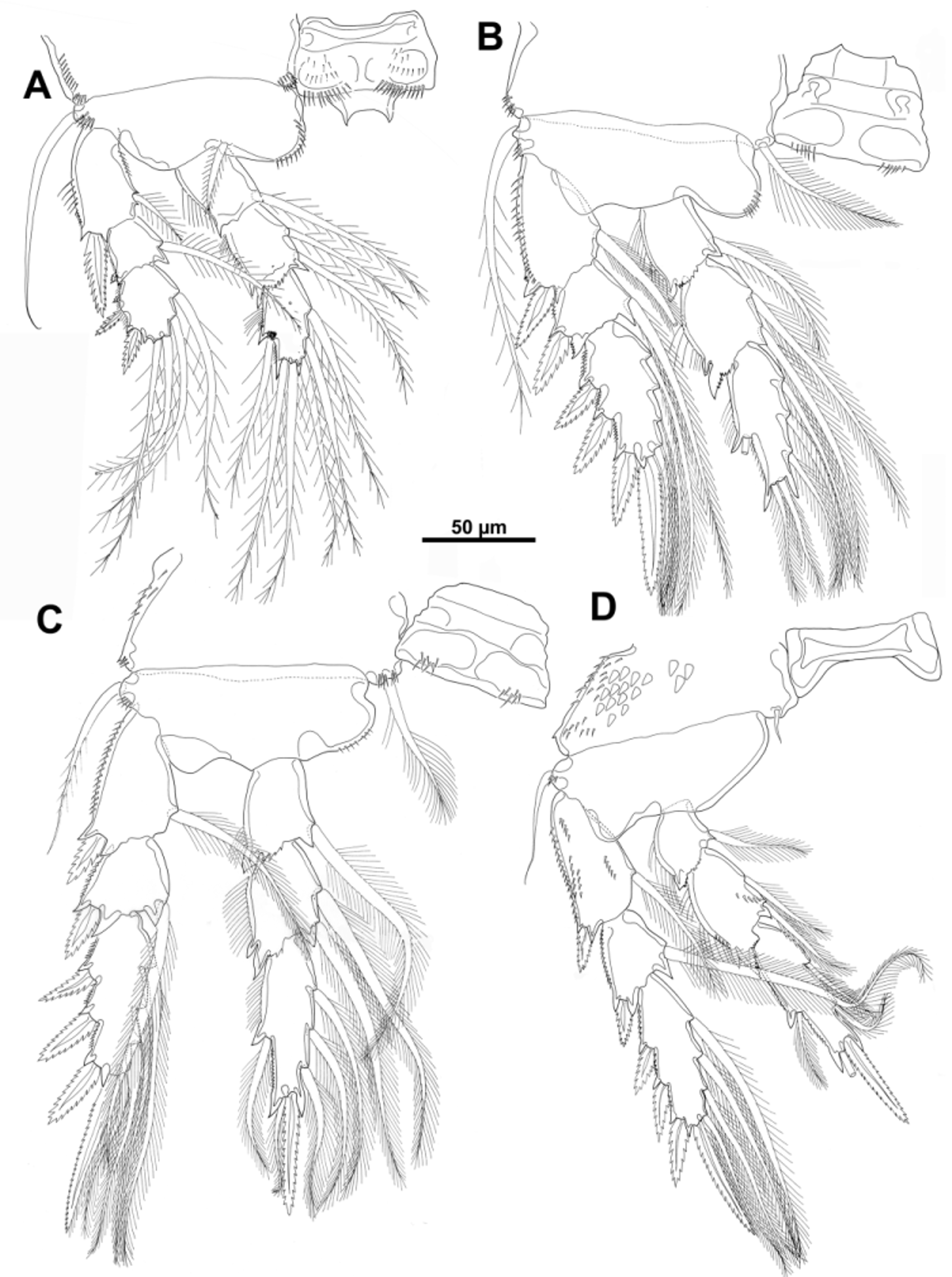
TABLE 3. Spine and seta formula of swiming legs for Asterocheres sarsi Bandera & Conradi 2009.
| Coxa | Basis | Exopod | Endopod | |
|---|---|---|---|---|
| Leg 1 | 0-0 | 1-1 | I-1; I-1; III,2,2 | 0-1; 0-2; 1,2,3 |
| Leg 2 | 0-1 | 1-0 | I-1; I-1; III,I+1,3 | 0-1; 0-2; 1,2,3 |
| Leg 3 | 0-1 | 1-0 | I-1; I-1; III,I+1,3 | 0-1; 0-2; 1,1+I,3 |
| Leg 4 | 0-1 | 1-0 | 1-1; I-1; III,I+1,3 | 0-1; 0-2; 1,1+I,2 |
No known copyright restrictions apply. See Agosti, D., Egloff, W., 2009. Taxonomic information exchange and copyright: the Plazi approach. BMC Research Notes 2009, 2:53 for further explanation.
|
Kingdom |
|
|
Phylum |
|
|
Class |
|
|
Order |
|
|
Family |
|
|
Genus |
Asterocheres sarsi Bandera & Conradi, 2009
| Bandera, Eugenia & Conradi, Mercedes 2014 |
Ascomyzon latum
| Sars 1915 |