
Procerodella cervix Sluys and Kawakatsu, 2005
|
publication ID |
https://doi.org/ 10.1080/00222930410001671309 |
|
persistent identifier |
https://treatment.plazi.org/id/039387D4-E50B-927A-4253-A3C6DAE43753 |
|
treatment provided by |
Felipe |
|
scientific name |
Procerodella cervix Sluys and Kawakatsu |
| status |
sp. nov. |
Procerodella cervix Sluys and Kawakatsu View in CoL , sp. nov.
( Figures 13 View Figure 13 , 22–27 View Figures 22–24 View Figures 25–27 )
Material examined
HOLOTYPE: ZMA V.Pl. 953.1, Ishigaki-jima Island (24 ° 209–24 ° 379N, 124 ° 209–124 ° 409E), Yaeyama Islands, Southwest Islands, Japan, 11 March 1986, sagittal sections on one slide.
PARATYPES: ZMA V.Pl. 953.2–12, ibid., sagittal sections of 11 specimens, each on one slide ; V.Pl. 953.13–21, ibid., sagittal sections of nine specimens, each on one slide ; V.Pl. 953.22, ibid., one whole mount on one slide .
Diagnosis
Procerodella cervix differs from its congeners by having a strongly muscularized male atrium, provided with a short neck projecting into the common atrium, and a knob-shaped tip at the penis papilla.
Ecology and distribution
The animals were collected from a brackish culture pond of the crab Scylla serrata (Forskål, 1775) in Ishigaki-jima Island, one of the Yaeyama Islands in the Southwest Islands of Japan ( Figure 13 View Figure 13 ). In the winter of 1986 a very large population of planarians was present in the pond.
Etymology
The specific epithet is based on the Latin noun ‘ cervix ’, neck. It refers to the muscular nozzle of the male atrium pointing into the common atrium, reminding one of the cervix uteri of the female human.
Description
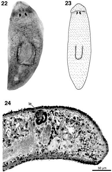
Preserved specimens approximately 2× 1 mm, with obtusely pointed body ends ( Figures 22, 23 View Figures 22–24 ). Dorsal surface uniformly pale brown with a somewhat darker transverse band directly anterior to the eyes. The eye cups contain three retinal cells; there is a rounded, relatively large, granular structure to the eyes that probably represents a lens ( Figure 24 View Figures 22–24 ). The pharynx measures between one-quarter and one-fifth of the body length; mouth opening at the posterior of the pharyngeal pocket.
Relatively few and large testes situated between the ovaries and the root of the pharynx; there are about six follicles on either side of the body. The testis follicles occupy most of the dorso-ventral space. Ovaries located directly behind the brain.
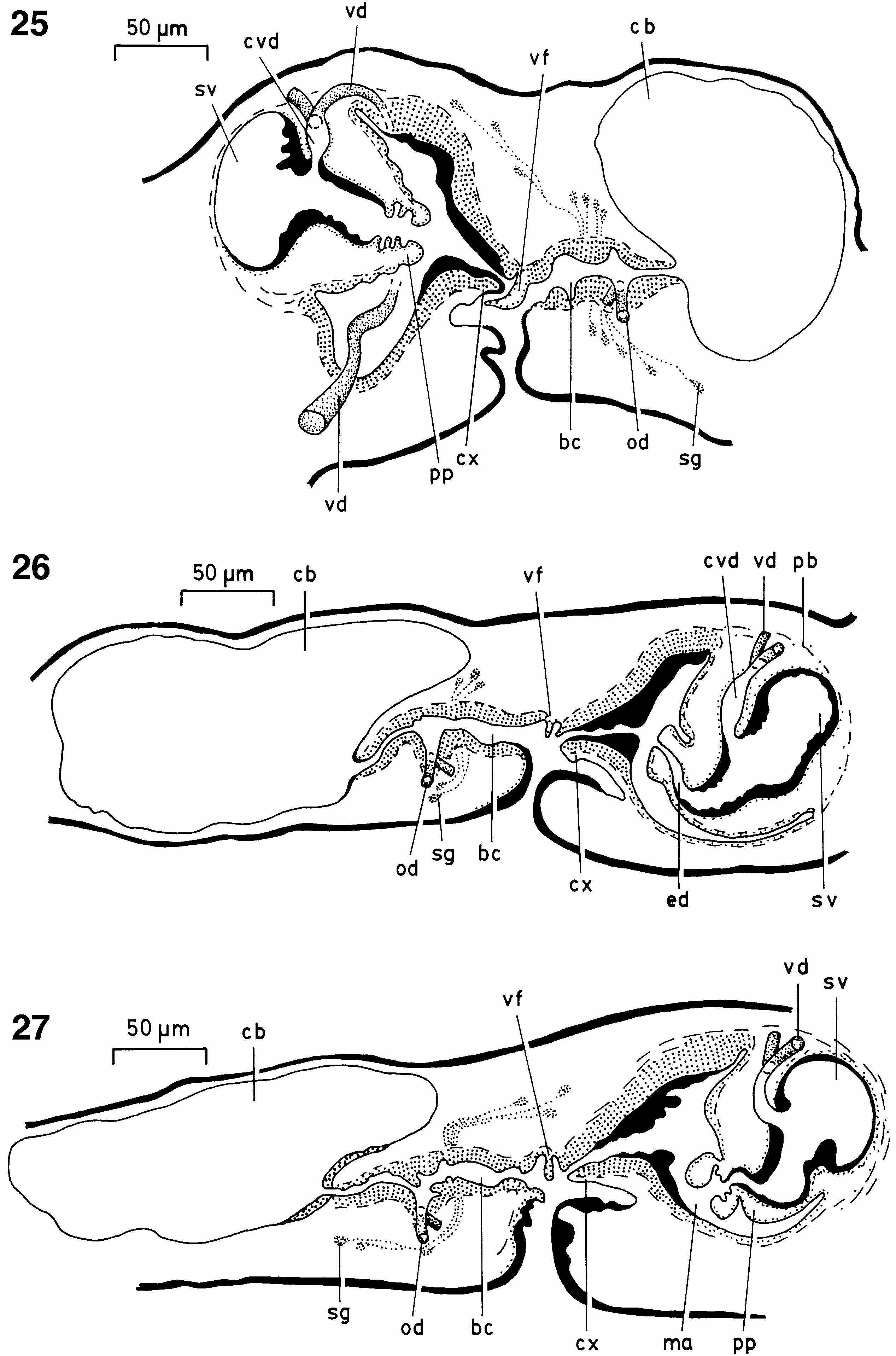
The vasa deferentia form well-developed spermiducal vesicles; at the level of the male copulatory apparatus the ducts curve dorso-medially, decrease in diameter and penetrate the dorsal section of the penis bulb. Within the penial bulb the vasa deferentia fuse to form a common vas deferens, with a slightly larger diameter than the separate ducts, that communicates with the mid-dorsal section of the penis lumen. This penial lumen consists of a spacious vesicle in the penis bulb that gradually narrows to form a broad ejaculatory duct, opening at the knob-shaped tip of the broadly conical penial papilla. The penial lumen is provided with a layer of mostly circular muscle fibres. The nucleated epithelium of the penis papilla is underlain with a subepithelial layer of circular muscle, followed by longitudinal muscle fibres ( Figure 25 View Figures 25–27 ).
The male atrium is of a very peculiar nature since it is provided with a thick layer of subepithelial circular muscle, followed by a much thinner layer of longitudinal muscle. But the most characteristic feature is that the distal, posterior section of the atrium forms a short neck pointing into the common atrium. This neck, short blunt nozzle, or cervix is also provided with the well-developed muscle layers that surround the rest of the male atrium ( Figure 26 View Figures 25–27 ).
The female copulatory apparatus consists of a relatively large copulatory bursa, communicating with the common atrium through a bursal canal that runs more or less parallel to the body surface. The very distal end of the bursal canal (i.e. at the point of opening into the common atrium) is provided with more or less developed valvelike folds ( Figure 27 View Figures 25–27 ). The oviducts unite to form a very short common oviduct that opens into the ventral section of the bursal canal. The latter receives the secretion of shell glands just ectally to the opening of the common oviduct. The bursal canal is lined with an infranucleate epithelium and is surrounded by a thick, subepithelial layer of circular muscle and a much thinner layer of longitudinal muscle fibres. Distinct unicellular glands opening into the bursal canal were not clearly observed. However, it is not excluded that the presence of such glands is obscured by the numerous nuclei of the infranucleated epithelium of the canal that are located just outside the muscle coat.
Comparative discussion
The genus Procerodella Sluys, 1989 was erected for two Japanese species and one from Chile: P. japonica (Kato, 1955) , P. asahinai ( Kato, 1943) and P. macrostoma (Darwin, 1844) , respectively ( Sluys 1989). Characteristic features for the genus were considered to be (1) a common oviduct; (2) a thick zone of circular muscle around the bursal canal; (3) bursal canal with vestibulum ectally to the opening of the common oviduct and receiving the secretion of shell glands; and (4) numerous unicellular glands discharging their secretion into the bursal canal. Not all of these features are present in P. cervix . In particular, P. cervix may lack the unicellular glands opening into the bursal canal and a vestibulum appears to be absent. On the other hand, the morphology of the female copulatory apparatus of P. cervix much resembles the situation in P. japonica , notably the well-muscularized common oviduct. It is evident that with the discovery of new species generic diagnoses may have to be adapted. Furthermore, it is also because we wish to follow a conservative approach to taxonomy and to refrain from coining unnecessary monotypic genera that we have decided to assign the new species to the genus Procerodella .
The presence of an eye lens in P. cervix is not unexpected since it belongs to a group of species, the superfamily Bdellouroidea Diesing, 1862 , for which the presence of a lens is presumed to be an apomorphic feature ( Sluys 1989). However, it should be noted that in histological preparations eye lenses usually appear as densely and brightly staining structures, in contrast to the faintly staining and granular lenses in P. cervix .

| ZMA |
Universiteit van Amsterdam, Zoologisch Museum |
| V |
Royal British Columbia Museum - Herbarium |
No known copyright restrictions apply. See Agosti, D., Egloff, W., 2009. Taxonomic information exchange and copyright: the Plazi approach. BMC Research Notes 2009, 2:53 for further explanation.