
Miroplana paulula Sluys, 2005
|
publication ID |
https://doi.org/ 10.1080/00222930410001671309 |
|
persistent identifier |
https://treatment.plazi.org/id/039387D4-E51E-926E-4285-A186DB2D37B7 |
|
treatment provided by |
Felipe |
|
scientific name |
Miroplana paulula Sluys |
| status |
sp. nov. |
Miroplana paulula Sluys View in CoL , sp. nov.
( Figures 4–8 View Figures 4–6 View Figures 7, 8 )
Material examined
HOLOTYPE: QM G222882 , Coomera River , Coomera (27 ° 519000S, 153 ° 219000E), Queensland, Australia, 24 June 1998, sagittal sections on three slides.
PARATYPES: ZMA V.Pl. 949.1, ibid., whole mount on one slide ; QM G 222883 , ibid., transverse sections on three slides ; ZMA V.Pl. 949.2, ibid., transverse sections on five slides ; ZMA V.Pl. 949, preserved specimens.
Diagnosis
Miroplana paulula Sluys , sp. nov. is characterized by three pigmented transverse bands, with the anterior band located in front of the eyes, a genito-intestinal duct between copulatory bursa and the gut, and one pair of large testes located between the brain and the root of the pharynx. The species differs from its congener by the absence of spines in the ejaculatory duct and lack of a communication between bursal canal and male atrium.
Ecology and distribution
The animals were found in brackish water (11‰) sediments of the Coomera River.
Etymology
The specific epithet is derived from the Latin adjective ‘ paululus ’, few in number, and alludes to the small number of testes in this species.
Description
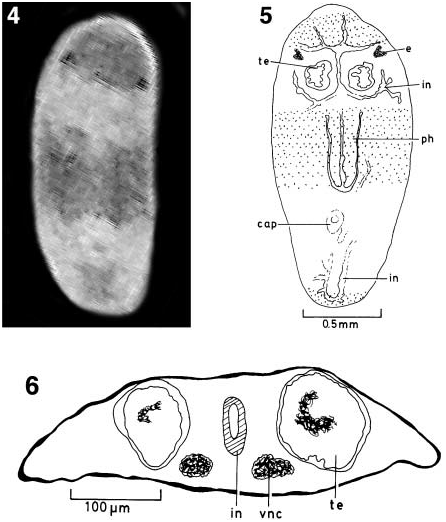
Living animals up to 1.5 mm in length; preserved specimens measuring up to 0.85× 0.4 mm. Body lanceolate-oblong in shape, with broadly rounded anterior end and a somewhat narrower tail end that is rounded or obtusely pointed.
Dorsal surface with three dark brown, pigmented bands: an anterior band extending from the level of the eyes to the anterior body margin, a broad band lying over the region of the pharynx, and a posterior band at the very tail end of the body ( Figure 4 View Figures 4–6 ).
The eyes are well developed, the pigment cup measuring about 37 Mm in diameter; the number of retinal cells present in each eye cup could not be determined. An eye lens is absent.
The pharynx is situated in the middle of the body and measures about one-quarter of the body length in preserved specimens. The mouth opening is located at the posterior end of the pharyngeal pocket.
The anterior ramus of the intestine extends forward anterior to the eyes and gives off preocellar diverticula. The posterior intestinal gut trunks communicate in the very hind end of the body ( Figure 5 View Figures 4–6 ).
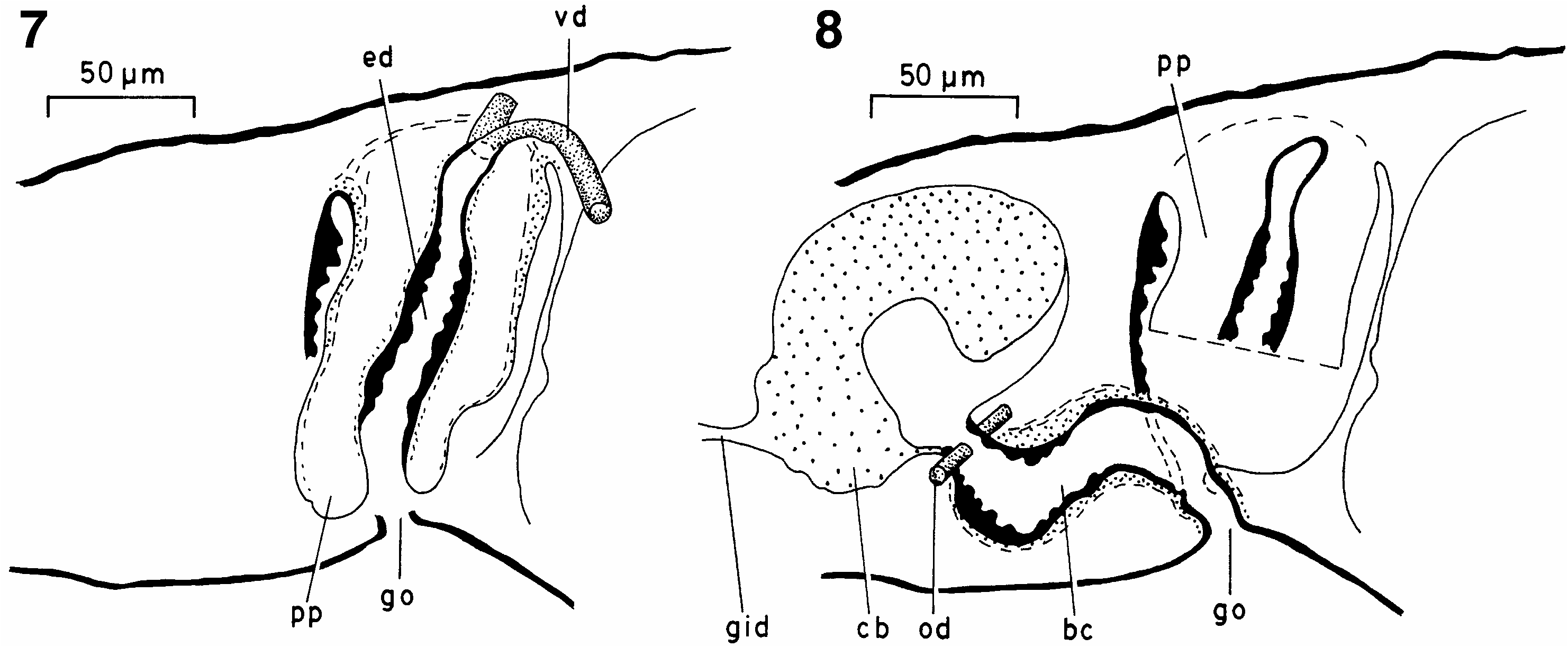
A pair of large testes is located between the brain and the root of the pharynx, the follicles occupying most of the dorso-ventral space ( Figure 6 View Figures 4–6 ). The vasa deferentia expand to form well-developed spermiducal vesicles in the pharynx region. At the posterior end of the pharyngeal pocket the sperm ducts curve dorso-medially and subsequently penetrate the penial bulb. Immediately after having penetrated the bulb, the vasa deferentia unite to form the ejaculatory duct. The latter is lined with a relatively tall, nucleated epithelium and is surrounded by a well-developed coat of muscle; the ejaculatory duct opens at the tip of the penial papilla ( Figure 7 View Figures 7, 8 ).
The penis bulb consists of a well-developed coat of muscle. The penial papilla is an obtusely pointed cone, covered with a flat, nucleated epithelium that is underlain with a subepithelial layer of circular muscle, followed by a thin layer of longitudinal muscle.
The male atrium communicates through a constriction with the common genital atrium, which is actually very small and immediately passes into the gonopore.
The paired ovaries are located medially to the testes and are also situated medially to the ventral nerve cords. The oviducts open separately into the bursal canal, close to the point of communication between the canal and the copulatory bursa. Distinct shell glands were not observed.
The bursal canal is lined with an infranucleate epithelium and is surrounded by a welldeveloped, subepithelial layer of circular muscle, followed by a layer of longitudinal muscle. The copulatory bursa of the specimens examined is filled with a finely granular substance. Through a genito-intestinal duct the bursa communicates with the intestine ( Figure 8 View Figures 7, 8 ).
Comparative discussion
Although the present species differs in several respects from the type species of the genus, Miroplana trifasciata Kato, 1931 (cf. Kato 1931), we have nevertheless assigned it to Miroplana for several reasons. It should be recognized that the current diagnosis of the genus (cf. Sluys 1989) is based only on the type species and therefore may be highly biased. Furthermore, we are of the opinion that in cases of limited knowledge a conservative approach to taxonomy should be followed as much as possible and therefore we refrain from creating a new genus for the present species.
The three transverse bands of M. paulula immediately remind one of the similar bands reported for M. trifasciata . In M. trifasciata , however, the anterior band is positioned behind the eyes, whereas in M. paulula it occurs anterior to the eyes. Furthermore, the posterior band in M. trifasciata is located more anteriorly than in M. paulula .
In contrast to the type species, M. paulula does not have any spines in the ejaculatory duct, nor does it exhibit the communication between the male atrium and the bursal canal. Both species show a connection between the bursa and the intestine. Miroplana trifasciata has two pairs of testes, one pair situated in the same location as the testes of M. paulula , the other pair located at about the posterior quarter of the pharynx.
In M. trifasciata the posterior gut trunks give rise to about four medial diverticula, forming commissures between the two main branches. In M. paulula the posterior gut trunks are only connected at their most posterior ends.
Miroplana trifasciata was collected from a brackish habitat (see below) and also the biotope of M. paulula differs from that of a standard marine planarian in that it lives in brackish water.
| QM |
Queensland Museum |
| ZMA |
Universiteit van Amsterdam, Zoologisch Museum |
| V |
Royal British Columbia Museum - Herbarium |
No known copyright restrictions apply. See Agosti, D., Egloff, W., 2009. Taxonomic information exchange and copyright: the Plazi approach. BMC Research Notes 2009, 2:53 for further explanation.