
Neostygarctus grossmeteori, Tchesunov, 2018
|
publication ID |
https://doi.org/ 10.5852/ejt.2018.479 |
|
publication LSID |
lsid:zoobank.org:pub:EA678C5B-2E5B-4F73-B254-B9A524307E70 |
|
DOI |
https://doi.org/10.5281/zenodo.3845837 |
|
persistent identifier |
https://treatment.plazi.org/id/0047D8B7-BEAE-4751-B098-86EA728FABA3 |
|
taxon LSID |
lsid:zoobank.org:act:0047D8B7-BEAE-4751-B098-86EA728FABA3 |
|
treatment provided by |
Valdenar |
|
scientific name |
Neostygarctus grossmeteori |
| status |
sp. nov. |
Neostygarctus grossmeteori View in CoL sp. nov.
urn:lsid:zoobank.org:act:0047D8B7-BEAE-4751-B098-86EA728FABA3
Figs 2–6 View Fig View Fig View Fig View Fig View Fig , Table 1 View Table 1 (morphometrics)
Diagnosis
Neostygarctus with five middorsal spines on the cephalic and each body plate and on the caudal plate, the spines decreasing in length backwards. Ocelli present. One or two pairs of ventral cervical spines present. Each lateral body proсess provided basally on the ventral side with a transversal row of two to five short but strong spikes. Only inner claws of each foot provided with a fine dorsal accessory spine; external claws may carry vestigial accessory spines.
Etymology
The species name is derived from the name of locality (Gross Meteor in German).
Type material
Holotype
ATLANTIC OCEAN: ♀, Great Meteor Seamount plateau, 29º48.974 N, 28º25.941 W, depth 299 m, RV Poseidon , expedition P397 GROMET, location #17, stn 106-6, 1 Mar. 2010 ( SMF 51 About SMF ).
GoogleMapsParatypes
ATLANTIC OCEAN: 3 ♀♀ ( SMF 52-54), 1 Ƌ ( SMF 58), one specimen of unidentified gender ( SMF 60) ( Table 1 View Table 1 ), same data as for the holotype. The holotype and the paratypes are all embedded in permanent glycerin slides.
General description
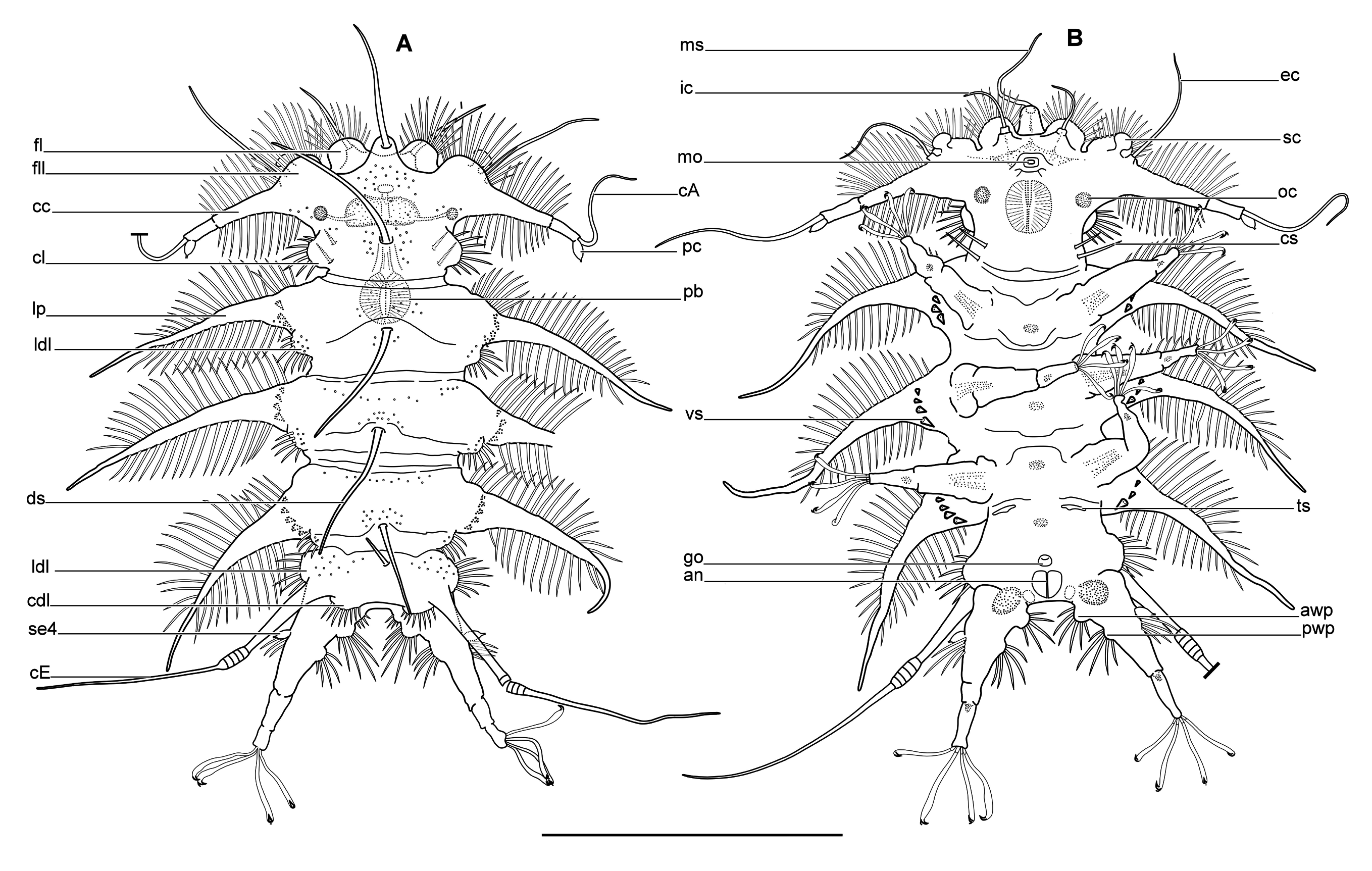
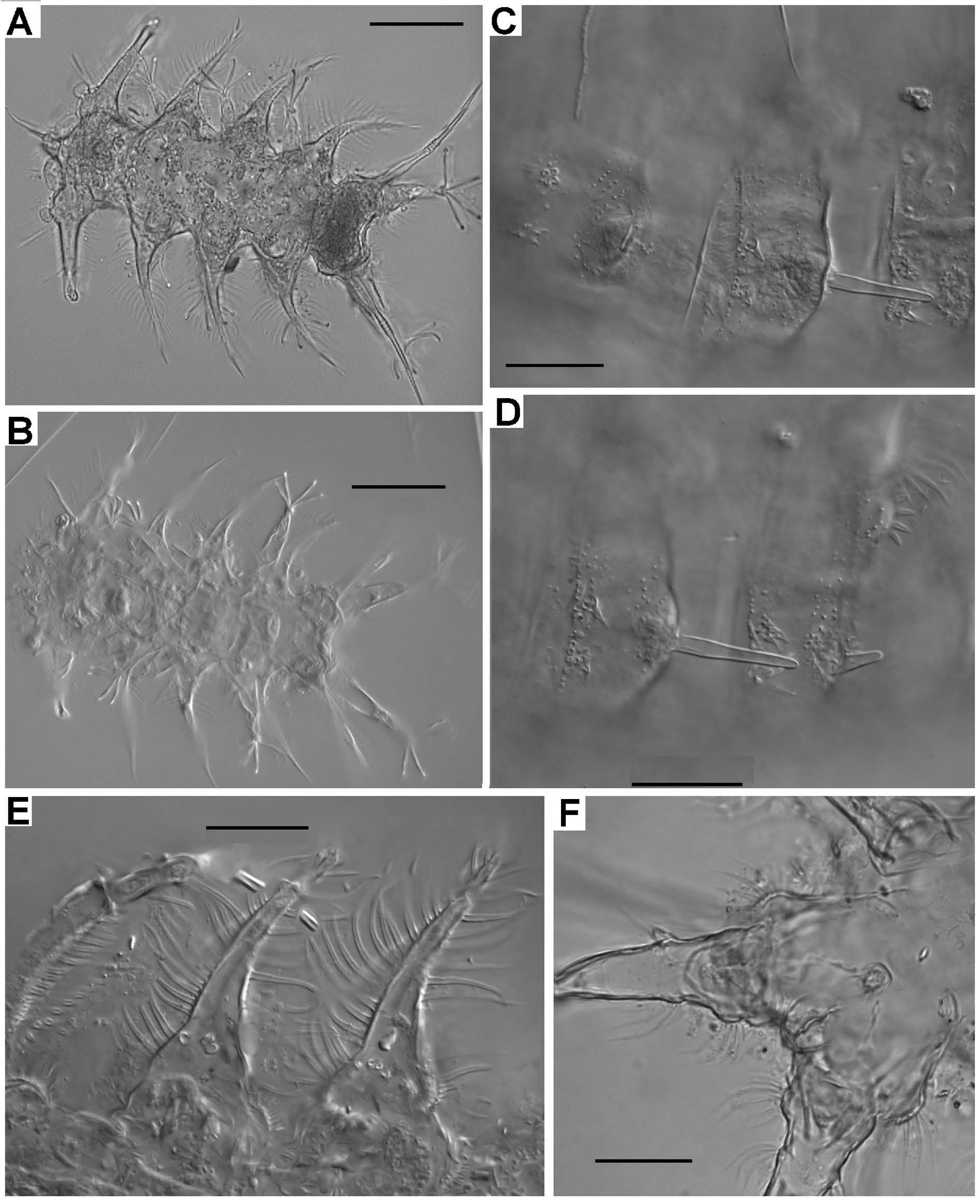
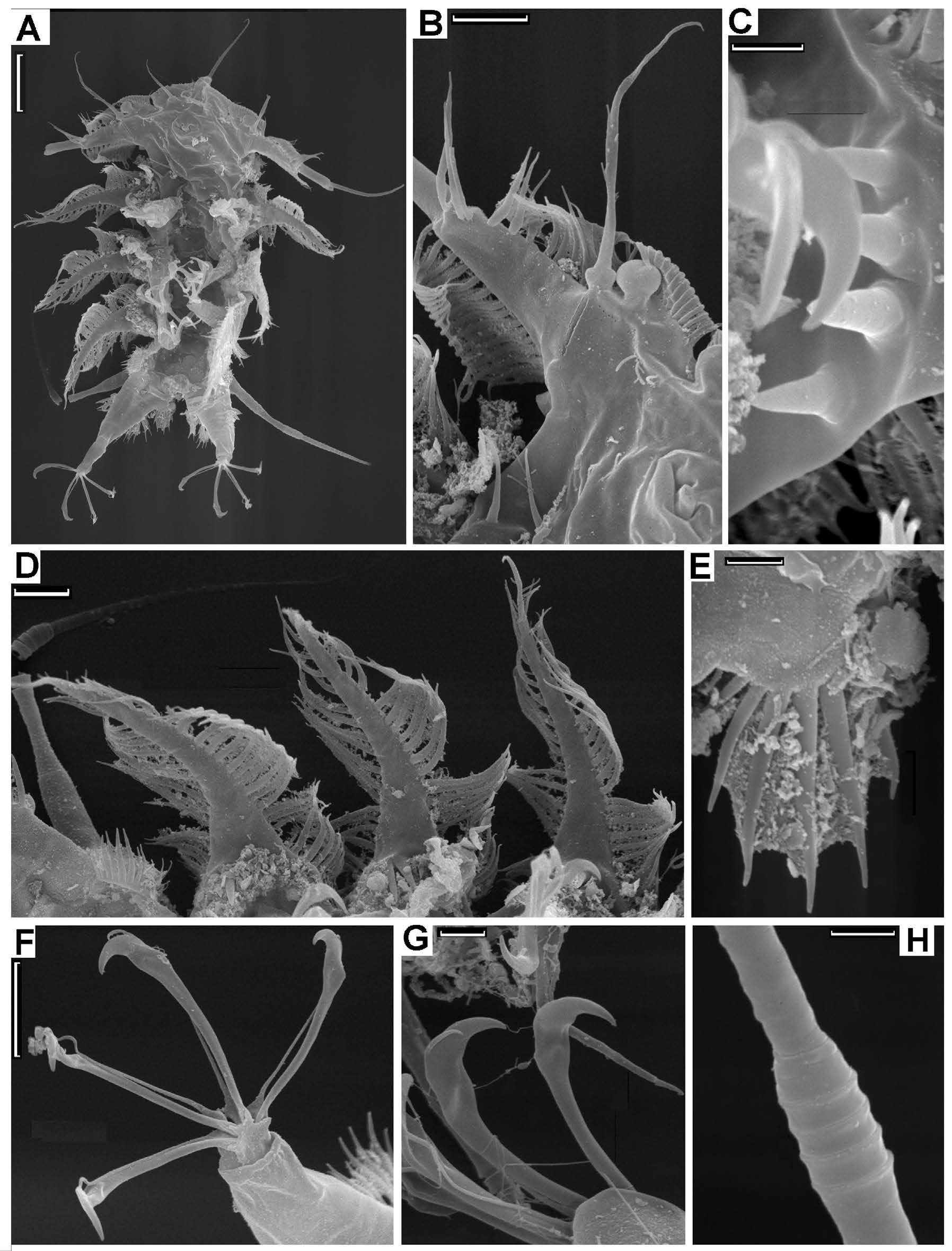
BODY SURFACE AND SCULPTURES. Dorsal cuticle consists of five transversal dorsal plates (one cephalic, three body and one caudal plates) and intermediate areas between them ( Fig. 2A View Fig ). Ventral cuticular plates not developed as such; the only transversal cuticular folds may be discernible ( Fig. 2B View Fig ). Edges of dorsal plates discernible as discontinuous cuticular folds which may be obscured here and there. Cuticle of plates hardly differs from that of intermediate areas – but sparse and irregular coarse punctuations on dorsal plates ( Fig. 2A View Fig ), which are, in fact, seemingly epicuticular pillars not detectable in SEM on surface but visible in optical microscope laterally as short projecting bars. Five robust middorsal spines present, which are elongated and conical, with acute or slightly rounded tips. Each plate carries one median spine attached in pit in middle on cephalic and on body plates I and II, close to posterior edge on body plate III, and close to anterior edge of caudal plate. Middorsal spines tend to be reducing in length from cephalic to caudal plate ( Fig. 2A View Fig , ds). Set of middorsal spines may be incomplete in some specimens ( Table 1 View Table 1 ), possibly because of loss.
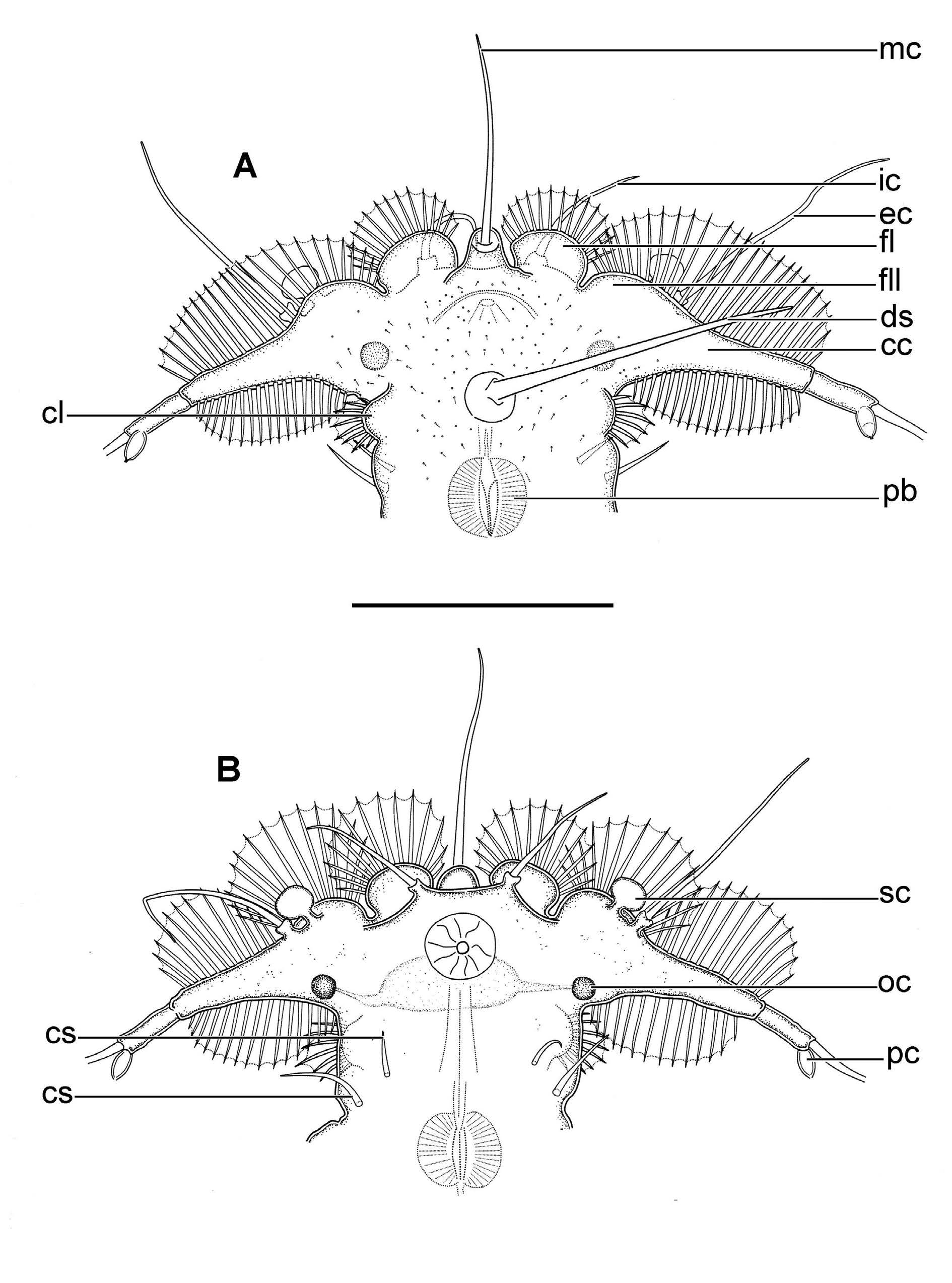
CEPHALIC REGION ( Figs 2 View Fig A–B, 3A–B). Anteriorly subdivided into two pairs of lobes densely feathered by fine hollow cuticular spines: two frontal semicircular lobes (fl), two fronto-lateral lobes (fll), also semicircular but less prominent, and two long lateral common cirrophores of cirrus A and primary clava (cc). Pair of not very prominent but distinct latero-dorsal wart-like cervical lobes (cl) on narrowed neck region just posterior to lateral cirrophores. Dorsal cuticle of cephalic region marked by sparse and irregularly distributed coarse punctuations which are actually projections of short rod-like epicuticular pillars ( Fig. 3A View Fig ). Long middorsal spine at level of cervical lobes. Anterior edges of frontal and frontolateral lobes as well as both anterior and posterior edges of lateral cirrophores on head flanked by tight rows of finest hollow spines: about 15 spines on frontal lobe, 10-12 spines on fronto-lateral lobe, 9-11 spines on anterior and about 15 spines on posterior edge of lateral lobe. Spines of anterior frontal lobes very fine, spines of fronto-lateral lobes and then to anterior and posterior edges of body lateral processes become gradually more and more robust. Cervical lobes bear about eight medium-sized, slightly curved spines each. Fine spines arranged in tight rows on all head and body processes joined with thin film or membrane ( Fig. 2 View Fig A–B); rags of the membranes observable in SEM ( Fig. 6E View Fig ). About two pairs of straight medium-sized ventro-lateral cervical spines (cs) on neck region just posterior to cervical lobes. Anterior cervical spines a bit smaller and located more antero-ventrally than posterior cervical spines (the latters are paired on either side in one specimen). Soft mouth cone on ventral side situated a bit posterior to anterior head edge.
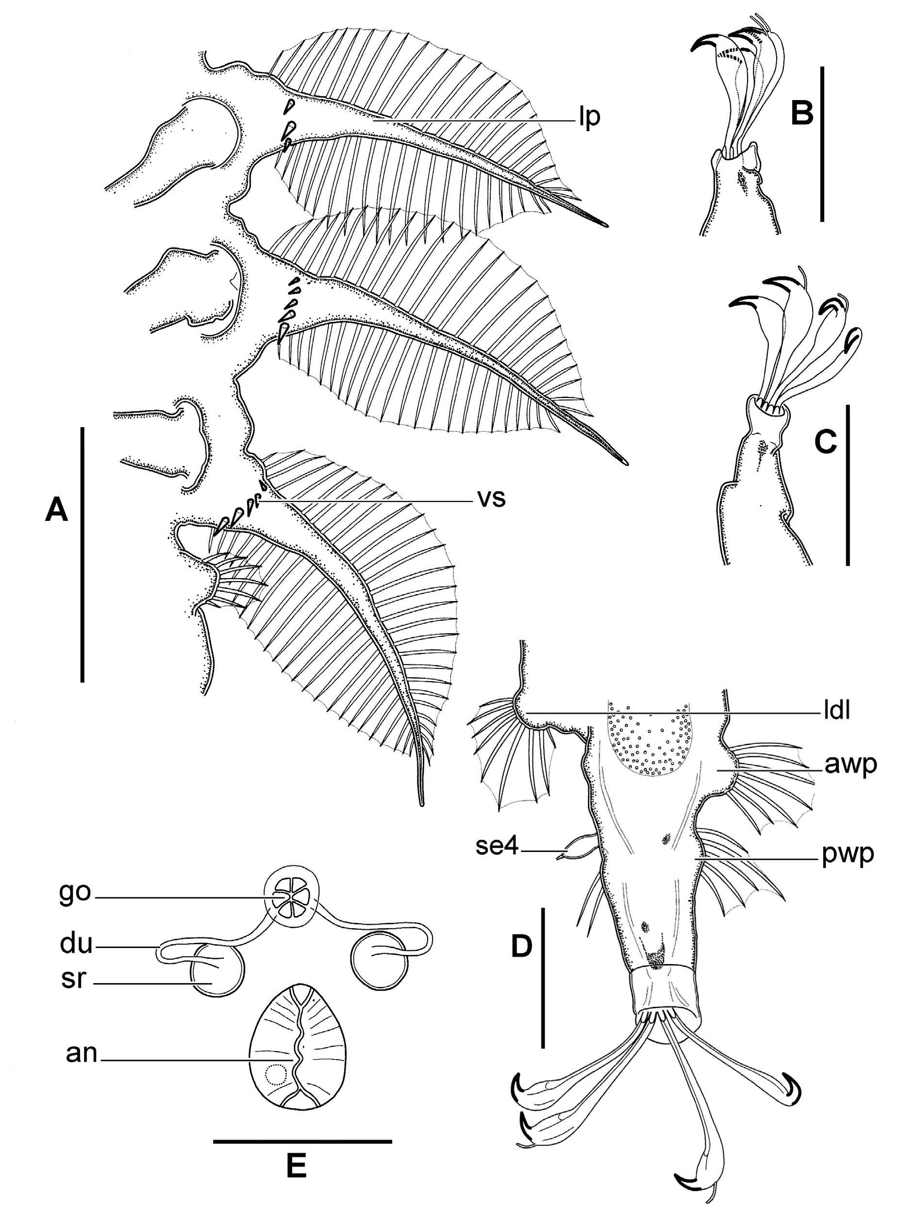
BODY SEGMENTS I–III. With long lateral processes (lp) almost normal to long body axis, ornamented by rows of long hollow spines on anterior and posterior edges; terminal narrowed region of processes lack spines and slightly curved rearward ( Figs 2 View Fig A–B, 4E, 5A, 6A–B, D). Spines make processes feather-like. Numbers and position of spines on body lateral projections: (1) about 10 medium-sized spines arranged in arch dorso-posteriorly near bases of processes; (2) about 19–22 long marginal spines along anterior edge of process; (3) about 14-16 also long marginal spines along posterior edge of process (both anterior and posterior rows of marginal sets of dorso-lateral process give impression of feather). Two to five short and stout but acute ventral conical spikes (vs) arranged in row parallel to long body axis on ventral side of each lateral body process ( Figs 2B View Fig , 5A View Fig , 6C View Fig ). Pair of wart-like latero-dorsal lobes (ldl) on each body segment just posterior to long lateral processes on segments I–III and in the middle on caudal segment. In shape, they resemble cervical latero-dorsal lobes. Caudal segment bears another pair of caudal dorsal wart-like lobes (cdl) on posterior edge. All wart-like lobes armed with five to seven medium-sized spines.
SENSORY ORGANS. Long anterior flagellar median cirrus (mc) rooted on prominent cirrophore hanging over anterior edge of head ( Figs 2 View Fig A–B, 3A–B). Internal cirri (ic) consist of short stub-shaped cirrophore and whip-like flagellum situated ventrally to anterior frontal lobes ( Fig. 3 View Fig A–B). External cirri (ec) situated ventrally to fronto-lateral lobes and also consist of short stub-shaped cirrophore and longer whip-like flagellum; similar in shape to internal cirri but a bit longer ( Figs 2 View Fig A–B, 3A–B, 6B). All cephalic cirri (flagellar parts) possess minute and sparse irregular lateral sprouts visible in SEM ( Fig. 6B View Fig ). External cirri located close to secondary clava just ventrally of fronto-lateral lobe edge. Globular secondary clava on short narrow stalk situated at inner position to external cirrus ( Figs 3 View Fig A–B, 6B). Cirrus A (lateral cirrus) and small primary clava rooted together on common cylindrical cirrophore (cc) which is continuation of lateral head lobe ( Figs 3 View Fig A–B, 6B); primary clava ovoid to lemon-shaped, with pointed terminal end. In some female and male specimens, paired ventral transversal slits (ts) observed just posterior to legs III ( Fig. 2B View Fig ); they are considered as possible sensory organs. No sensory organs present on legs I–III. Cirri E (cE) on caudal segment growing on body postero-laterally. Each cirrus E consists of long robust cirrophore without any discernible tubercles, cylindrical accordion-like thickening composed of three to five rings, and long whip-like flagellum ( Figs 2 View Fig A–B, 6H). Legs IV provided with latero-dorsal sense organs (se4) on external side of coxae. Sense organs small, ovoid, with tiny apical papillum ( Figs 2 View Fig A–B, 5D).
LEGS I–IV. Rather long, consisting of long truncate conical coxa/femur, telescoping cylindrical tibia and palm-shaped (hand-shaped in Kristensen et al. 2015) tarsus with four equal digits. Border between coxa and femur indistinct. Legs I–III have neither sensory organs nor bristles. Legs IV provided with three groups of spines on coxa: (1) about ten short to medium-sized spines on anterior wart-like projection (awp) on internal side of coxa basally, (2) five to six medium-sized spines on posterior wart-like projection (pwp) on internal side of coxa right opposite to ovoid papilla, (3) about four medium-sized spines on external side of leg just ventrally to ovoid latero-dorsal sense organ ( Fig. 5D View Fig ).
DIGITS. Digits of legs IV bit longer than those of legs I–III. All digits of same leg about equal in length to one another. Digits long, slim and flat, with slight subterminal ventral bulge and terminal solid beak-like claws. Only internal claws provided with fine but distinct dorsal spine (accessory spine); external claws may bear very tiny appendage visible only in SEM and probably presenting vestigial spine. Peduncles (cuticular supports) not visible ( Figs 5 View Fig B–D, G, 6F–G).
GENITO- ANAL AREA. In females, gonopore (go) is six-lobed rosette-like structure within circle; two lateral sinuous seminal ducts (du) open posteriorly into spherical seminal receptacles (sr) ( Fig. 5E View Fig ). In males, transversally-oval gonopore situated more close to anal opening ( Fig. 2B View Fig ). Anal opening in both genders is longitudinal slit on anal plate posterior to gonopore.
INTERNAL ORGANS. Internal organs obscure in some specimens, possibly because of maceration. Pair of wide-set internal spheric ocelli (oc) at level of lateral lobes of head; ocelli pallid and indistinct in some specimens. Pharyngeal bulb obscure. Stylets not seen. Midgut and gonad not observed.
No known copyright restrictions apply. See Agosti, D., Egloff, W., 2009. Taxonomic information exchange and copyright: the Plazi approach. BMC Research Notes 2009, 2:53 for further explanation.
|
Kingdom |
|
|
Phylum |
|
|
Class |
|
|
Order |
|
|
Family |
|
|
SubFamily |
Stygarctinae |
|
Genus |