
Singaporemma takensis, Yan & Lin, 2018
|
publication ID |
https://doi.org/ 10.11646/zootaxa.4392.2.6 |
|
publication LSID |
lsid:zoobank.org:pub:E0D597C5-194B-4EF0-AD6D-3A178F8DD2F7 |
|
DOI |
https://doi.org/10.5281/zenodo.5983986 |
|
persistent identifier |
https://treatment.plazi.org/id/039487ED-FFDC-493D-35BC-F98CFDF6AAB5 |
|
treatment provided by |
Plazi |
|
scientific name |
Singaporemma takensis |
| status |
sp. nov. |
Singaporemma takensis sp. n.
Figures 3A–H View FIGURE 3 , 4A–E View FIGURE 4 , 5A–D View FIGURE 5 , 6G–g View FIGURE 6
Examined material. Holotype ♂, paratypes 4♀ ( NHMSU), THAILAND: Tak, Tha Song Yang District, Bam Thung Tham subdistrict, an anonymous cave, 17°16.603'N, 98°11.497'E, altitude 169 m, 19 November 2016, H. Zhao and Y. Li leg. GoogleMaps
Etymology. The specific name refers to the type locality; adjective.
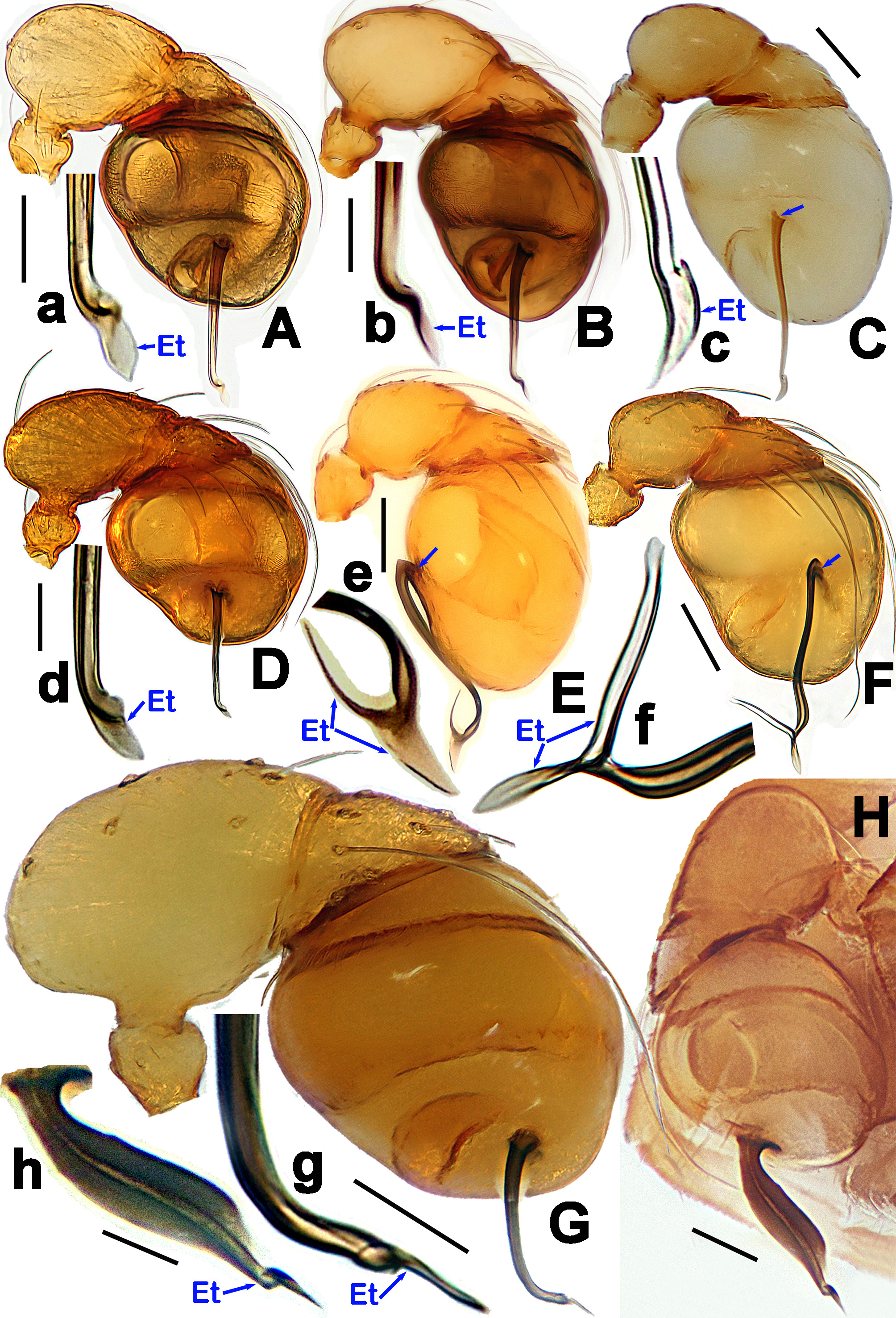
Diagnosis. Male of this new species can be distinguished from S. bifurcata and S. wulongensis by the embolic tip without any bifurcation, but furcate in the latter two ( Fig. 6g View FIGURE 6 vs. Fig. 6e, 6f View FIGURE 6 ); from S. adjacens by the narrower embolus, but wider, belt-shaped in the latter ( Fig. 6g View FIGURE 6 vs. Fig. 6h View FIGURE 6 ); from S. halongense , S. banxiaoensis , S. singulare , and S. lenachanae by the nearly right-angle bending embolus at distally one third position, and the sharply pointed embolic tip ( Figs 4B, D–E View FIGURE 4 , 6G, g View FIGURE 6 vs. Fig. 6A–D, 6a–d View FIGURE 6 ), but straight embolus except distal tip in the latter four ( Fig. 6A–D View FIGURE 6 ), and knife-shaped embolic tip in S. singulare ( Fig. 6c View FIGURE 6 ). Female of this new species differs from S. singulare and S. lenachanae by the presence of central process, but absent in the latter two ( Fig. 5C–D View FIGURE 5 vs. Fig. 9A–B View FIGURE 9 ); from S. adjacens and S. halongense by the presence of inner vulval plate, but absent in the latter two ( Fig. 5C–D View FIGURE 5 vs. Fig. 7A–B View FIGURE 7 ); from S. banxiaoensis by the longer central process, but shorter in the latter ( Fig. 5D View FIGURE 5 vs. Fig. 7C View FIGURE 7 ); from S. bifurcata and S. wulongensis by having a nearly oval-shaped inner vulval plate, but “Ω”-shaped in the latter two ( Fig. 5C–D View FIGURE 5 vs. Fig. 8A–B View FIGURE 8 ).
Description. Male (holotype). Measurements: total length 1.12; carapace 0.45 long, 0.38 wide, 0.36 high; abdomen 0.79 long, 0.60 wide, 0.60 high; clypeus 0.14 high; sternum 0.28 long, 0.30 wide. Legs yellowish-brown Length of legs: I 1.35 (0.45, 0.13, 0.32, 0.22, 0.23); II 1.25 (0.40, 0.12, 0.30, 0.21, 0.22); III 1.15 (0.34, 0.12, 0.26, 0.21, 0.22); IV 1.48 (0.48, 0.13, 0.37, 0.26, 0.24).
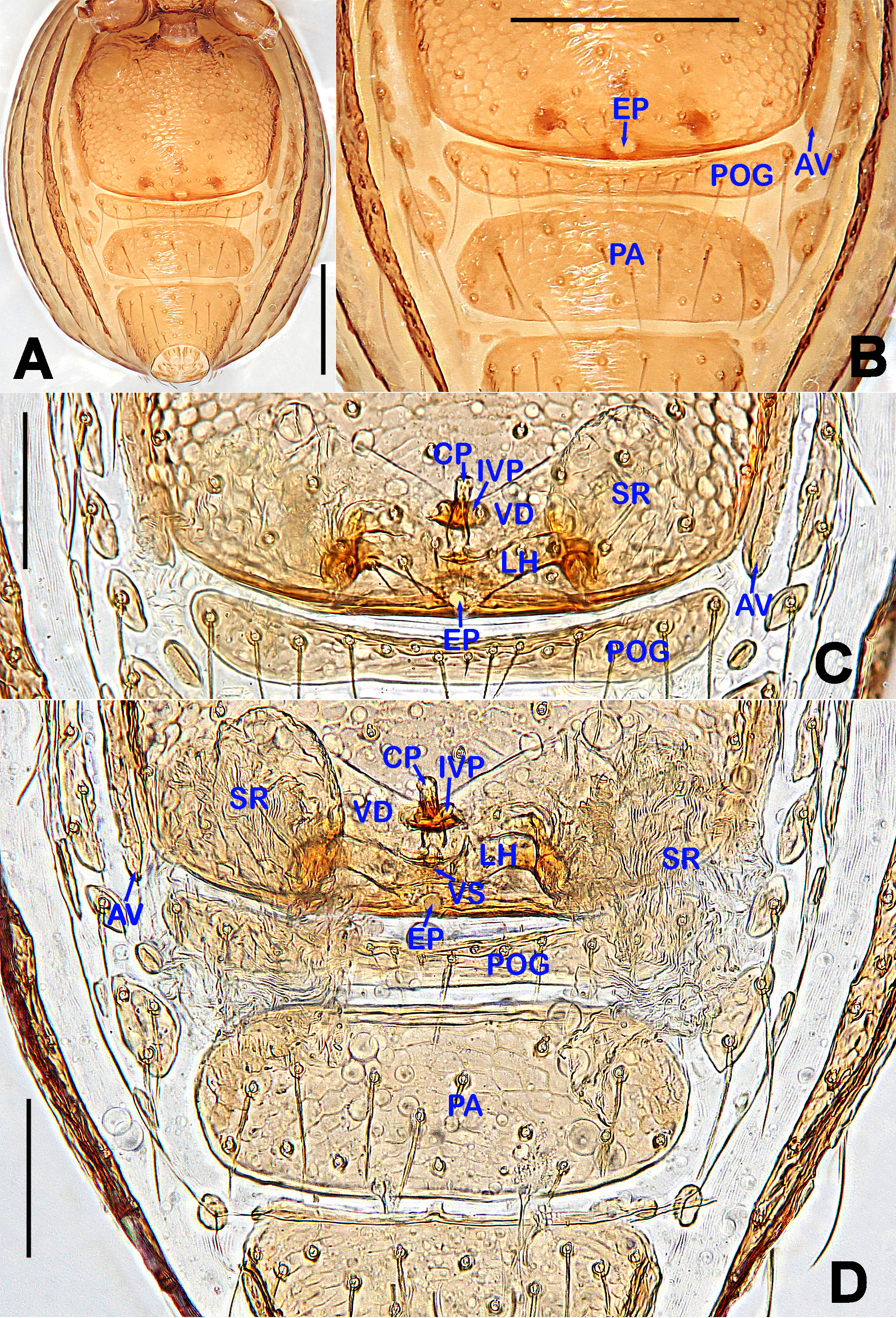
Prosoma ( Fig. 3A–B, E, G View FIGURE 3 ): reddish-yellow, carapace finely reticulated, except for the radial grooves in thoracic area, marginally rugose ( Fig. 3A, E View FIGURE 3 ); six eyes, white with black ocular base, ALE>PLE>PME, ALE and PLE adjacent, AER slightly recurved ( Fig. 3A View FIGURE 3 ); cephalic part raised, top flat ( Fig. 3E View FIGURE 3 ); clypeus high, sloping forward, with sparse setae, marginally rectangular; chelicerae lacking any horns or processes, cheliceral lamina well developed ( Fig. 3G View FIGURE 3 ); endites basally wide, distally narrow, labium triangular, distally blunt ( Fig. 3B View FIGURE 3 ); sternum finely reticulated, scutellate, marginally rugose, posterior corner truncated ( Fig. 3B View FIGURE 3 ). Legs: cuticle striated; all tibiae with 3 trichobothria, and one on metatarsi I–IV. Opisthosoma ( Fig. 3A–B, E View FIGURE 3 ): cuticular slightly pale, dorsal scutum oval, finely reticulated, posteriorly truncated ( Fig. 3A View FIGURE 3 ); ventral scutum reticulated ( Fig. 3B View FIGURE 3 ); lateral scutum I long, and exceeding beyond the posterior margin of preanal scutum ( Fig. 3E View FIGURE 3 ); perigenital scutum absent; postgential scutum slightly precurved, mesially narrow, laterally wide ( Fig. 3B View FIGURE 3 ); preanal scutum approximately oblong ( Fig. 3B View FIGURE 3 ).
Palp ( Figs 4A–E View FIGURE 4 , 6G, g View FIGURE 6 ): femoral cuticle granulated, about 3 times longer than patella; patella short, proximally narrow, distally wide; tibia large, extremely swollen, about 3 times wider than femur ( Figs 4A–B View FIGURE 4 , 6G View FIGURE 6 ); cymbium triangular from lateral view, bearing long setae ( Fig. 4C–D View FIGURE 4 ); bulb egg-shaped, surface smooth ( Figs 4A–B View FIGURE 4 , 6G View FIGURE 6 ); embolus long, tubular, distinctly sclerotized, starting from the subapical position of bulb ( Figs 4A, C, D View FIGURE 4 , 6G View FIGURE 6 ), almost formed a right angle and bend at distal one third part ( Figs 4B, D View FIGURE 4 , 6G View FIGURE 6 ); embolic tip sharply pointed, lamellar ( Fig. 4B View FIGURE 4 ).
Female (one of paratypes). Measurements: total length 1.18; carapace 0.48 long, 0.40 wide, 0.38 high; abdomen 0.77 long, 0.60 wide, 0.54 high; clypeus 0.14 high; sternum 0.29 long, 0.30 wide. Length of legs: I 1.35 (0.44, 0.13, 0.32, 0.23, 0.23); II 1.27 (0.41, 0.13, 0.29, 0.22, 0.22); III 1.16 (0.36, 0.12, 0.26, 0.21, 0.21); IV 1.49 (0.47, 0.14, 0.38, 0.26, 0.24).
Prosoma ( Fig. 3C–D, F, H View FIGURE 3 ) as in male, but colour deeper than in male. Clypeus smooth, palps distinctly reduced. Legs as in male. Opisthosoma ( Figs 3C–D, F View FIGURE 3 , 5A View FIGURE 5 ): dorsal and ventral scuta as in male, except for darker coloration ( Fig. 3C–D View FIGURE 3 ); lateral scutum I long, extending beyond posterior margin of preanal scutum; perigenital scuta small, oval; postgenital scutum long, bearing a row of setae, mesially narrow, laterally wide ( Fig. 5B View FIGURE 5 ); preanal scutum weakly rugose, wider than long, subretangular, with sparse serrated setae ( Fig. 5A View FIGURE 5 ).
Epigyne and vulva ( Fig. 5A–D View FIGURE 5 ): epigynal pit distinct, oval, opening at the margin of the pulmonary scutum ( Fig. 5A–B View FIGURE 5 ); vulval posterior margin strongly sclerotized ( Fig. 5C, D View FIGURE 5 ); vulval dorsal plate rhombic, fused to vulval posterior margin ( Fig. 5C, D View FIGURE 5 ); vulval stem triangular ( Fig. 5C View FIGURE 5 ); vulval ducts wide, translucent; lateral horns weakly sclerotized, forming a “V”-shape, distally reflexed ( Fig. 5C View FIGURE 5 ); seminal receptacula rugose, transparent, and membranous; inner vulval plate transversely oblong, strongly sclerotized, wider than central process ( Fig. 5C–D View FIGURE 5 ); central process straight, clavate, basally contracted, distally beyond inner vulval plate ( Fig. 5C–D View FIGURE 5 ).
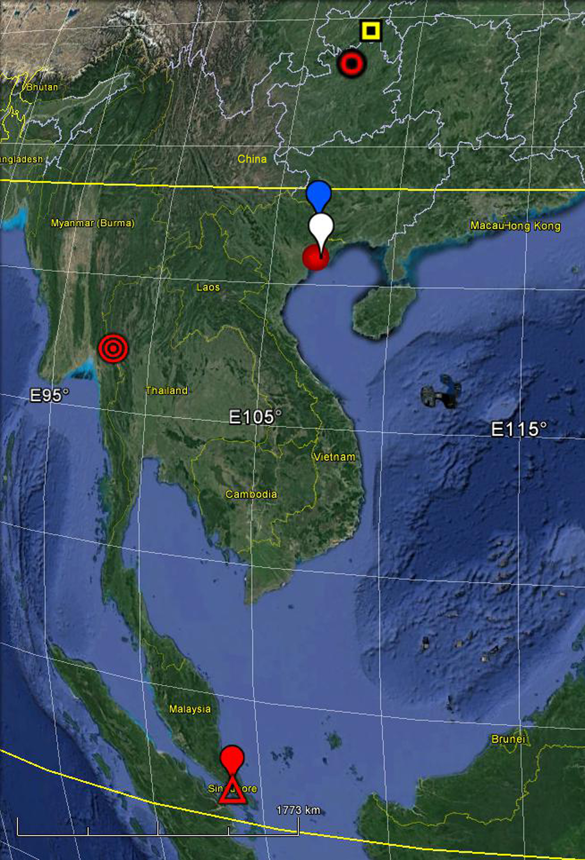
Distribution. Thailand ( Fig. 10 View FIGURE 10 ).


No known copyright restrictions apply. See Agosti, D., Egloff, W., 2009. Taxonomic information exchange and copyright: the Plazi approach. BMC Research Notes 2009, 2:53 for further explanation.
|
Kingdom |
|
|
Phylum |
|
|
Class |
|
|
Order |
|
|
Family |
|
|
Genus |