
Macrolygistopterus subparallelus Pic, 1930
|
publication ID |
https://doi.org/ 10.5281/zenodo.281850 |
|
DOI |
https://doi.org/10.5281/zenodo.3502234 |
|
persistent identifier |
https://treatment.plazi.org/id/0394F836-FF85-FFF5-FF64-41A64786FC5C |
|
treatment provided by |
Plazi |
|
scientific name |
Macrolygistopterus subparallelus Pic, 1930 |
| status |
|
Macrolygistopterus subparallelus Pic, 1930
( Figs. 1 View FIGURE 1 – 2 –19)
Description of mature larva. Body campodeiform, almost cylindrical, subparallel (fig. 3), integument creamy with sclerites yellowish brown to dark-brown, urogomphi yellowish.
Head prognathous, small, hexagonal, transverse (figs 9–10), completely sclerotized excepting posterior region which is retractable into pronotum, without nasal. One large stemma on each side; lateral sides of head converging gradually backwards behind stemmata. Dorsal setae: two long above each stemma and one in front; one long on each anterior angle; four anteromedian, posterior pair longer. Ventral setae: two anterolateral on each side. Antennae 2 segmented, basal connecting membrane large; antennomere 1 ring-shaped, about four times wider than long; antennomere 2 elongated slightly narrowed to apex, sclerotized area with three dorsal pegs and three setae.
Mandibles complex (figs.11–12), tripartite, consisting of: a) the mandible proper or sheath (fig. 12–13), falciform, with dorsal condyle, received by an acetabular fossa on the shutter, and ventral condyle, articulated with stipes of mandible, forming a sheath to lodge the stiletto and labral lobe or shutter; b) stiletto (fig.14) slender, articulated basally with hypopharyngeal lobe (fig.11), hidden within cavity formed by sheath and shutter; c) shutter (fig.15) segmented, with wide basal sclerite constituting basal portion of exterior surface and connected by condyle to sheath, its exterior surface bearing three setae aligned forming a longitudinal row, median much longer, and two microsetae in the inner side; and distal sclerite narrow and slender, tapering to apex.
Ventral mouthparts retracted forming a maxillolabial complex (fig.10); large pentagonal plate formed by the fused maxillary stipes and postmentum with a pair of mediolateral setae; maxillary cardo represented by a pair of elongate cardines. Palpifer slightly shorter than maxillary palpi, dorsally with a long and a short setae; maxillary palpi 3-segmented, first palpomere transversal, with a long ventral seta, second and third palpomeres elongate, second bisetose (one dorsal and one ventral setae); mala elongate-oval (fig.10), apex reaching distal margin of first palpomere, weakly sclerotized dorsally and ventrally, ventral side with two short and two long apical setae, one long seta on posterior outer margin, and one long seta placed near middle; basal three fifths of mala laterally fused with palpifer. Prementum transverse (fig.10), anterior margin sinuous, with a pair of long anterior setae; labial palpi 2-segmented. Hypopharynx (fig.11) with hypopharyngeal apodemes divergent posteriorly, lobe acuminate.
Thorax slightly narrower than abdomen (fig.3–5). Pronotum (only basally), meso- and metanotum divided in two parts by very narrow, middle, longitudinal ecdysial lines (fig.4). Pronotum transverse, lateral margins nearly rounded, with one pair of sclerotized plates and two pairs of setae on each side, one pair anteriorly and one pair posteriorly; mesonotum and metanotum with three sclerotized plates and short and large setae on each side.
Ventral part of thoracic segments (fig.5). Prosternum with bisetose triangular, elongate sclerite; proepisternum triangular; proepimerum crescent shaped; mesosternum and metasternum with transversal sclerites with four setae; meso- and metaepimerum almost of same size. Meso-and metathorax pleural areas (fig.5) with two glabrous sclerites, anterior epipleurite (= episternum) and posterior epipleurite (=epimeron); mesothorax epipleurite or spiracular plate bearing biforous spiracle. Legs (fig.7) short and stout, coxae ring-like, trochanters undivided.
Abdominal terga (fig. 4) A1–A6 with one seta and a large lateral sclerite on each side; A7 and A8 with lateral sclerites subdivided; lateral margin of A1-A8 produced forming a short unisetose blunt process (fig.5–6). Pleural area A1–A8 with two unisetose epipleurites, upper one bearing biforous spiracle; spiracles of abdomen and mesothorax subequal, with large atrium and ecdysial scar and ecdysial tube well evident (fig. 8). Sternal region A1–A8 with a large quadrangular bisetose sclerite. Tergum A9 with one anterolateral dark sclerite bearing about 11 setae and 3 setae near base of each urogomphus. Urogomphi (fig. 6) fixed, robust, curved, with blunt apex, each one with three erect setae. A10 (fig.6) pygopodium-like, ventrally located and with five pairs of setae.
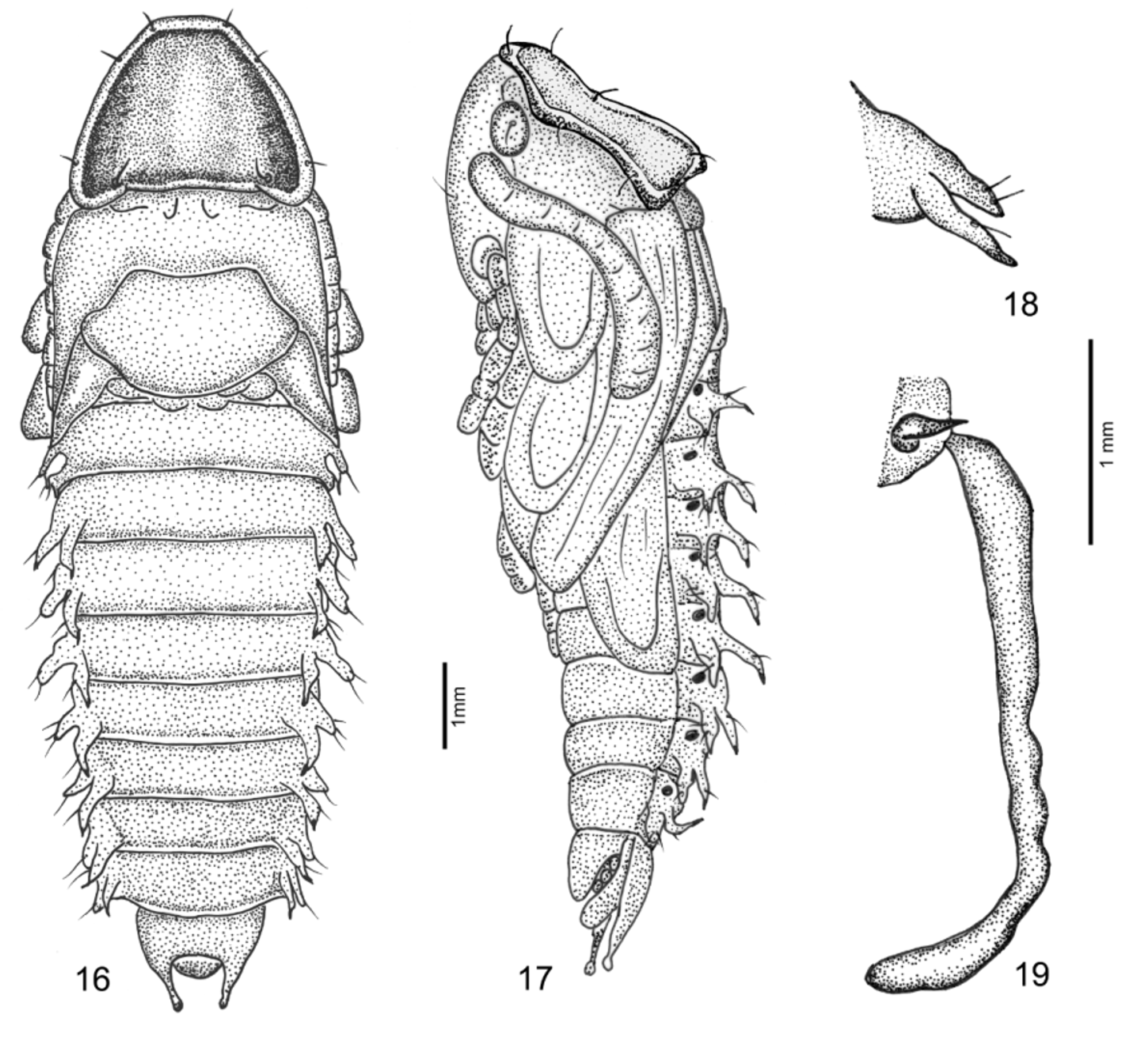
Pupa description (figs.16–18). Adecticous and exarate. Body slightly depressed, cream and almost smooth. Head completely covered with pronotum in dorsal view. Eyes small, slightly convex. Mouthparts visible in ventral view. Antennae lying parallel to ventral sides of prothorax. Pronotum marginated, with four pairs of small setae each lateral side. Pterothecae extending up to fourth ventrite. Abdomen with nine visible tergites and six visible sternites. Tergum A9 with lateral margins rounded and fixed urogomphi. Lateral A1–A8 (fig.17, 18) with a pair of short peg-like process, inner lobe slightly longer and unisetose, outer lobe bisetose. Spiracles A1–A 8 in the pleural membrane, visible from lateral view.
Material examined. Macrolygistopterus subparallelus . BRAZIL. São Paulo, Salesópolis, Estação Biológica de Boracéia, 09-10. VII. 1992, Exc. BIZ-739 col., 1 larva fixed ( MZSP); 13. VI. 1996, S. Ide & R. dos Santos cols. 1 larva reared to adult; 1 larva reared to pupa and 1 larva fixed in alcohol, dissected ( MZSP). Rio de Janeiro, Rio de Janeiro (Corcovado), 1 male, IX.1967, Alvarenga & Seabra, cols ( MZSP).
Macrolygistopterus sp. BRAZIL. Mato Grosso, Chapada dos Guimarães (Buriti), 06.II.1986, C. Costa col., 4 larvae fixed, 1 prepupa reared to adult and 1 pupal exuvia ( MZSP).
Biological notes. Larvae were collected alive inside dead trunk in the Atlantic forest at Estação Biológica de Boracéia, Salesópolis, São Paulo, Brazil. Larvae were maintained at laboratory conditions, the pupal period was 12 days (one observation).
Geographic distribution. Macrolygistopterus subparallelus was originally described from Paraguay and subsequently recorded for the states of São Paulo and Rio de Janeiro, Brazil.
Discussion. The larva of Macrolygistopterus subparallelus is quite similar to the larva of Macrolygistopterus sp. described by Costa et al (1988). They share the exterior surface of mandibles with three setae aligned forming a longitudinal row and the median seta much longer. It should be stressed that in the figure of the head of Macrolygistopterus sp. presented by Costa et al. (1988, pl. 66, fig. 7) the posterior limits of the maxillolabial complex were not represented, and the cardines omitted. Larvae of both species differ by the campodeiform body, almost cylindrical and subparallel in M. subparallelus and slightly depressed in Macrolygistopterus sp., and by the lateral margin of segments A1–A8 produced forming a short unisetose blunt process in M. subparallelus , as well as with stick-like process in Macrolygistopterus sp. The urogomphi are fixed, robust, curved, with blunt apex in both species, but in M. subparallelus it is longer.
The peg-like processes of A1–A8 are very different in the pupae of M. subparallelus and Macrolygistopterus sp. (observed in the pupal exuvia). In the former ( Fig. 18 View FIGURE 16 – 18 ), the peg-like processes are subequal in length, the inner are unisetose and the outer are bisetose. In the latter (Fig.19), the processes are asetose, the inner are very small and spiniform and the outer are about four to five times longer than in M. subparallelus .
The larva of Macrolygistopterus subparallelus differs from Lygistopterus sanguineus (Linnaeus, 1758) mainly by the smooth integument, color pattern and longer urogomphi; both species share the lateral margin of A1–A8 produced forming a short unisetose blunt process.

| MZSP |
Sao Paulo, Museu de Zoologia da Universidade de Sao Paulo |
No known copyright restrictions apply. See Agosti, D., Egloff, W., 2009. Taxonomic information exchange and copyright: the Plazi approach. BMC Research Notes 2009, 2:53 for further explanation.
|
Kingdom |
|
|
Phylum |
|
|
Class |
|
|
Order |
|
|
Family |
|
|
Genus |