
Proislandiana pallida ( Kulczyński, 1908 )
|
publication ID |
https://doi.org/ 10.11646/zootaxa.4743.2.7 |
|
publication LSID |
lsid:zoobank.org:pub:F4FF78DB-1568-457B-9A95-0B8915FE879C |
|
DOI |
https://doi.org/10.5281/zenodo.3688022 |
|
persistent identifier |
https://treatment.plazi.org/id/03955567-FFBC-C713-13E1-3B09FBC2FEBC |
|
treatment provided by |
Plazi |
|
scientific name |
Proislandiana pallida ( Kulczyński, 1908 ) |
| status |
|
Proislandiana pallida ( Kulczyński, 1908) View in CoL
Figs 17–24 View FIGURES 17–20 View FIGURES 21–24
Microneta View in CoL (?) pallida Kulczyński, 1908: 36 View in CoL , pl. 1, Figs 34–35
Proislandiana pallida: Tanasevitch, 1985: 56 View in CoL , Figs 10–12 View FIGURES 9–12
Proislandiana pallida: Tanasevitch & Khruleva, 2017: 355 View in CoL
Material examined. 2♂, 2♀. Russia, Arkhangelsk area, Nenets AO, env. of Amderma , 69.760° N, 61.659 ° E, 23.07.– 06.08.2016, leg. A. Tanasevitch, O. Khruleva ( MHNG) GoogleMaps .
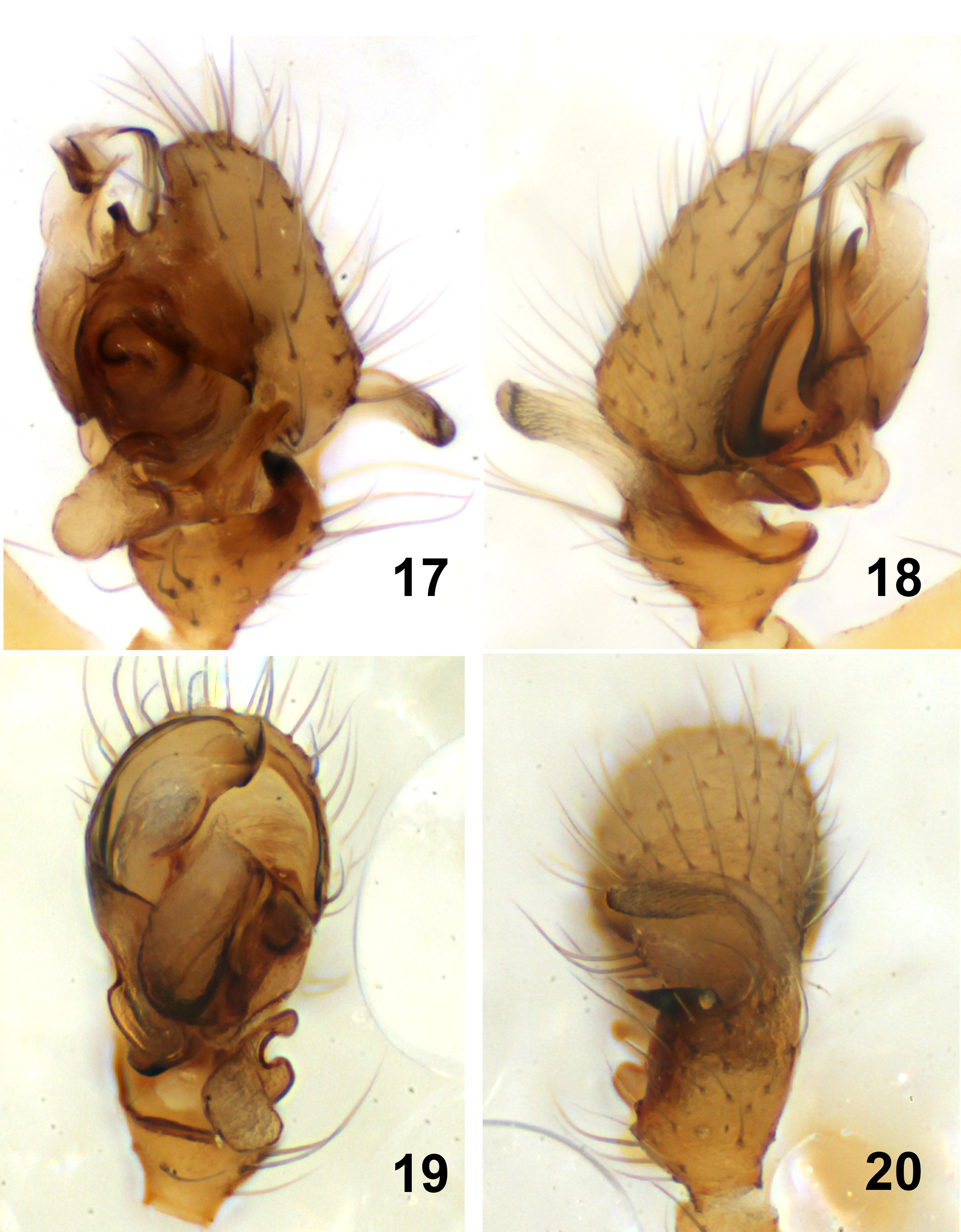
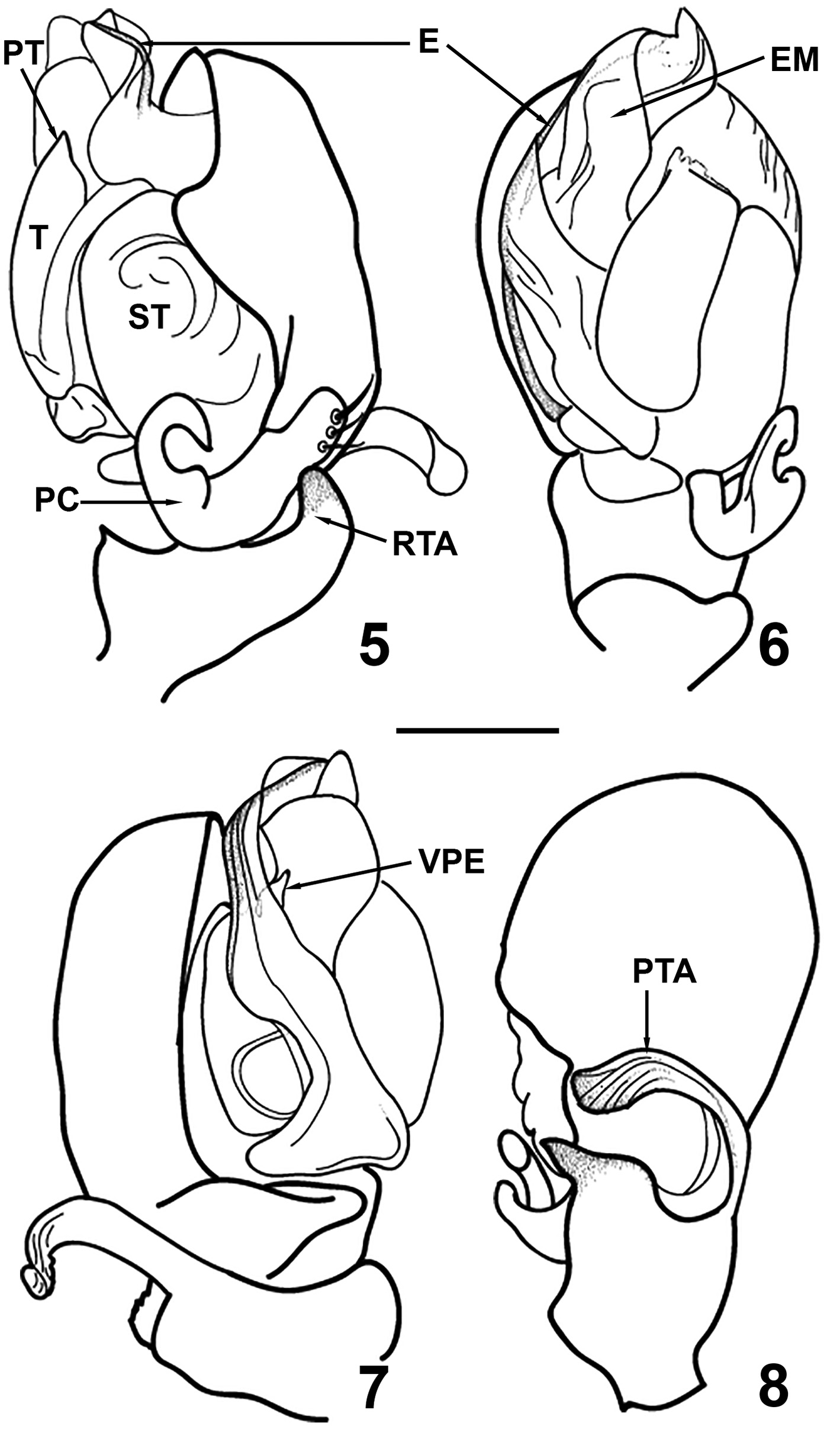
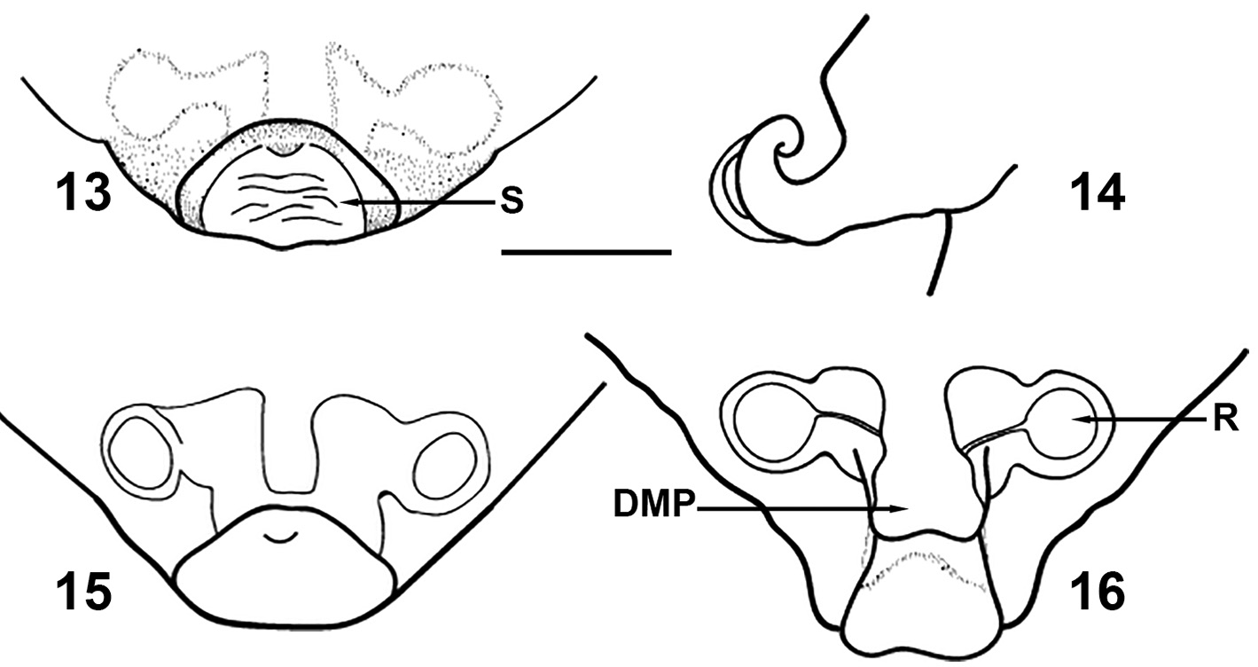
Diagnosis. The species is close to P. beroni n. sp. Males can be distinguished by the bulged paracymbium ( Fig 17 View FIGURES 17–20 ), as well as by the shape of the embolic membrane ( Fig 17–19 View FIGURES 17–20 ) and the shape of the prolateral tibial apophyse. In P. pallida the prolateral tibial apophyse is bent in the middle, the distal part is strait, situated perpendicularly to the cymbium ( Fig 20 View FIGURES 17–20 ), while in P. beroni n. sp. it is arc-shaped ( Figs 4 View FIGURES 1–4 , 8 View FIGURES 5–8 ). The females differ from those of P. beroni n. sp. by the longer and narrower epigynal scape ( Figs 21, 23 View FIGURES 21–24 ). Seen from lateral view, the scape of P. pallida is ringshaped ( Fig 22 View FIGURES 21–24 ), while in P. beroni n. sp. it looks like semi-ring ( Figs 10 View FIGURES 9–12 , 14 View FIGURES 13–16 ).
Redescription. Male. Measurements. Total length 1.66; carapace length 0.76, width 0.55; sternum length 0.45, width 0.40; abdomen length 0.90, width 0.72; clypeus length 0.11; chelicerae length 0.35; eye diameters AME 0.035, ALE 0.050, PME 0.050, PLE 0.050; leg measurements I—2.57 (0.30, 0.65, 0.20, 0.54, 0.45, 0.43), II—2.28 (0.32, 0.45, 0.18, 0.50, 0.43, 40), III—2.02 (0.29, 0.54, 0.15, 0.36, 0.36, 0.32), IV—2.56 (0.29, 0.66, 0.18, 0.60, 0.54, 0.40); palpal cymbium length 0.26.
Carapace unmodified. Chelicerae with four large, sharp promarginal and four small retromarginal teeth. The retromarginal teeth located in the distal part of the chelicerae, near the cheliceral fang.
Coloration. Carapace, chelicerae and legs light yellow. Sternum a little darker than the carapace.Abdomen gray, without pattern, covered with small dark hairs.
Dorsal tibial spination 2, 2, 2, 1. The position of TmI—0.45.
Palp ( Figs 17–20 View FIGURES 17–20 ). Tibia with one prolateral and one retrolateral apophyses ( Fig 20 View FIGURES 17–20 ). The retrolateral one small, curved, narrowing apically. The prolateral one long, curved at the middle. Its apical part straight, positioned perpendicularly to the cymbium ( Fig 20 View FIGURES 17–20 ). Cymbium simple, covered with thin black hairs. Paracymbium bulged. Its distal part notched on the inner side. The base of paracymbium bears 3 stout spines near the tip ( Fig 17 View FIGURES 17–20 ). Embolus long, thin, curved out apically ( Figs 18–19 View FIGURES 17–20 ). Ventral part of the embolic division thin, thorn-shaped, well chitinized ( Fig 18 View FIGURES 17–20 ). Embolic membrane large and complex ( Fig 19 View FIGURES 17–20 ).
Female. Measurements. Total length 2.35; carapace length 0.85, width 0.60; sternum length 0.54, width 0.40; abdomen length 1.50, width 0.94; clypaeus length 0.11; chelicerae length 0.35; eye diameters AME 0.036, ALE 0.055, PME 0.55, PLE 0.055; leg measurements I—2.78 (0.32, 0.70, 0.22, 0.58, 0.54, 0.42), II—2.66 (0.30, 0.68, 0.22, 0.58, 0.48, 0.40), III—2.31 (0.25, 0.58, 0.20, 0.45, 0.47, 36), IV—2.87 (0.30, 0.75, 0.22, 0.76, 0.44, 0.40).
Chelicerae with four large teeth on promargin and four small ones on retromargin. The promarginal teeth bigger than in male.
Epigynum ( Figs 21–24 View FIGURES 21–24 ). Scape short and relatively narrow, with an upturned distal part, making a full ring, clearly seen from lateral view ( Figs 21–23 View FIGURES 21–24 ). The dorsal median plate rounded at the end ( Fig 24 View FIGURES 21–24 ). Receptacles oval, ducts simple and strait. The genital openings are on the dorsal side, near the tip.
Coloration as in male. Dorsal tibial spination 2, 2, 2, 1. The position of TmI—0.43.
Distribution. Arctic region of Russia, from the polar Ural Mts. to the Far East.


| MHNG |
Museum d'Histoire Naturelle |
No known copyright restrictions apply. See Agosti, D., Egloff, W., 2009. Taxonomic information exchange and copyright: the Plazi approach. BMC Research Notes 2009, 2:53 for further explanation.
|
Kingdom |
|
|
Phylum |
|
|
Class |
|
|
Order |
|
|
Family |
|
|
Genus |
Proislandiana pallida ( Kulczyński, 1908 )
| Dimitrov, Dragomir 2020 |
Proislandiana pallida: Tanasevitch & Khruleva, 2017: 355
| Tanasevitch, A. V. & Khruleva, O. A. 2017: 355 |
Proislandiana pallida:
| Tanasevitch, A. V. 1985: 56 |
Microneta
| Kulczynski, W. 1908: 36 |