
Turleania balli ( McLaughlin & Haig, 1996 )
|
publication ID |
https://doi.org/10.5281/zenodo.181757 |
|
DOI |
https://doi.org/10.5281/zenodo.6230735 |
|
persistent identifier |
https://treatment.plazi.org/id/03958703-3921-FFE0-83AD-FF232A88197E |
|
treatment provided by |
Plazi |
|
scientific name |
Turleania balli ( McLaughlin & Haig, 1996 ) |
| status |
|
Turleania balli ( McLaughlin & Haig, 1996) View in CoL
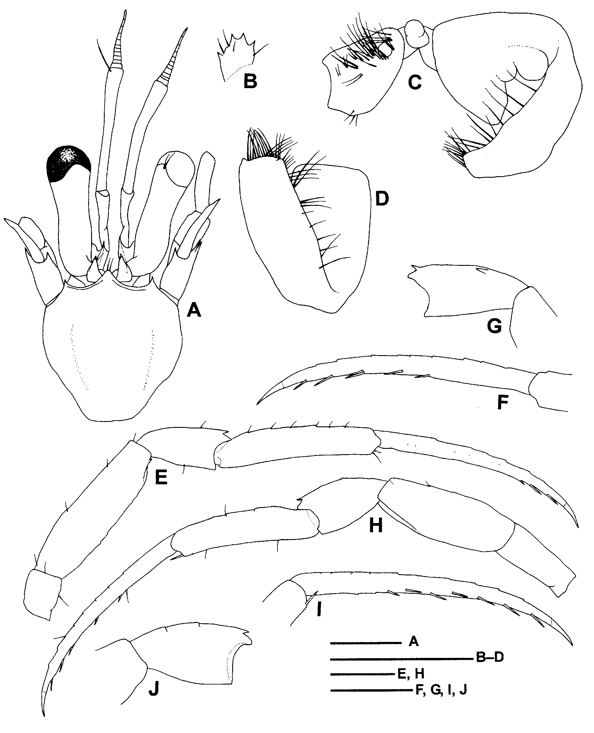
( Figs. 1 View FIGURE 1 , 2 View FIGURE 2 )
Anapagrides View in CoL sp. — Haig & Ball, 1988: 177, fig.8. Laurentia balli McLaughlin & Haig, 1996: 84 , fig. 5. Turleania balli View in CoL . — McLaughlin, 1997: 477.
Type material. Holotype: male (sl 2.1 mm), off Pulau Saparua, Indonesia, 3°37.9’ S, 128°38.6–39’ E, “Alpha Helix” Saparua St. 3, 0–20 m, cliff face of rock and coral, coll. E. Ball, 29 March 1975 ( NIOJ).
Redescription. Nine pairs of quadriserial gills; no arthrobranchs on third maxillipeds.
Shield ( Fig. 1 View FIGURE 1 A) 1.1 times longer than broad; anterior margin between rostrum and lateral projections concave; anterolateral margins sloping; posterior margin roundly truncate. Rostrum triangular, well developed, overreaching lateral projections. Lateral projections also well developed, each terminating in small spine.
Ocular peduncles ( Fig. 1 View FIGURE 1 A) 0.8 length of shield, with corneas slightly dilated; lateral surfaces concave medially; ocular acicles narrowly subtriangular, each with small marginal spine.
Antennular peduncles ( Fig. 1 View FIGURE 1 A) overreaching distal margins of corneas by 0.6 length of ultimate segment.
Antennal peduncles ( Fig. 1 View FIGURE 1 A) also overreaching distal margins of corneas by 0.3 length of fifth segments. Second segment with small spine at dorsomesial distal angle; dorsolateral distal angle produced, moderately long, terminating in single or bifid spine. Antennal acicle slightly arcuate, barely reaching proximal margin of cornea, terminating in small spine.
Mouthparts not dissected. Third maxilliped with well developed crista dentata of small teeth but without accessory tooth on ischium.
Chelipeds ( Fig. 2 View FIGURE 2 A–H) subequal in length but right ( Fig. 2 View FIGURE 2 A–E) distinctly stouter than left ( Fig. 2 View FIGURE 2 F–H). Right chela elongate oval in general outline, 2.6 times as long as broad. Dactylus approximately as long as palm, dorsomesial margin with small proximal spines and short slightly elevated ridges distally, cutting edge with 2 prominent teeth in proximal half and row of small corneous teeth distally. Palm approximately as long as carpus, dorsomesial margin with row of 4 strong spines, dorsolateral margin not delimited, dorsal surface with small but distinct spines laterally, cutting edge of fixed finger with 2 prominent calcareous teeth medially and some smaller teeth on remaining margin. Carpus slightly longer than merus, dorsomesial margin with row of 3 spines decreasing in size proximally, dorsodistal margin with median spine, ventrolateral margin with small distal spine, ventromesial margin unarmed. Merus also with spine at each ventrolateral and ventromesial distal angle.
Left chela elongate, 3.4 times longer than broad. Dactylus 1.2 times longer than palm, unarmed on surfaces, cutting edge with row of short corneous teeth. Palm 0.7 length of carpus, unarmed; cutting edge of fixed finger with row of small calcareous and corneous teeth. Carpus approximately as long as merus, dorsolateral margin with row of 3 spines, dorsomesial margin with only strong distal spine, ventrolateral margin with small distal spine, ventromesial margin unarmed. Merus with small spine at ventromesial distal angle and 2 subdistal spines on ventrolateral margin.
Ambulatory legs ( Fig. 1 View FIGURE 1 E–I) slender [left second pereopod missing, but illustrated by Haig & Ball (1988, fig. 8E)]. Dactyli 1.4–1.5 times longer than propodi, slightly curved distally in lateral view, terminating in long corneous claws; dorsal surfaces each with row of small pits; ventral margins each with row of 6 or 7 slen- der corneous spines. Propodi not tapering distally, ventrodistal margins each with slender corneous spine. Carpi each with small subdistal spine on dorsal surface, right second pereopod with additional small spine in proximal half. Meri each with very small subdistal spine (right second) or unarmed (third) on ventral margin. Ischia unarmed on surfaces.
Fourth pereopods [now missing, but right fourth pereopod was illustrated by Haig & Ball (1988, fig. 8G) and described by McLaughlin & Haig (1996)] semichelate; dactylus without preungual process; propodal rasp composed of single row of corneous scales; carpus unarmed on dorsal surface.
Coxae of fifth pereopods ( Fig. 1 View FIGURE 1 C) unequal, left coxa larger than right, with well developed, stout and flattish sexual tube directed exteriorly and recurved posteriorly, and then upward, with broad tip obscured by dense fringe of short setae, surfaces also with scattered setae; right coxa with gonopore partially obscured by fringe of short setae.
Sixth thoracic sternite with anterior lobe ( Fig. 1 View FIGURE 1 B) roundly subquadrate and bearing 4 small marginal spines. Eighth thoracic sternite ( Fig. 1 View FIGURE 1 C) developed as single, small subovate lobe.
Telson [missing, but figured by Haig & Ball (1988, fig. 8H)] with lateral indentations indistinct; posterior lobes markedly asymmetrical, with terminal margins oblique, each with 3 small spines and stronger apical spine; lateral margin of left lobe with narrow chitinous plate.
Habitat. Haig & Ball (1988) mentioned that the sole specimen was collected on a cliff face of rock and coral, at a depth of 5 m or less. On the label of the holotype, the depth is given as 0– 20 m.
Distribution. Confirmed records of T. balli are restricted Saparua Island in Maluku, Indonesia. A specimen lacking chelipeds and ambulatory legs from “Siboga” station “Banda” was tentatively assigned to T. balli (as Laurentia balli ) by McLaughlin & Haig (1996).
Remarks. McLaughlin & Haig (1996) described the present specimen as a new species of their genus Laurentia , L. balli . Subsequently, McLaughlin (1997) proposed the replacement name Turleania for Laurentia because the latter proved to be a junior homonym and established an allied genus Enneopagurus for E. garciagomezi McLaughlin, 1997 from deep waters off Indonesia.
The holotype of Turleania balli is now in poor condition. The specimen is largely decalcified in the integument and has much fewer setae on the chelipeds and ambulatory legs than those illustrated by Haig & Ball (1988, fig. 8 B–G, as Anapagrides sp.) and McLaughlin & Haig (1996, fig. 5B, C, as Laurentia balli ). The reexamination of the holotype has revealed that it possesses only nine pairs of quadriserial gills (arthrobranchs are absent from the third maxilliped) and the left sexual tube being comparatively broad and flattish. The number of gills is consistent with that of the two new species described below and with the diagnosis of the allied genus Enneopagurus . However, these three species fit much better in Turleania than Enneopagurus in the other characters such as the elongate ocular peduncles with slightly dilated corneas, the right cheliped being much stouter than the left, the right chela with some distinct spines, and the dactyli of the ambulatory legs each armed with a row of well developed corneous spines on the ventral margin. In the sole species of Enneopagurus , E. garciagomezi , the ocular peduncles are stout and have strongly dilated corneas, the palm of the right cheliped lacks spines on the dorsomesial margin, and the dactyli of the ambulatory legs are unarmed or have only tiny spinules on the ventral margin. Thus, the generic diagnosis of Turleania is emended to include nine to eleven pairs of quadriserial gills to accommodate McLaughlin & Haig’s species and the two new species from the Ryukyu Islands.
In the holotype of T. balli , the dactyli of the ambulatory legs each have a row of small pits on the dorsal surface, which represent setal insertions. The pits show that they had bristles as observed in the T. saliens n. sp. and T. tenebrosa n. sp.

No known copyright restrictions apply. See Agosti, D., Egloff, W., 2009. Taxonomic information exchange and copyright: the Plazi approach. BMC Research Notes 2009, 2:53 for further explanation.
|
Kingdom |
|
|
Phylum |
|
|
Class |
|
|
Order |
|
|
Family |
|
|
Genus |
Turleania balli ( McLaughlin & Haig, 1996 )
| Osawa, Masayuki & Fujita, Yoshihisa 2008 |
Anapagrides
| McLaughlin 1997: 477 |
| McLaughlin 1996: 84 |
| Haig 1988: 177 |