
Protolichus strangulatus Favette et Trouessart, 1904
|
publication ID |
https://doi.org/ 10.5281/zenodo.196425 |
|
DOI |
https://doi.org/10.5281/zenodo.5686093 |
|
persistent identifier |
https://treatment.plazi.org/id/039587A1-5327-5430-FF14-DED4E512FAE9 |
|
treatment provided by |
Plazi |
|
scientific name |
Protolichus strangulatus Favette et Trouessart, 1904 |
| status |
|
Protolichus strangulatus Favette et Trouessart, 1904
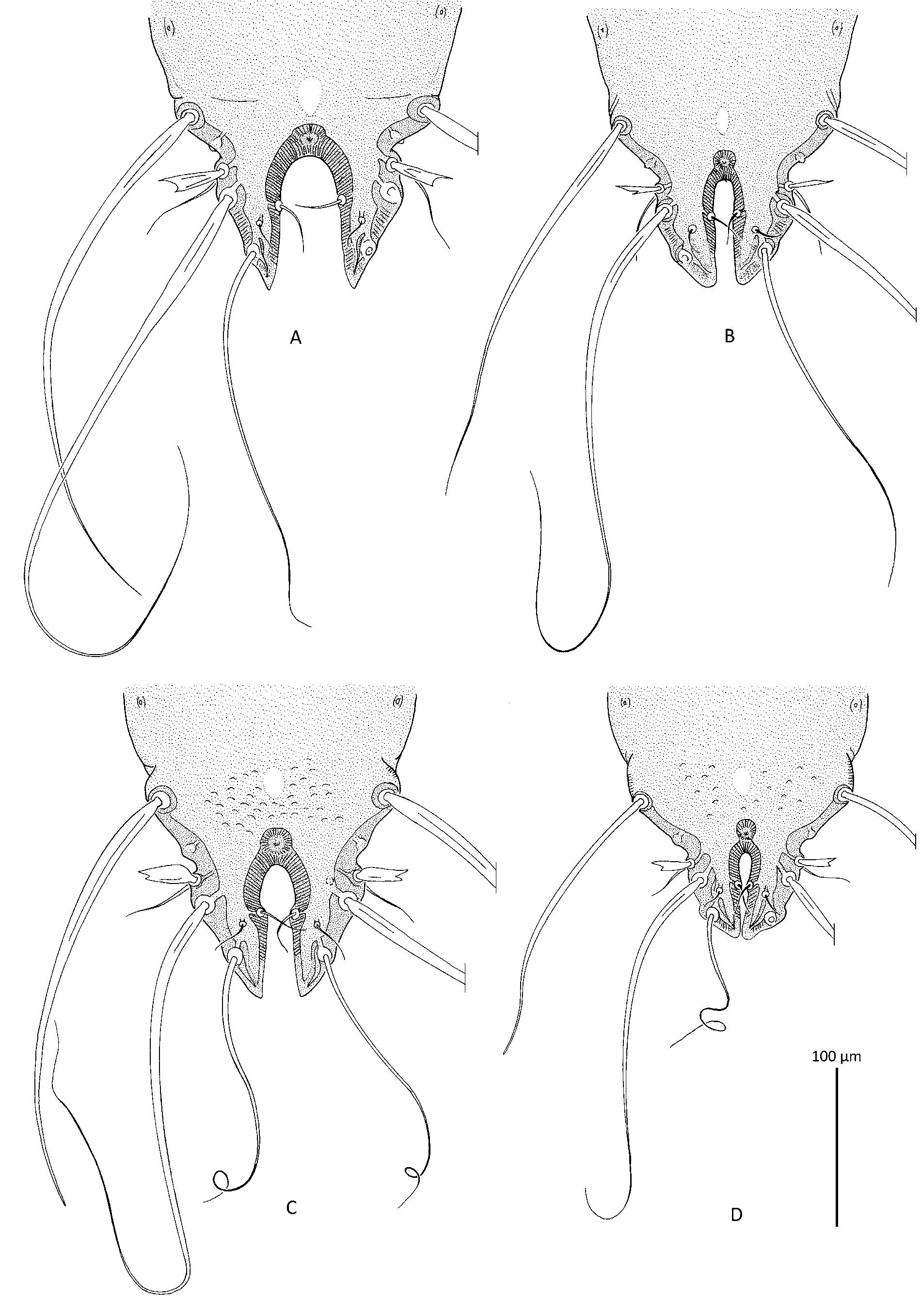
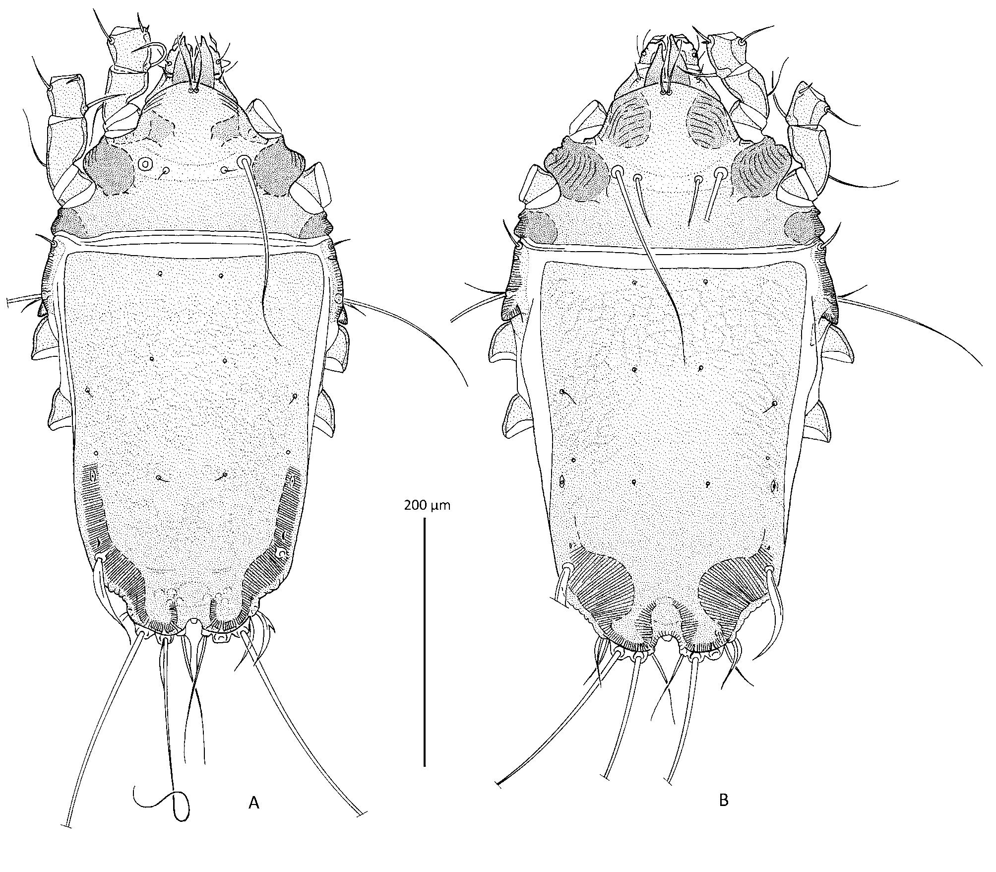
( Figs 5 View FIGURE 5 C, D, 6, 11A)
Material examined. Heteromorph male lectotype, homeomorph male and 2 female paralectotypes (3 slides with number TRT 34F1) ex Charmosyna pulchella Gray G.R., 1859 ( Psittacidae : Loriinae ), New Guinea, no other data; lectotype designated here. 1 female ( TRT 34F14, labeled as P. brachiatus ) ex Loriculus sclateri Wallace, 1863 , Sulawesi (contamination).
Description. Heteromorph male (lectotype). Idiosoma, length x width, 590 x 305. Subcapitulum with posterior margin strongly convex, widest part crossed by single transverse fold, area with setae subc outlined by short folds forming lateral and posterior sides ( Fig. 6 View FIGURE 6 G). Prodorsal shield: posterior margin straight, length along midline 150, surface monotonously punctate, transverse bar across scapular setae poorly sclerotized. Setae si missing (probably spiculiform). Distance between scapular setae: se-se 65, si-si 28. Hysterosoma 420 long. Length of hysteronotal shield 405, width 240, anterior margin slightly convex, most of surface monotonously punctate, posterior part near bases of opisthosomal lobes with numerous poorly expressed pits ( Fig. 5 View FIGURE 5 C). Opisthosomal lobes with acute apices, without transverse crest posterior to base of setae h3. Terminal cleft as a narrow inverted U, slightly enlarged in anterior third, length 82, width at level of setae e1 15, width in anterior part 22. Setae c2 spiculiform, 42 long; setae d2, setiform, about 5 long, setae e2 represented by macrosetae extending far beyond lobar apices, terminal part thick without filiform end, 255– 260 long; setae e1 setiform, situated on margin of terminal cleft, slightly posterior to level of macrosetae h2; setae f2 flattened, spatuliform with bidentate apex. Distance between dorsal setae: c2:d2, 135, d2:e2 150, e2:h3 125, e2: e1 70, e2:e2 140, h2:h2 90, h3:h3 58, ps1:ps1 48, e2:h2 62, h2:h3 32, h2: e1 8, ps1:h3 17. Genital apparatus 27 x 22, its base at level of trochanters IV. Anterior ends of paragenital apodemes connected with inner tips of epimerites IIIa, middle parts of apodemes connected by large and poorly sclerotized transverse bridge ( Fig. 6 View FIGURE 6 A). Distance between ventral setae: 3b:3a 8, 3a:g 30, g:4a 68, 4a:ps3 118. Cupules ih at level of setae ps2. Diameter of anal suckers 21.
Legs II approximately 1.5 times longer than legs I. Tarsus I with crest-like ventral extension stretching along all segment, proximal end of this extension widely rounded ( Fig. 6 View FIGURE 6 B). Tibia I, II with small verrucous ventral tubercle, solenidia φ of these segments with verrucae ( Figs 6 View FIGURE 6 B, C). Genu I and femora I, II without any apophyses. Tarsus II with small ventral extensions bearing setae s and wa. Genu II with dorso-apical tubercle; seta cG II strongly thickened along its entire length, smooth, nearly 3 times longer than segment. Setae d, e of tarsus IV cone-shaped, with inflated base, poorly sclerotized ( Fig. 6 View FIGURE 6 D).
Homeomorph male (1 paralectotype). Subcapitulum with posterior margin strongly convex, median part with several transverse striae ( Fig. 6 View FIGURE 6 H). Terminal cleft as a very narrow U, approximately 6 times longer than wide. Anterior ends of paragenital apodemes not connected with epimerites IIa, IIIa; transverse bridge between middle parts of paragenital apodemes absent.
Legs II about 1.3 times longer than legs I. Tarsi I, II with blunt-angular ventral margin. Tibiae II with scarcely distinct ventral tubercles. Setae cG of genu II thickened, approximately 1.4 times longer than segment.
Measurements: Idiosoma, length x width, 490 x 260. Prodorsal shield 135 long. Setae si 22 long. Distance between scapular setae: se:se 65, si:si 35. Hysterosoma 350 long. Length of hysteronotal shield 330, width 200. Terminal cleft 52 x 5. Lateral setae c2 28 long; setae d2 5 long; setae e2 thick represented by macrosetae extending beyond lobar apices, terminal part thick without filiform tip, about 180 long. Distances between dorsal setae: c2:d2 120, d2:e2 130, e2:h3 75, e2: e1 55, e2:e2 122, h2:h2 55, h3:h3 32, ps1:ps1 28, e2:h2 50, h2:h3 22, h2: e1 8, ps1:h3 17. Genital apparatus 28 x 18. Distance between ventral setae: 3b:3a 12, 3a:g 18, g:4a 58, 4a:ps3 100. Diameter of anal suckers 20.
Female (2 paralectotypes). Gnathosoma as in homeomorph male. Idiosoma, length x width, 440–450 x 242–250. Prodorsal shield with posterior margin slightly concave; length along midline 118–128. Setae si setiform, 10–12 long. Distance between scapular setae: se:se 75–78, si:si 45–50. Hysterosoma 320–325 long. Opisthosoma with short bluntly rounded opisthosomal lobes, terminal cleft as a small inverted U ( Fig. 11 View FIGURE 11 A). Length of hysteronotal shield 300–315, width 205–210, anterior margin slightly concave, most of surface monotonously punctate with faint net-like striations, medial area posterior to level of setae e2 with several (6– 10) lacunae; subtegumental sclerotized bars in postero-lateral parts of opisthosoma narrow. Setae c3 setiform, 15–18 long. Setae c2, d2, setiform, short, setae e2 flattened, saber-shaped; setae f2, ps2 slightly flattened and enlarged in middle part, f2 usually with additional spine on outer margin; setae e1 situated between levels of openings gl and cupules im. Length of hysteronotal setae: c 2 10–12, d2 5, e2 65 –68, f2 40. Distance between setae: c2:d2 115–125, d2:e2 128–130, e2:h3 68–70, e2:e2, 140–145, h2:h2 75–80, ps1:ps 1 20–22. Epigynum bow-shaped, 13–15 x 40 –42. Seta mG of genu I thin, spiculiform.
Type host and locality. Charmosyna pulchella (designated here), New Guinea.
Remark. Favette and Trouessart (1904) described this species as a subspecies of P. brachiatus and mentioned two hosts, Lorius garrulus and Charmosyna pulchella , from New Guinea. There are formally no specimens labeled as “ strangulatus ” in the Trouessart collection. Nevertheless, this collection contains slides with a specimen from Charmosynopsis pulchella (recent name Charmosyna pulchella ) from New Guinea and identified by Trouessart as “ P. brachiatus ”. Probably it was a preliminary identification, because neither the original description of P. brachiatus nor the subsequent revision of Protolichus listed Ch. pulchella as a host of Protolichus species other than P. b. strangulatus . Based on this indirect evidence, we consider that specimens in three slides (34F1) from Ch. pulchella represent a part of type series of P. strangulatus . Additional evidence is a very clear correspondence of the homeomorph male in this slides to the photo of P. b. strangulatus given by Favette and Trouessart (1904: Pl. V, Fig. 4 View FIGURE 4 .), particularly in respect to a very narrow terminal cleft (compare Fig. 5 View FIGURE 5 B with 5D), a feature that was specifically stressed in the original description of this species as the character discriminating it from P. brachiatus . It is necessary to add that one male in this slide is actually a heteromorph one. Based on the facts above, we mark the lectotype of P. strangulatus , in the specimen 34F1 from Ch. pulchella .
Trouessart’s (1884) report of P. brachiatus from Loriculus sclateri is quite obviously the result of contamination. Examination of the slide 34F14 determined as P. brachiatus , has shown that it actually contains a female of P. strangulatus .


| TRT |
Royal Ontario Museum - Herbarium |
No known copyright restrictions apply. See Agosti, D., Egloff, W., 2009. Taxonomic information exchange and copyright: the Plazi approach. BMC Research Notes 2009, 2:53 for further explanation.