
Hyalascus farallonesensis, Reiswig, 2018
|
publication ID |
https://doi.org/ 10.11646/zootaxa.4466.1.11 |
|
publication LSID |
lsid:zoobank.org:pub:5410B0DF-67BA-4D9A-B891-3ADFAB79A8EC |
|
DOI |
https://doi.org/10.5281/zenodo.5970391 |
|
persistent identifier |
https://treatment.plazi.org/id/039587B3-BE21-FFEF-FF51-FC7A5C7D79D8 |
|
treatment provided by |
Plazi |
|
scientific name |
Hyalascus farallonesensis |
| status |
sp. nov. |
Hyalascus farallonesensis View in CoL n. sp.
( Figs 11 View FIGURE 11 & 12 View FIGURE 12 , Table 6)
Material examined. Type material: Holotype: CAS 218807, ROV Hercules from EV Nautilus , dive H 1566, 26 Aug 2016, Wreck of USS Independence, side of midship gun turret, off Farallones Is., Greater Farallones National Marine Sanctuary, off San Francisco, California, U.S.A., 37.4776°N, 123.1346632°W, 805.8 m, Fix 95% ethanol GoogleMaps . Paratype: CAS 218809, same ship, same dive, same date, same general location, edge of flight deck by aft elevator, 37.4775 °N, 123.1345499 °W, 802.6 m.
Not seen: Holotype fragment: MCZ IZ 141480, data as above. Paratype fragment: MCZ IZ 141485, data as above.
Species Diagnosis. Hyalascus with bare surface (no prostalia), hypodermalia with tangential rays to 4.6 mm long, dermalia are about equal numbers of rough stauractins and pentactins, atrialia as mostly non-pinular hexactins. Microscleres include oxyhexactins, hemioxyhexasters, full oxyhexasters and microdiscohexasters of small size (19.1–32.9 µm).
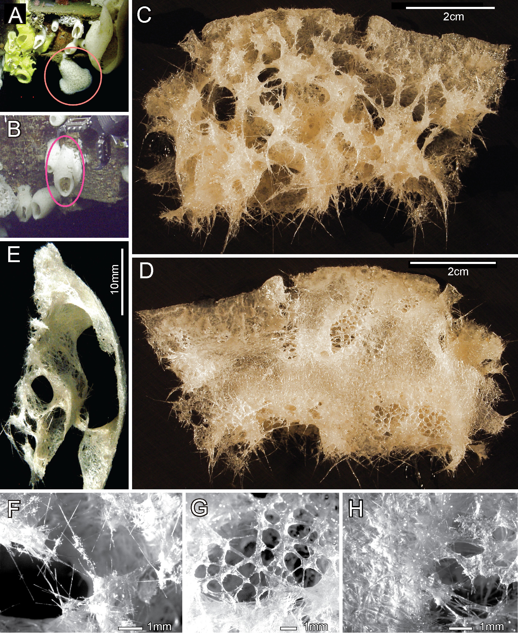
Description. Body form of the holotype is that of a hanging trumpet with the distal body wall turned out to 90° at the oscular margin ( Fig. 11A View FIGURE 11 ), while the paratype is a cylindrical tube without any diversion of the distal body wall ( Fig. 11B View FIGURE 11 ). Bodies of both specimens are soft, smooth (without prostalia) and so cavernous that upon removal from water the body and especially the dermal side collapses ( Fig. 11C View FIGURE 11 ) leaving only strands of tissues where support is adequate. The atrial surface has firm subsurface support and thus retains its form ( Fig. 11D View FIGURE 11 ). The extent of the large water-filled spaces is clearly shown in a thick wall section of the turned-out margin of the holotype ( Fig. 11E View FIGURE 11 ). The dermal lattice of the outer surface remains only as patches on strands of diactins left standing after collapse of most of the dermal surface ( Fig. 11F View FIGURE 11 , dermal side on left). The atrial surface of the holotype is traversed by pore fields of ca 8 x 17 mm diameter scattered across the surface ( Fig. 11D View FIGURE 11 ); the individual pores are about 0.3– 0.9 – 1.5 mm in diameter ( Fig. 11G View FIGURE 11 ). The larger area of the atrial surface appears imperforate but fine spaces probably remain between spicules; the atrialia here form a compact felt-like layer on a close-packed layer of supporting diactins ( Fig. 11H View FIGURE 11 ). In the paratype, the relative areas of pore fields and aporous surface are reversed; the pore fields covered almost the entire surface and the aporous areas were relatively small. The holotype is calculated to have been 40.6 cm long by 38.5 cm wide at the flared oscular margin with collection restricted to a significant part (nearly half) of the margin; the fragment available for study was an 8.1 by 4.5 cm marginal subsample of that, ( Figs 11C–D View FIGURE 11 ). Total size of the paratype could not be determined due to orientation of the specimen of in situ images but it is similar in size to the holotype. The preserved fragment available for study was again a subsample of the margin, 7.0 x 3.9 cm, Color of the fragments preserved in ethanol is light tan. The known distribution of the species is the type location on the USS Independence sunken in what is now the Farallones Sanctuary off San Francisco, California at a depth of 802.6-805.8 m.
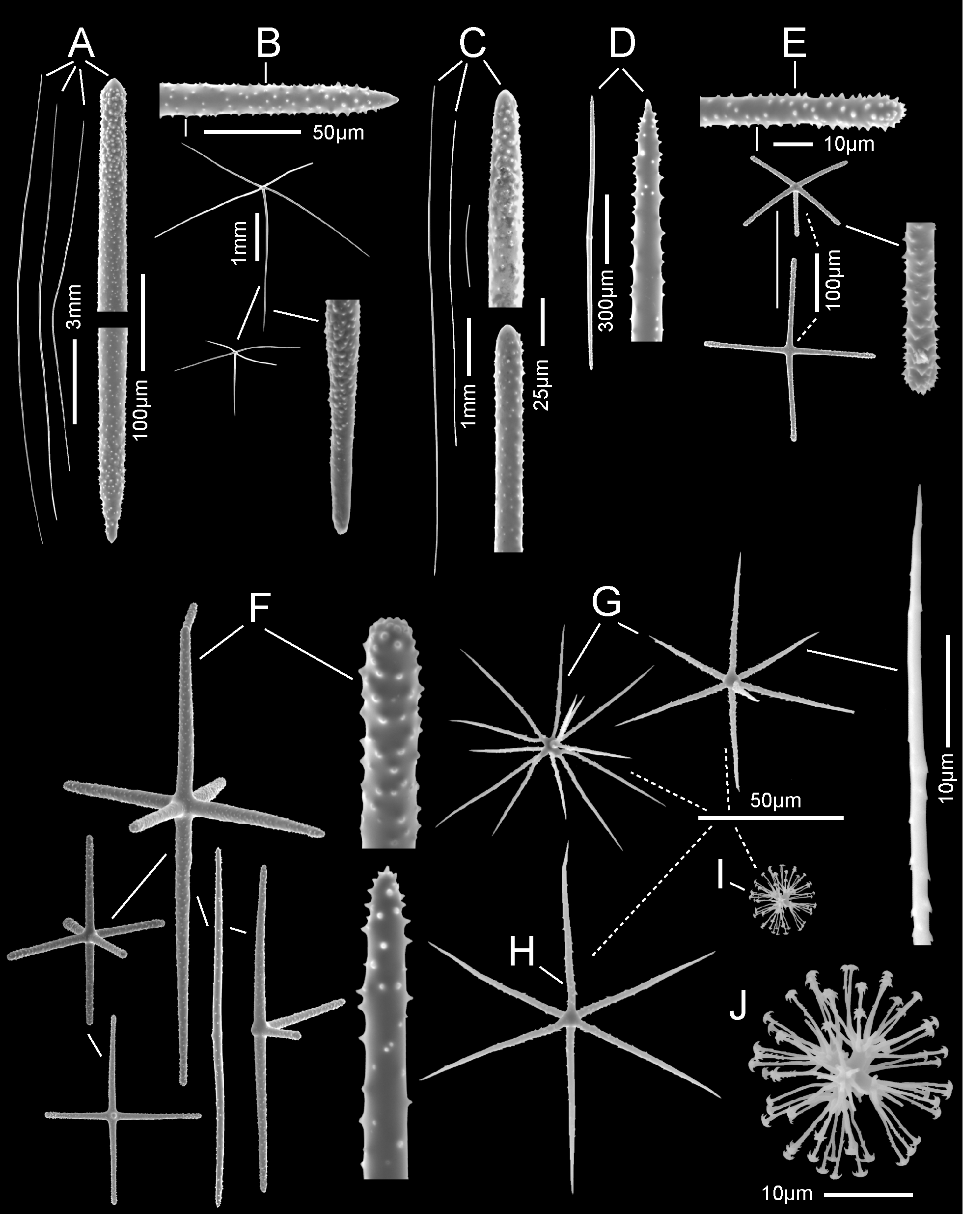
Megascleres (for measurements see Table 6) a variety of diactins (primary, choanosomal, small atrial), hypoatrial pentactins, dermalia and atrialia. Primary diactins ( Fig. 12A View FIGURE 12 ) are the longest and thickest spicules, usually gently curved and reaching nearly 17 mm; their tips are rough, either rounded or tapered to a small parabolic tip. Hypodermal pentactins ( Fig. 12B View FIGURE 12 ) are regular crucial forms with rays approximately equal in length; all ray ends are subterminally rough but the tangential tips are tapered to a smooth point and the proximal tips are slowly tapered to a long drawn-out smooth round tip. Choanosomal diactins ( Fig. 12C View FIGURE 12 ) are the most common spicules of the body; they are smooth. straight to gently curved, mostly smooth with rough rounded ends. Short diactins of the atrial surface ( Figs 12D & F View FIGURE 12 ) differ from typical body diactins in having sharp tips and being entirely rough. Their size and morphology suggest that the smaller members of this group may act to support atrialia and some may be atrialia, thus the two groups widely overlap in Table 6. Dermalia ( Fig. 12E View FIGURE 12 ) are mostly stauractins (53% of 100) and pentactins (43%) and a few tauactins; hexactins occur but are rare (<1%). These spicules are entirely rough, have cylindrical rays and have rounded rough tips without the smooth terminal end found in most diactins and hypodermal pentactins. Atrialia ( Fig. 12F View FIGURE 12 ) are patchy as to form; overall 1244 spicules from three locations were grouped with the most common being hexactins (50%), followed by diactins (22%), pentactins (19%), stauractins (6%), tauactins (1%), and paratetractins (0.6%).
Microscleres (for measurements see Table 6) are mainly oxy- and hemioxyhexasters (78% of 138), oxyhexactins (22%) and microdiscohexasters, not enumerated but their abundance is approximately the same as oxyhexasters. Oxyhexasters and hemioxyhexasters ( Fig. 12G View FIGURE 12 ) have short primary rays and 1-3 relatively stumpy robust secondary rays on each primary ray; terminal rays are sparsely covered with very small reclined spines. Oxyhexactins ( Fig. 12H View FIGURE 12 ) are similar to oxyhexasters but lack ray branching. Microdiscohexasters ( Figs 12I –J View FIGURE 12 ) are spherical; each smooth and stout primary ray carries a tuft of 11– 14.2 –17 (n = 12) thin, crooked, sparsely–spined secondary rays which are about twice the length of the primary rays. Disks at ray tips have 6–9 marginal teeth.
Remarks. This lyssacine species with choanosomal megascleres as diactins and with hypodermal pentactins is clearly a member of Rossellidae ; without strobiloplumicomes or discoctasters it falls into the subfamily Rossellinae . Keys to genera of Rossellinae and generic diagnoses in Systema Porifera ( Tabachnick 2002) show it is compatible only with the genus Hyalascus : hypodermalia are crucial (= orthotropal); lacks large choanosomal hexactine megascleres; microsclere ends are oxyoid and discoid forms; discoid microscleres are only micro-size [not defined], dermalia are mainly pentactins and stauractins. Hyalascus presently contains nine accepted species. Among other differences the new form differs from H. anisoactinus Tabachnick & Lévi, 2004 , in longer tangential rays of hypodermal pentactins (to 4.56 vs 0.6 mm) and much smaller microdiscohexasters (19.1–32.9 vs 25–72 µm); from H. attenuatus Okada, 1932 in longer tangential rays of hypodermal pentactins (to 4.56 vs 1.5 mm) and smaller microdiscohexasters (19.1–32.9 vs 40–45 µm); from H. baculifer ( Schulze, 1886) in having dermalia of different form (pentactins and stauractins vs diactins) and smaller discohexasters (19.1–32.9 vs 120 µm calculated from figure); from H. giganteus Ijima, 1898 , in longer tangential rays of hypodermal pentactins (to 4.56 vs 0.7 mm) and commonness of full oxyhexaster microscleres (~ 16% of oxy-tip microscleres vs none); from H. mitsukurii ( Ijima, 1898) in absence of prostal diactins (none vs present) and absence of a class of larger of discohexasters (none vs 80-120 µm class); from H. pinulohexactinus Tabachnick & Lévi, 2004 , in longer tangential rays of hypodermal pentactins (to 4.56 vs to 0.53 mm) and form of atrialia (non-pinular (regular) vs pinular); from H. sagamiensis Ijima, 1898 , in longer tangential rays of hypodermal pentactins (to 4.56 vs to 1.2 mm) and smaller microdiscohexasters (19.1–32.9 vs 80–90 µm); from H. similis Ijima, 1904 , in longer tangential rays of hypodermal pentactins (to 4.56 vs to 1.2 mm as in H. sagamiensis ) and smaller microdiscohexasters (19.1–32.9 vs 2 classes, spherical 76 µm and stellate 46–50 µm); from H. stellatus Schulze, 1886 , in mixed form of dermalia (near equal pentactins and stauractins vs almost all stauractins) and smaller microdiscohexasters (19.1–32.9 vs 50 µm calculated from figure). These and other differences lead to the conclusion that the Farallones specimens represent a new species of Hyalascus , here designated as H. farallonesensis .
Etymology. The species name is formed from the location of collection, the Greater Farallones National Marine Sanctuary.
| ROV |
Museo Civico di Rovereto |
No known copyright restrictions apply. See Agosti, D., Egloff, W., 2009. Taxonomic information exchange and copyright: the Plazi approach. BMC Research Notes 2009, 2:53 for further explanation.
|
Kingdom |
|
|
Phylum |
|
|
Class |
|
|
Order |
|
|
Family |
|
|
Genus |