
Domorganus suecicus, Holovachov, Oleksandr, 2012
|
publication ID |
https://doi.org/ 10.5281/zenodo.215493 |
|
DOI |
https://doi.org/10.5281/zenodo.5686079 |
|
persistent identifier |
https://treatment.plazi.org/id/039587B9-0D3C-FF87-0CA2-FDC3FA2B7778 |
|
treatment provided by |
Plazi |
|
scientific name |
Domorganus suecicus |
| status |
sp. nov. |
Domorganus suecicus sp. n.
( Fig. 1 View FIGURE 1 ; Table 1 View TABLE 1 )
Type material. Holotype female (slide # 8308), as well as one female and one male paratype (slides # 8309 and # 8310) each deposited in the type collection of the Department of Invertebrate Zoology, Swedish Museum of Natural History, Stockholm, Sweden.
Additional material. Three females and two males deposited in general collection of the Department of Invertebrate Zoology, Swedish Museum of Natural History, Stockholm, Sweden.
Type locality. Coarse gravel with detritus from 30–50 m deep, Skagerrak off the west coast of Sweden (N 58 17' 58'', E 11 10' 05''), 0 9 August 2011, legit O. Holovachov (two females and one male).
Additional localities. Sand and shells from 15–22 m deep, Bonden island in Skagerrak off the coast of Sweden (N 58 12' 37'', E 11 18' 53''), 0 9 August 2011, legit O. Holovachov (two females and two males); sediment, mud and gravel from 40–60 m deep, Skagerrak off the coast of Sweden (N 58 18' 06'', E 11 05' 22''), 0 9 August 2011, legit O. Holovachov (one female).
Etymology. The specific epithet refers to the country in which this species was discovered.
Description. Adult. Body more or less spindle-shaped, tapering anteriorly in pharyngeal region and posteriorly on tail; straight or ventrally curved upon fixation, more strongly curved ventrad in posterior part in males. Cuticle annulated; annuli 2.1–2.8 µm wide at mid-body, without ornamentation. Lateral field present, consist of two alae (three incisures), 3.5–16.5 µm wide at mid-body; originating just posterior to the amphid and extending to posterior 1/3rd–1/5th of the tail. Epidermal glands and somatic sensilla absent. Labial region bluntly rounded, continuous with the body contour, lips fused. First annulus appearing just posterior to the amphid. Inner and outer labial sensilla indistinct. Cephalic sensilla setiform, directed outward. Subcephalic and cervical sensilla and ocelli absent. Amphidial fovea circular, 3.5–4.0 µm wide, located immediately posterior to the level of stoma. Nerve ring surrounding pharynx just posterior to the median pharyngeal bulb. Secretory-excretory system present; renette cell elongate, with granular cytoplasm. It is located posterior to the pharynx base, extending along the right side of intestine 220–260 µm from pharyngo-intestinal junction. Excretory pore located at the level of basal pharyngeal bulb, 5–28 µm anterior to pharyngo-intestinal junction. Excretory canal cuticularised, extending from pore along right sublateral region of intestine towards renette cell, enveloped by extension of renette cell. Two large elongate ovoid pseudocoelomocytes with large nuclei and transparent cytoplasm located on the ventral side adjacent to the renette cell. They are followed posteriorly by a cluster of smaller pseudocoelomocytes.
Buccal cavity narrow tubular; cheilostom with small bar-shaped rhabdia; gymnostom cylindrical, with parallel, thin walls; stegostom short, its slender muscular lining continuous with that of corpus. Stoma and anterior part of pharynx are arched to the ventral side by the pressure from fusus amphidialis. Narrow radial tubes originate at base of stoma and extend posteriorly. Pharynx cylindrical anteriorly, with elongate-ovoid median bulb anterior to nerve ring, and gradually widening again posteriorly to pear-shaped basal bulb; muscularised with uniformly thickened lumen throughout; without valves. Pharyngeal lumen penetrated by dorsal gland orifice in the anterior part of the median bulb. A second set of radial tubes originate in the middle of the median bulb (71–97 µm from anterior end) and extend posteriorly. Nucleus of dorsal pharyngeal gland distinct in some specimens, located within the dorsal sector of the basal bulb. Cardia cylindrical, embedded in intestine. Tail elongate-conoid, arcuate ventrad. Three caudal glands present, their nuclei are incaudal. Spinneret functional.
Female. Reproductive system didelphic, amphidelphic; ovary branches reflexed antidromously. Anterior genital branch 97–328 µm long (equal to 17–20% of total body length), located on right-hand side of intestine, posterior genital branch 98–310 µm long (equal to 14–19% of total body length), located on left-hand side of intestine. Oviduct a narrow tube. Spermatheca as separate structure absent. Uterus a wide tube composed of large cells. There is a sphincter between uterus and ovijector. Vagina straight, 0.2–0.3 times vulval body diameters long (0.6 in immature female); pars proximalis vaginae encircled by single sphincter muscle. Epiptygmata and sensitive structures around vulva (advulval sensilla) absent. Intrauterine eggs measuring 90– 91 x 28–29 µm. Rectum 1.3–1.7 anal body diameters long; surrounded by three gland-like cells at intestine-rectum junction.
Male. Reproductive system diorchic; anterior testis outstretched; posterior one reflexed. Spicules paired, symmetrical, with straight shaft and arcuate manubrium. Gubernaculum absent. Single midventral precloacal sensillum present, located anterior to cloacal opening, i.e. at level of spicule manubrium. Other sensilla absent. Lateral field widens at the level of cloaca.
Character Holotype, Females Males female (n=5, incl. holotype) (n=3)
Diagnosis. Domorganus suecicus sp. n. is particularly characterised by its long body (1.3–1.6 mm), lateral field originating immediately posterior to amphid, excretory pore located at the level of basal pharyngeal bulb, absence of advulval sensilla, straight spicules and absence of gubernaculum.
Relationships. The new species differs from all other species of Domorganus by the position of excretory pore anterior to the pharyngo-intestinal junction, at the level of basal pharyngeal bulb (vs. at or posterior to the pharyngo-intestinal junction), and absence of gubernaculum (vs. present). Furthermore, D. suecicus sp. n. differs from D. macronephriticus , D. delgadoi Hernández & Jordana, 1990 and D. navarrensis Hernández & Jordana, 1990 in much longer body (1307–1612 µm vs. less than 1000 µm in all three species). The new species can be separated from D. oligochaetophilus by generally longer (1307–1612 µm vs. 522–1448 µm in D. oligochaetophilus ) and slenderer (a = 39–83 vs. a = 21–53 in D. oligochaetophilus ) body, shorter cephalic setae (1.5 µm vs. 2.5–4.5 µm in D. oligochaetophilus ) and longer spicules (17.0–18.5 µm vs. 10.0–13.5 µm in D. oligochaetophilus ). From D. beklemishevi Valovaya, 1989 , it differs in relatively longer pharynx (b = 7.9–9.1 vs. b = 15–22 in D. beklemishevi ) and shorter cephalic setae (1.5 µm vs. 3.5–4.5 µm in D. beklemishevi ), and from D. bathybius ( Schneider, 1943) Lorenzen, 1981 in shorter body (1307–1612 µm vs. 1950–2110 µm in D. bathybius ), relatively longer tail (c' = 7.0– 8.5 vs. c' = 4.5–5.3 in D. bathybius ) and shorter spicules (17.0–18.5 µm vs. 32 µm in D. bathybius ). D. suecicus sp. n. most closely resembles D. acutus ( Tsalolikhin, 1977) Lorenzen, 1981 and D. subtilis ( Tchesunov, 1978) Lorenzen, 1981 in body size and most measurements, but can be easily separated from both by relatively longer tail (c' = 7.0–8.5 vs. c' = 4.0– 6.4 in D. acutus and c' = 4.8–5.3 in D. subtilis ), shorter spicules (17.0–18.5 µm vs. 36–41 µm in D. acutus and 30 µm in D. subtilis ), position of the anterior end of the lateral field near amphid (vs. at middle of pharynx in D. acutus and at base of pharynx in D. subtilis ), position of the excretory pore anterior to pharyngointestinal junction (vs. posterior in both species), and absence of gubernaculum (vs. present in both species). Further characters summarising the differences between all species of the genus Domorganus are given in Table 2.
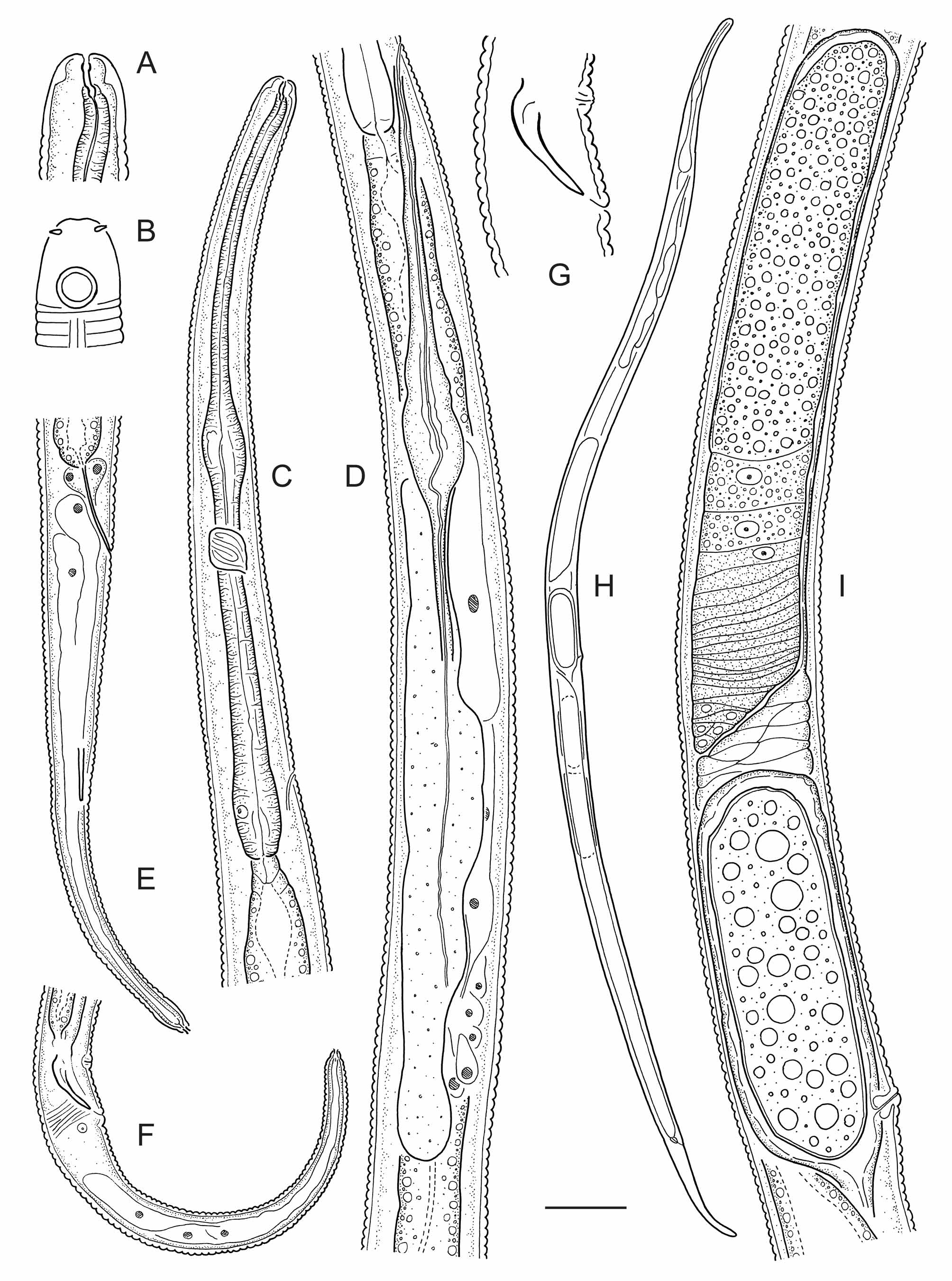
TABLE 1. Morphometrics of Domorganus suecicus sp. n. (all measurements are in µm, except for the ratios a, b, c, c', V and T).
| Body length | 1543 | 1535 (1307–1612) | 1365–1479 |
|---|---|---|---|
| a | 38.6 | 47.3 (38.6–73.2) | 74.0–83.1 |
| b | 8.1 | 8.5 (7.9–9.1) | 8.0–9.1 |
| c | 11.9 | 13.0 (11.9–13.5) | 12.4–13.4 |
| c' | 7.5 | 7.8 (7.2–8.5) | 7.0–7.6 |
| V or T (%) | 52.0 | 53.1 (52.0–54.9) | 50.4–55.7 |
| Body diameter | 40.0 | 35.0 (18.0–40.0) | 16.5–20.0 |
| Pharynx length | 191 | 180 (166–191) | 157–178 |
| Tail length | 129 | 119 (95–129) | 109–110 |
| Anal or cloacal body diameter | 17. | 15.0 (11.5–17.0) | 14.0–16.0 |
| Labial region diameter | 6.0 | 7.0 (6.0–8.0) | 6.5–8.0 |
| Cephalic setae length | 1.5 | 1.5 | 1.5 |
| Amphid position from ant. end | 6.5 | 6.9 (6.5–7.0) | 7.0 |
| Stoma length | 6.0 | 6.3 (6.0–7.0) | 6.0–6.5 |
| Excretory pore from ant. end | 169 | 161 (157–169) | 152–169 |
| EP (%) | 88.3 | 89.6 (85.2–94.8) | 94.7–96.7 |
| Vagina length | 8.0 | 11.5 (8.0–14.0) | - |
| Rectum length | 23.5 | 22.0 (16.5–25.0) | - |
| Spicule length | - | - | 17.0–18.5 |
| Precloacal sensilla from cloaca | - | - | 13.0–14.0 |
No known copyright restrictions apply. See Agosti, D., Egloff, W., 2009. Taxonomic information exchange and copyright: the Plazi approach. BMC Research Notes 2009, 2:53 for further explanation.